. 1999 Aug 15;13(16):2087-97.
doi: 10.1101/gad.13.16.2087.

The cytoplasmic Purkinje onconeural antigen cdr2 down-regulates c-Myc function: implications for neuronal and tumor cell survival

Affiliations

The cytoplasmic Purkinje onconeural antigen cdr2 down-regulates c-Myc function: implications for neuronal and tumor cell survival

H J Okano et al. Genes Dev. .

Abstract

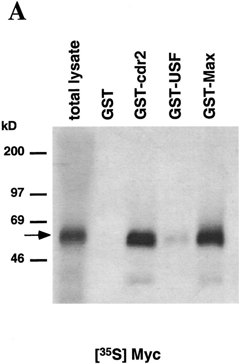
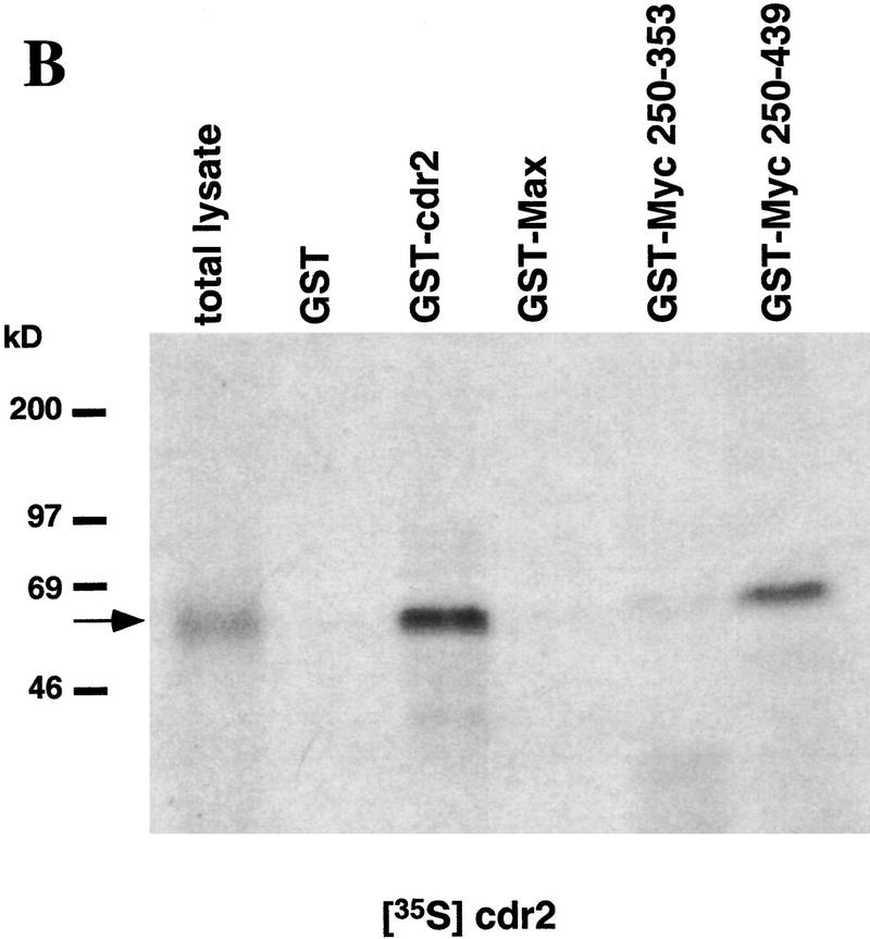
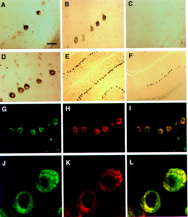
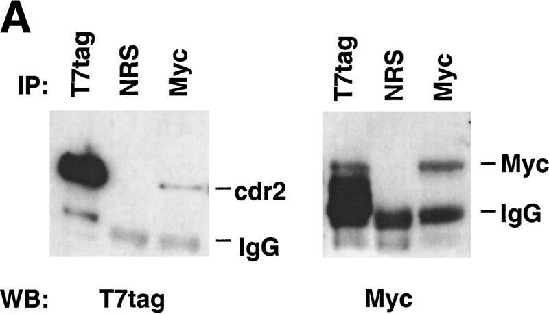
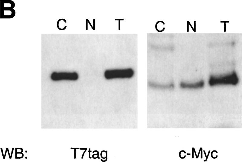
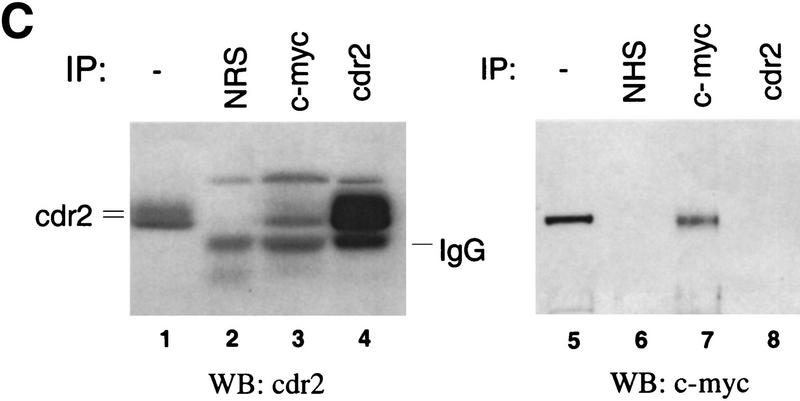
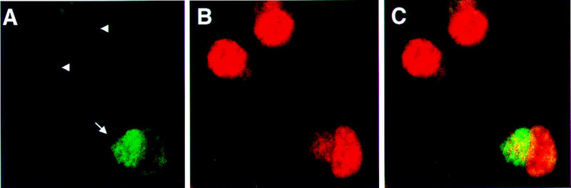
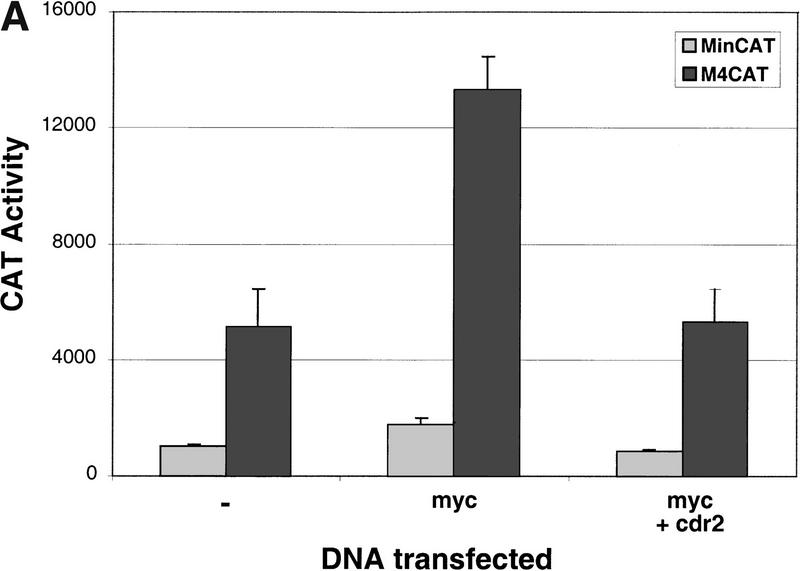
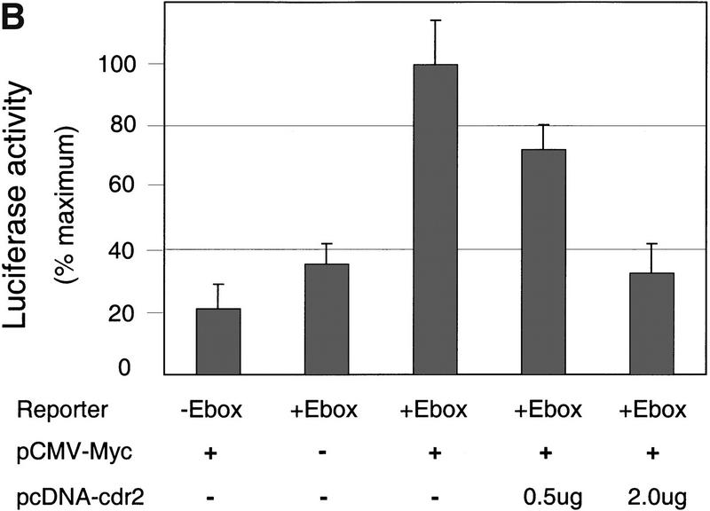
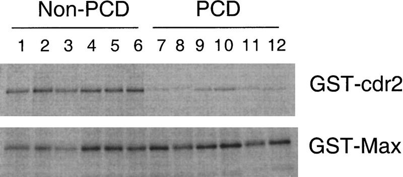
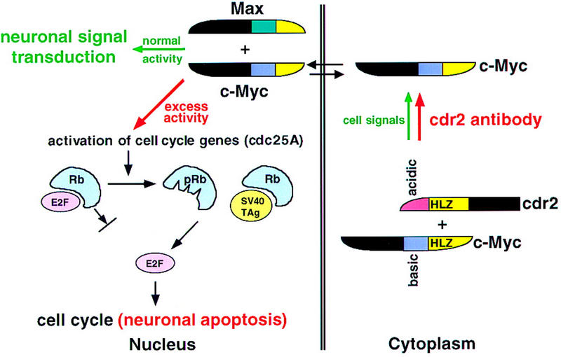
Paraneoplastic cerebellar degeneration (PCD) is a disorder in which breast or ovarian tumors express an onconeural antigen termed cdr2, which normally is expressed in cerebellar Purkinje neurons. This leads to an immune response to cdr2 that is associated with tumor immunity and autoimmune cerebellar degeneration. We have found that cdr2, a cytoplasmic protein harboring a helix-leucine zipper (HLZ) motif, interacts specifically with the HLZ motif of c-Myc. Both proteins colocalize in the cytoplasm of adult cerebellar Purkinje neurons, and coimmunoprecipitate from tumor cell lines and cerebellar extracts. cdr2 down-regulates c-Myc-dependent transcription in cotransfection assays, and redistributes Myc protein in the cytoplasm. Disease antisera from six of six PCD patients specifically blocked the interaction between cdr2 and c-Myc in vitro. These data indicate that cdr2 normally sequesters c-Myc in the neuronal cytoplasm, thereby down-regulating c-Myc activity, and suggest a mechanism whereby inhibition of cdr2 function by autoantibodies in PCD may contribute to Purkinje neuronal death.


Figures

Figure 1
Figure 1
cdr2 and c-Myc interact in vitro. A GST pull-down assay was used to examine the in vitro binding of cdr2 and c-Myc. Immobilized GST fusion proteins were incubated with full–length in vitro translated c-Myc and cdr2. (A) Full–length 35S-labeled c-Myc binds to GST–cdr2 in vitro. c-Myc did not bind to GST alone, and weakly bound to GST–USF, another HLZ protein. GST–Max was used as a positive control. (B) Full–length 35S-labeled cdr2 interacted with a truncated c-Myc fusion protein that contained the HLZ domain (Myc439) and also bound to itself (GST–cdr2). Cdr2 did not bind to GST alone, GST–Max, or Myc353, which lacks the HLZ region. The arrows indicate the size of the full-length 35S-labeled proteins.
Figure 1
Figure 1
cdr2 and c-Myc interact in vitro. A GST pull-down assay was used to examine the in vitro binding of cdr2 and c-Myc. Immobilized GST fusion proteins were incubated with full–length in vitro translated c-Myc and cdr2. (A) Full–length 35S-labeled c-Myc binds to GST–cdr2 in vitro. c-Myc did not bind to GST alone, and weakly bound to GST–USF, another HLZ protein. GST–Max was used as a positive control. (B) Full–length 35S-labeled cdr2 interacted with a truncated c-Myc fusion protein that contained the HLZ domain (Myc439) and also bound to itself (GST–cdr2). Cdr2 did not bind to GST alone, GST–Max, or Myc353, which lacks the HLZ region. The arrows indicate the size of the full-length 35S-labeled proteins.
Figure 2
Figure 2
Immunohistochemical colocalization of cdr2 and c-Myc in the cytoplasm of rat cerebellar Purkinje neurons. (A) A section of adult rat cerebellar cortex stained with anti-c-Myc mouse polyclonal antibody N262 shows strong reactivity with some but not all cerebellar Purkinje neurons. (B) Purkinje neuronal reactivity with a different anti-c-Myc antibody, C-19. (C) Reactivity with C-19 after preincubation with immunizing peptide (2 μg/ml). (D) Reactivity with PCD CSF (anti-cdr2) reveals cdr2 immunoreactivity in the cytoplasm of Purkinje cells. (E), Low power view of cerebellum reacted with PCD CSF, reveals that Purkinje neurons are uniformly labeled. (F) A section obtained near (∼24 μm) the section in (E) stained with anti-c-Myc antibody C-19, illustrating isolated groups of c-Myc expressing Purkinje neurons. (G) Immunofluorescent photograph of rat Purkinje neurons reactive with PCD CSF visualized with Cy-2-conjugated anti-human antibody. (H) Same neurons as (G) visualized with anti-c-Myc antibody and with Cy-3-conjugated anti-rabbit antibody. (I) Fusion of images in G and H showing overlap in the expression of both proteins within individual neurons; some neurons in the molecular layer (presumably stellate neurons) stain lightly with cdr2 antibody but not with c-Myc antibody. (J), Confocal laser image of cdr2 expression in a single 2–μm optical section, imaged with Cy-2-conjugated anti-human antibody. (K) Same 2–μm section as J visualized with anti-c-Myc polyclonal C-19 antibody and with Cy-5-conjugated anti-rabbit antibody. (L) Fusion of images in Jand K showing colocalization of both cdr2 and c-Myc within the Purkinje neuronal cytoplasm. (AD, GIBar, 20 μm; (E,F) bar, 80 μm; (J–L) bar, 3.4 μm.
Figure 3
Figure 3
Cytoplasmic cdr2 coimmunoprecipitates with c-Myc in vitro and in mouse cerebellum. (A) c-Myc coprecipitates with T7-tagged cdr2 in HTC-75 cells. Expression of T7-tagged cdr2 protein was induced by removing doxycyclin from the media. Cell extracts were run on Western blot before (data not shown) or after immunoprecipitation with the indicated antibodies. Blots were then probed with monoclonal antibodies to c-Myc or the T7-tag, as indicated. Similar coimmunoprecipitations were obtained after cotransfection of Rat-1A cells (data not shown). (B) Western blot analysis of cell extracts after induction of cdr2 expression in HTC-75 cells. Total cell lysates (T), cytoplasmic fractions (C), or nuclear fractions (N) were run on Western blots and probed with either cdr2 antibody or c-Myc antibody as indicated. cdr2 expression is restricted to the cytoplasm; c-Myc is expressed in both compartments. (C) In vivo coimmunoprecipitation of cdr2 and c-Myc. (Lanes 1–4) Mouse cerebellar homogenate precipitated with normal rabbit sera (lane 2), anti-c-Myc rabbit polyclonal antibody (lane 3) or anti-cdr2 PCD patient’s CSF (lane 4). Immune complexes were analyzed by Western blot using PCD (anti-cdr2) patient’s serum. (Lane 1) Western blot of the cerebellar lysate. Two cdr2 immunoreactive proteins are indicated, the lower of which may preferentially be coimmunoprecipitated by c-Myc (lane 3). (Lanes 5–8) Homogenates immunoprecipitated as for lanes 1–4; Western blot probed with anti-c-Myc antibody.
Figure 3
Figure 3
Cytoplasmic cdr2 coimmunoprecipitates with c-Myc in vitro and in mouse cerebellum. (A) c-Myc coprecipitates with T7-tagged cdr2 in HTC-75 cells. Expression of T7-tagged cdr2 protein was induced by removing doxycyclin from the media. Cell extracts were run on Western blot before (data not shown) or after immunoprecipitation with the indicated antibodies. Blots were then probed with monoclonal antibodies to c-Myc or the T7-tag, as indicated. Similar coimmunoprecipitations were obtained after cotransfection of Rat-1A cells (data not shown). (B) Western blot analysis of cell extracts after induction of cdr2 expression in HTC-75 cells. Total cell lysates (T), cytoplasmic fractions (C), or nuclear fractions (N) were run on Western blots and probed with either cdr2 antibody or c-Myc antibody as indicated. cdr2 expression is restricted to the cytoplasm; c-Myc is expressed in both compartments. (C) In vivo coimmunoprecipitation of cdr2 and c-Myc. (Lanes 1–4) Mouse cerebellar homogenate precipitated with normal rabbit sera (lane 2), anti-c-Myc rabbit polyclonal antibody (lane 3) or anti-cdr2 PCD patient’s CSF (lane 4). Immune complexes were analyzed by Western blot using PCD (anti-cdr2) patient’s serum. (Lane 1) Western blot of the cerebellar lysate. Two cdr2 immunoreactive proteins are indicated, the lower of which may preferentially be coimmunoprecipitated by c-Myc (lane 3). (Lanes 5–8) Homogenates immunoprecipitated as for lanes 1–4; Western blot probed with anti-c-Myc antibody.
Figure 4
Figure 4
Transfected cdr2 redistributes c-Myc to the cytoplasm of N2A cells where it colocalizes with cdr2 protein. N2A cells were transfected transiently with pcDNA3-cdr2 plasmid, fixed and stained with anti-cdr2 patient CSF (A, green, Cy2) and anti-Myc polyclonal C-19 antibody (B, red, Cy5), and imaged by confocal microscopy (Zeiss). Fusion of images in A and B shows overlap in the cytoplasmic expression of both proteins (C, yellow). Nontransfected cells (arrowheads) show little or no cdr2 staining and nuclear c-Myc expression; transfected cells (arrow) show strong cytoplasmic cdr2 staining and a redistribution of c-Myc reactivity to the cytoplasm, where it colocalizes with cdr2. The same result was seen when a different anti-c-Myc antibody (N262) was used (data not shown).
Figure 5
Figure 5
cdr2 represses c-Myc transcriptional activity. (A) Rat 1A fibroblasts were transfected transiently with the minCAT or M4minCAT reporter plasmids. Cells were cotransfected with either no additional plasmid (−), a c-Myc expressing plasmid (myc; 0.5 μg SpMyc plasmid), or C-Myc together with a cdr2-expressing plasmid (myc + cdr2; 0.5 μg of SpMyc plasmid plus 0.75 μg of pcDNA3-cdr2). Transfection with c-Myc alone resulted in an average 2.6-fold induction of CAT activity. Cotransfection with cdr2 inhibited the c-Myc-induced CAT activity to near baseline levels. No effect of cdr2 alone on M4CAT expression was seen with up to 1 μg of pcDNA 3-cdr2 (data not shown). The results shown represent the average of two independent experiments, each performed in triplicate (n = 6). Error bars indicate 2 s.d. (B) NIH-3T3 cells were transfected transiently with 1.5 μg of reporter plasmid that did not harbor Myc E-box-binding elements (−E-box; pGL3, Promega) or reporter plasmid that did harbor E-box binding elements (+E-box; pM4luc, a pGL3 derivative), or pM4luc in the presence of 1.0 μg of pCMV-Myc and increasing amounts of pcDNA3-cdr2 plasmid as indicated. Cotransfected cdr2 inhibited c-Myc-dependent luciferase activity in a titratable manner. The results shown represent the average of transfections performed in triplicate, and error bars indicate the standard deviation. Relative transfection efficiency was determined in all experiments by measuring activity from a cotransfected reporter plasmid (see Materials and Methods). No effect of cdr2 alone on pM41uc expression was seen with up to 2 μg of pcDNA3-cdr2 (data not shown).
Figure 5
Figure 5
cdr2 represses c-Myc transcriptional activity. (A) Rat 1A fibroblasts were transfected transiently with the minCAT or M4minCAT reporter plasmids. Cells were cotransfected with either no additional plasmid (−), a c-Myc expressing plasmid (myc; 0.5 μg SpMyc plasmid), or C-Myc together with a cdr2-expressing plasmid (myc + cdr2; 0.5 μg of SpMyc plasmid plus 0.75 μg of pcDNA3-cdr2). Transfection with c-Myc alone resulted in an average 2.6-fold induction of CAT activity. Cotransfection with cdr2 inhibited the c-Myc-induced CAT activity to near baseline levels. No effect of cdr2 alone on M4CAT expression was seen with up to 1 μg of pcDNA 3-cdr2 (data not shown). The results shown represent the average of two independent experiments, each performed in triplicate (n = 6). Error bars indicate 2 s.d. (B) NIH-3T3 cells were transfected transiently with 1.5 μg of reporter plasmid that did not harbor Myc E-box-binding elements (−E-box; pGL3, Promega) or reporter plasmid that did harbor E-box binding elements (+E-box; pM4luc, a pGL3 derivative), or pM4luc in the presence of 1.0 μg of pCMV-Myc and increasing amounts of pcDNA3-cdr2 plasmid as indicated. Cotransfected cdr2 inhibited c-Myc-dependent luciferase activity in a titratable manner. The results shown represent the average of transfections performed in triplicate, and error bars indicate the standard deviation. Relative transfection efficiency was determined in all experiments by measuring activity from a cotransfected reporter plasmid (see Materials and Methods). No effect of cdr2 alone on pM41uc expression was seen with up to 2 μg of pcDNA3-cdr2 (data not shown).
Figure 6
Figure 6
Inhibition of cdr2:c-Myc interaction by PCD patients’ sera. The effect of PCD antisera on the cdr2:c-Myc interaction was assessed using GST pull-down assays. GST–cdr2 or GST–Max fusion proteins in solution were immobilized on glutathione–Sepharose beads, preincubated with sera obtained from six different PCD patients or non-PCD control sera, washed, and then mixed with in vitro-translated [35S]-methionine–labeled c-Myc protein. Specifically bound c-Myc protein was assessed by SDS-PAGE and fluorography. Non-PCD sera (from patients with irrelevant paraneoplastic neurologic disorders) do not affect the interactions of c-Myc with either cdr2 or Max. However, PCD patients’ sera inhibited the interaction of c-Myc with cdr2, but not with Max. Quantitation of the ratio of cdr2 to Max protein precipitated in the presence of each serum sample indicated that PCD sera inhibited 35S-labeled c-Myc pull-downs by an average of 5.5-fold (range, 4.9–7.1) relative to the average effect of non-PCD sera.
Figure 7
Figure 7
A model for the role of cdr2 antibodies in PCD pathogenesis. Cdr2 and c-Myc form a complex in Purkinje cell cytoplasm. It is presumed that this interaction is normally regulated by signals that allow c-Myc entry into the nucleus, where it acts to promote transcription and transduce neuronal signaling. In PCD, cdr2 antibodies are proposed to disrupt normal regulation of the cdr2:c-Myc interaction, leading to unregulated entry of c-Myc into the nucleus and aberrant c-Myc-induced gene transcription. c-Myc can drive cell cycle pathways or induce apoptosis in dividing cells (Evan and Littlewood 1998). For example, c-Myc activates cdc25A (Galaktionov et al. 1996), and cdc25 family members act on a number of downstream proteins leading to the phosphorylation of the retinoblastoma gene product (pRb) and the release of the transcription factor E2F, a common final step in S–phase entry. c-Myc also can induce ARF (Zindy et al. 1998), a protein that induces p53-mediated apoptosis (Prives 1998). Excess nuclear c-Myc in neurons is proposed to induce inappropriate cell cycle signaling and Purkinje cell apoptosis in a manner analogous to that observed in Purkinje promoter SV40 TAg transgenic mice (Feddersen et al. 1992, 1995). In these mice, T antigen binds Rb, leading to the release of E2F, S–phase entry, and Purkinje apoptosis. We suggest that cdr2 antibody-mediated disruption of the cdr2:c-Myc interaction leads to aberrant nuclear c-Myc activity, activation of these cell cycle and apoptotic pathways, and contributes to the pathogenesis of PCD.

References

    1. Albert ML, Darnell JC, Bender A, Francisco LM, Bhardwaj N, Darnell RB. Tumor-specific killer cells in paraneoplastic cerebellar degeneration. Nature Med. 1998;4:1321–1324. - PubMed
    1. Ayer DE, Kretzner L, Eisenman RN. Mad: A heterodimeric partner for Max that antagonizes Myc transcriptional activity. Cell. 1993;72:211–222. - PubMed
    1. Bai MK, Costopoulos JS, Christoforidou BP, Papadimitriou CS. Immunohistochemical detection of the c-myc oncogene product in normal, hyperplastic and carcinomatous endometrium. Oncology. 1994;51:314–319. - PubMed
    1. Bao J, Zervos AS. Isolation and characterization of Nmi, a novel partner of Myc proteins. Oncogene. 1996;12:2171–2176. - PubMed
    1. Blackwood EM, Luscher B, Eisenman RN. Myc and Max associate in vivo. Genes & Dev. 1992;6:71–80. - PubMed

Publication types

MeSH terms

LinkOut - more resources