. 1999 Aug 15;13(16):2148-58.
doi: 10.1101/gad.13.16.2148.

The yeast exosome and human PM-Scl are related complexes of 3' --> 5' exonucleases

Affiliations

The yeast exosome and human PM-Scl are related complexes of 3' --> 5' exonucleases

C Allmang et al. Genes Dev. .

Abstract

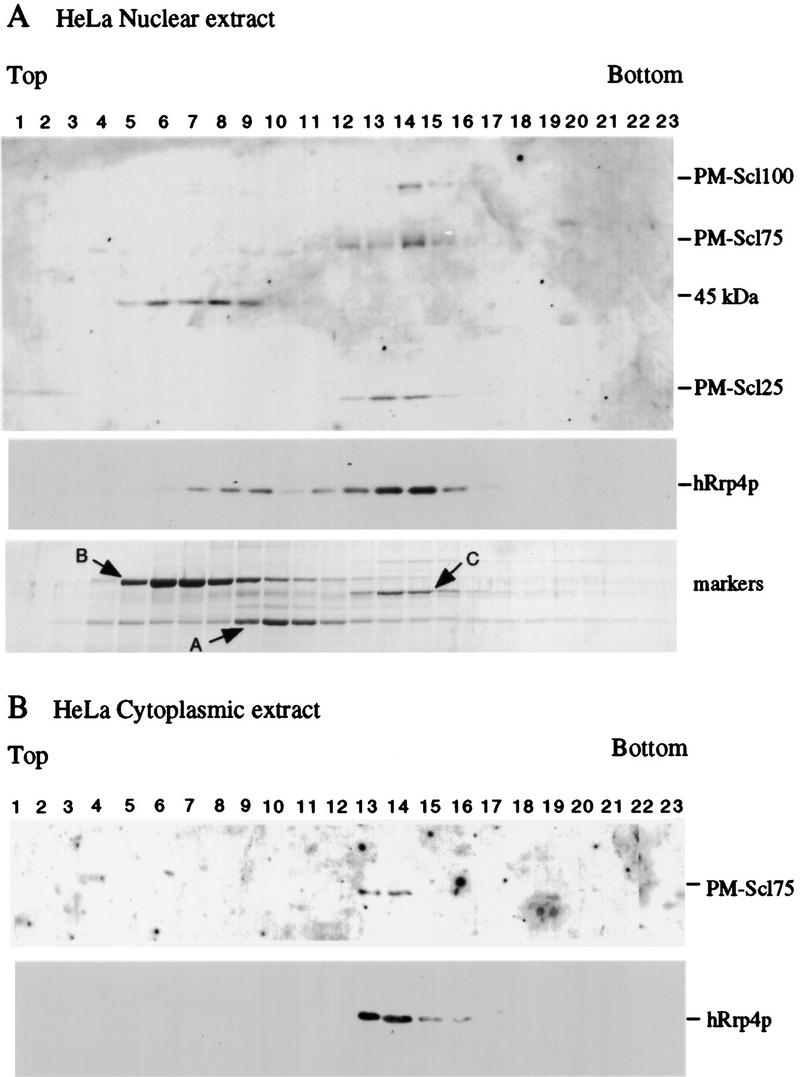
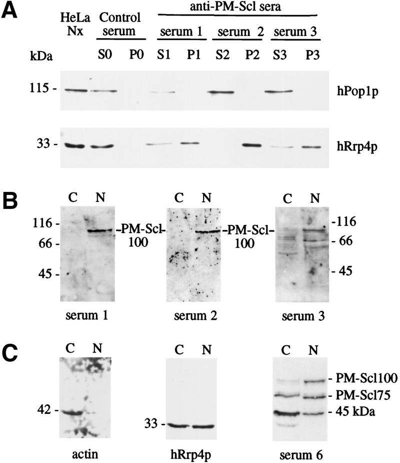
We previously identified a complex of 3' --> 5' exoribonucleases, designated the exosome, that is expected to play a major role in diverse RNA processing and degradation pathways. Further biochemical and genetic analyses have revealed six novel components of the complex. Therefore, the complex contains 11 components, 10 of which are predicted to be 3' --> 5' exoribonucleases on the basis of sequence homology. Human homologs were identified for 9 of the 11 yeast exosome components, three of which complement mutations in the respective yeast genes. Two of the newly identified exosome components are homologous to known components of the PM-Scl particle, a multisubunit complex recognized by autoimmune sera of patients suffering from polymyositis-scleroderma overlap syndrome. We demonstrate that the homolog of the Rrp4p exosome subunit is also a component of the PM-Scl complex, thereby providing compelling evidence that the yeast exosome and human PM-Scl complexes are functionally equivalent. The two complexes are similar in size, and biochemical fractionation and indirect immunofluorescence experiments show that, in both yeast and humans, nuclear and cytoplasmic forms of the complex exist that differ only by the presence of the Rrp6p/PM-Scl100 subunit exclusively in the nuclear complex.


Figures

Figure 1
Figure 1
The newly identified components of the exosome complex are required for viability. Growth curves of GAL-regulated constructs following transfer to glucose medium. Strains were pregrown in permissive, RSG medium and transferred to repressive, glucose medium for the times indicated. Strains were maintained in exponential growth by dilution with prewarmed medium. Cell densities measured by OD600 are shown corrected for dilution. (⋄) Wild type; (○) GAL::rrp45; (□) GAL::rrp46; (♦) GAL:csl4; (█) GAL::rrp40.
Figure 2
Figure 2
The newly identified components of the exosome complex are required for pre-rRNA processing. Northern analysis of processing of the 5.8S and degradation of the 5′ETS region of the pre-rRNA in exosome mutants. RNA was extracted from strains carrying GAL-regulated constructs following transfer from permissive, RSG medium to repressive, glucose medium for the times indicated, or from the mtr3-1 strain following transfer from 25°C to 37°C for the times indicated. RNA was separated on an 6% polyacrylamide gel and hybridized with: (a) oligonucleotide 020 (complementary to the 5.8S/ITS2 boundary), (b) oligonucleotide 017 (hybridizing to the mature 5.8S rRNA), (c) oligonucleotide 033 (hybridizing to the 5′ETS around position +278). (d) oligonucleotide 041 (hybridizing to the 5S rRNA). The position of migration of the pre-rRNA species is indicated. The species labeled 5′ ETS extends from the transcription start site to site A0 (+610). Also shown is a cartoon of the rDNA (not to scale) with the mature rRNA regions as rectangles and the transcribed spacers as lines. The 18S, 5.8S, and 25S rRNAs are cotranscribed, separated by the internal transcribed spacers (ITS1 and ITS2) and flanked by the external transcribed spacers (5′ETS and 3′ETS). The 5S rRNA is independently transcribed in the opposite direction. The mature 5.8S rRNA is synthesized from the 7S pre-rRNA, which is 3′ extended to site C2 in ITS2. The 5′ end of the 5.8S rRNA is generated by processing at sites at B1L and B1S, which lie about 8 nucleotides apart, generating 5.8SL and 5.8SS, respectively. Because this event precedes 3′ processing, the 7S, 6S, and 5.8S + 30 pre-rRNAs all show 5′ heterogeneity, generating, e.g., 6SL and 6SS.
Figure 3
Figure 3
Fractionation of the exosome complex and identification of new components. (A) Proteins associated with IgG–Sepharose via binding to ProtA–Rrp4p were eluted using a gradient of MgCl2 and analyzed by SDS-PAGE. (Lanes 1–20) Material eluted with a 100 mm step gradient of MgCl2 concentration from 100 mm (lane 1) to 2 m (lane 20). (HAc) Proteins eluted by the acid wash. Proteins are visualized by silver staining. The strong bands specifically seen in lane 8 were not observed in other experiments. (B) Purification scheme. A whole-cell extract (CXT) was batch-bound to DEAE–Sepharose FF. Bound material was eluted (E300) with TMN buffer containing 300 mm NaCl/10% glycerol (TMN-300). The eluate, in TMN-100, was passed through a Mono Q column and bound material was eluted stepwise with TMN-150, TMN-200 (E200), TMN-320 (E320), and TMN-500. Material that failed to bind to DEAE–Sepharose FF (FT) was passed through a Mono S column and bound material was eluted with TMN-500 (E500). Each sample was immunoprecipitated on IgG–Sepharose. (C) Proteins present in fractions 1, 2, and 3, obtained as outlined in B, were separated by SDS-PAGE. Approximately twofold more of the material recovered in fractions 2 and 3 was loaded onto the gel, as compared with fraction 1. Proteins positively identified by mass spectroscopy are indicated. Species in brackets were not identified in the preparations shown but are predicted to be present from other analyses. Molecular weight markers are also shown. Proteins are visualized by Coomassie staining.
Figure 4
Figure 4
Rrp4p and Rrp6p differ in their nuclear-cytoplasmic distribution. (A) Strains expressing ProtA–Rrp4p, ProtA–Rrp6p, or ProtA–Nop1p were examined by indirect immunofluorescence using an anti-protein A antibody coupled to Texas Red. Also shown is the position of the DNA, visualized by DAPI staining. The combined image is pseudocolored with DAPI in green and Texas Red in red. For each tagged strain an otherwise isogenic wild-type control strain was also analyzed. The wild-type strain shown (P51) is isogenic with the ProtA–Rrp4p strain (see Table 2). (B) Higher resolution images are shown for the ProtA–Rrp4p and ProtA–Rrp6p to show the punctate staining pattern.
Figure 5
Figure 5
Cosedimentation of hRrp4p and the PM–Scl complex. (A) HeLa cell nuclear extract. (B) HeLa cell cytoplasmic extract. Cell extracts were fractionated by glycerol gradient centrifugation. Samples were analyzed by Western blotting decorated with human autoimmune antisera reactive against PM–Scl100, PM–Scl75, and a previously uncharacterized human protein (PM–Scl25) or with rabbit antiserum raised against recombinant hRrp4p. The serum also cross-reacts with an unrelated 45-kD protein. Also shown is the sedimentation of molecular weight markers on a gradient run in parallel with the nuclear extract. Markers: (A) alcohol deyhdrogenase from yeast (7.4S); (B) bovine serum albumin (4.3S); (C) bovine catalase (11.3S; Siegel and Monty 1966).
Figure 6
Figure 6
Characterization of the PM–Scl complex. (A) Three different human autoimmune sera with specificity for PM–Scl100 were used for immunoprecipitation from a HeLa nuclear extract. The total HeLa nuclear extract, supernatant (S), and pellet (P) fraction are shown. Each lane represents an equivalent quantity of lysate. Western blots were decorated with rabbit sera raised against recombinant hRrp4p or hPop1p, a component of the RNase MRP complex. (B) Western blots of total HeLa nuclear (N) and cytoplasmic (C) extracts decorated with the anti-PM–Scl100 sera used for immunoprecipitation, demonstrating the specificity of the sera. (C) Western blots of total HeLa nuclear (N) and cytoplasmic (C) extracts decorated with rabbit sera raised against recombinant hRrp4p or human actin, or with a human autoimmune serum reactive against both PM–Scl100 and PM–Scl75. Cell equivalent volumes of the nuclear and cytoplasmic fractions were loaded.

References

    1. Alderuccio F, Chan EK, Tan EM. Molecular characterization of an autoantigen of PM–Scl in the polymyositis/scleroderma overlap syndrome: A unique and complete human cDNA encoding an apparent 75-kD acidic protein of the nucleolar complex. J Exp Med. 1991;173:941–952. - PMC - PubMed
    1. Anderson JSJ, Parker RP. The 3′ to 5′ degradation of yeast mRNAs is a general mechanism for mRNA turnover that requires the SKI2 DEVH box protein and 3′ to 5′ exonucleases of the exosome complex. EMBO J. 1998;17:1497–1506. - PMC - PubMed
    1. Baker RE, Harris K, Zhang K. Mutations synthetically lethal with cep1 target S. cerevisiae kinetochore components. Genetics. 1998;149:73–85. - PMC - PubMed
    1. Baudin A, Ozier-Kalogeropoulos O, Denouel A, Lacroute F, Cullin C. A simple and efficient method for direct gene deletion in Saccharomyces cerevisiae. Nucleic Acids Res. 1993;21:3329–3330. - PMC - PubMed
    1. Beltrame M, Tollervey D. Identification and functional analysis of two U3 binding sites on yeast pre-ribosomal RNA. EMBO J. 1992;11:1531–1542. - PMC - PubMed

Publication types

MeSH terms