. 1999 Aug 31;96(18):10039-44.
doi: 10.1073/pnas.96.18.10039.

Purification, molecular cloning, and expression of 2-hydroxyphytanoyl-CoA lyase, a peroxisomal thiamine pyrophosphate-dependent enzyme that catalyzes the carbon-carbon bond cleavage during alpha-oxidation of 3-methyl-branched fatty acids

Affiliations

Purification, molecular cloning, and expression of 2-hydroxyphytanoyl-CoA lyase, a peroxisomal thiamine pyrophosphate-dependent enzyme that catalyzes the carbon-carbon bond cleavage during alpha-oxidation of 3-methyl-branched fatty acids

V Foulon et al. Proc Natl Acad Sci U S A. .

Abstract

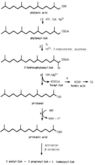
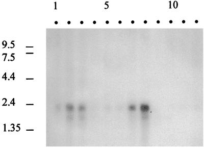
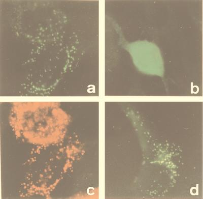
In the third step of the alpha-oxidation of 3-methyl-branched fatty acids such as phytanic acid, a 2-hydroxy-3-methylacyl-CoA is cleaved into formyl-CoA and a 2-methyl-branched fatty aldehyde. The cleavage enzyme was purified from the matrix protein fraction of rat liver peroxisomes and identified as a protein made up of four identical subunits of 63 kDa. Its activity proved to depend on Mg(2+) and thiamine pyrophosphate, a hitherto unrecognized cofactor of alpha-oxidation. Formyl-CoA and 2-methylpentadecanal were identified as reaction products when the purified enzyme was incubated with 2-hydroxy-3-methylhexadecanoyl-CoA as the substrate. Hence the enzyme catalyzes a carbon-carbon cleavage, and we propose calling it 2-hydroxyphytanoyl-CoA lyase. Sequences derived from tryptic peptides of the purified rat protein were used as queries to recover human expressed sequence tags from the databases. The composite cDNA sequence of the human lyase contained an ORF of 1,734 bases that encodes a polypeptide with a calculated molecular mass of 63,732 Da. Recombinant human protein, expressed in mammalian cells, exhibited lyase activity. The lyase displayed homology to a putative Caenorhabditis elegans protein that resembles bacterial oxalyl-CoA decarboxylases. Similarly to the decarboxylases, a thiamine pyrophosphate-binding consensus domain was present in the C-terminal part of the lyase. Although no peroxisome targeting signal, neither 1 nor 2, was apparent, transfection experiments with constructs encoding green fluorescent protein fused to the full-length lyase or its C-terminal pentapeptide indicated that the C terminus of the lyase represents a peroxisome targeting signal 1 variant.


Figures

Figure 1
Figure 1
α-Oxidation pathway. The figure represents the recently revised α-oxidation pathway for phytanic acid, including the results of this paper. The enzymes involved are: 1, phytanoyl-CoA synthetase; 2, phytanoyl-CoA hydroxylase; 3, 2-HPCL; 4, branched fatty aldehyde dehydrogenase; and 5, formyl-CoA hydrolase.
Figure 2
Figure 2
Subcellular distribution of 2-HPCL activity in rat liver. A fresh rat liver homogenate was fractionated by differential centrifugation into a nuclear (N), a heavy mitochondrial (M), a light mitochondrial (L), a microsomal (P), and a cytosolic (S) fraction. The fractions were incubated with 2-hydroxy-3-methyl-[1-14C]hexadecanoyl-CoA, and labeled formate was measured as reaction product. In these experiments, no TPP or Mg2+ was present in the assay mixtures. Marker enzymes and protein were determined in each fraction: catalase (peroxisomal matrix), glutamate dehydrogenase (GDH, mitochondria), glucose-6-phosphatase (G-6-Pase, endoplasmic reticulum), acid phosphatase (Acid Pase, lysosomes), and lactate dehydrogenase (LDH, cytosol). Relative specific activities are presented vs. cumulative percentage of total protein (relative specific activity is defined as the percentage of total recovered activity in a given fraction vs. the percentage of total recovered protein in that fraction). Overall recovery for lyase was 92%; overall recoveries for marker enzymes were between 99 and 113%. Total lyase activity was 112.8 mU (nmol/min) per gram of liver. A similar distribution pattern was found in the presence of TPP and Mg2+ (V.F., K.C., G.P.M., P.P.V.V. and M.C., unpublished data).
Figure 3
Figure 3
Purification of 2-HPCL. The lyase was purified from a peroxisomal matrix protein fraction by successive chromatographic steps as described in Methods. (Upper) The polypeptide pattern of fractions 30–45 from the final gel filtration column as analyzed by SDS/PAGE (100 μl fraction; 10–20% gradient gel; silver staining). The migration of molecular mass (expressed in kDa) markers is represented in lane M. (Lower) The elution of lyase activity, expressed in mU/ml, from the same gel filtration column. Elution of calibration proteins is indicated by arrows: 1, catalase (240 kDa); 2, lactate dehydrogenase (140 kDa).
Figure 4
Figure 4
Substrate dependency of 2-HPCL. Lyase activity was measured at increasing concentrations of 2-hydroxy-3-methylhexadecanoyl-CoA on the partially purified enzyme preparation after phosphocellulose chromatography. The velocity (V, expressed in mU/ml) vs. substrate concentration (S, expressed in μM) curve was linearly transformed according to the method of Lineweaver-Burk as shown (Inset). An apparent Km of 15 μM could be calculated.
Figure 5
Figure 5
cDNA and deduced amino acid sequence of Hs 2-HPCL. The cDNA shown is based on overlapping sequences, determined at least once in both directions of the HindIII/NotI inserts of the Lafmid BA vector containing either T77417 or R17722 and of the 1.8-kb amplicon. Accession numbers of other ESTs overlapping this sequence are AA306411, AA156133, W74012, AA056397, AA298588, F08168, W21374, R58040, H10428, AA150650, AA643224, AI380322, and Z41242. The start codon and the corresponding methionine are indicated in bold. The position of the stop codon is marked by an asterisk. Sequences corresponding to the tryptic peptides, obtained from rat enzyme, are underlined. The C-terminal peroxisome targeting sequence [R]SNM is printed in bold, and the TPP-binding region is underlined twice.
Figure 6
Figure 6
Northern blot analysis of the Hs 2-HPCL. A human 12-lane multiple tissue Northern blot was analyzed with the 1.8-kb amplicon, derived from the Hs 2-HPCL cDNA, as a probe. Lanes: 1, brain; 2, heart; 3, skeletal muscle; 4, colon; 5, thymus; 6, spleen; 7, kidney; 8, liver; 9, small intestine; 10, placenta; 11, lung; 12, peripheral blood leukocytes. RNA size markers are indicated in kb.
Figure 7
Figure 7
Subcellular localization of GFP fusion proteins in cultured mouse fibroblasts. Immortalized mouse PEX5+/− fibroblasts (a, c, and d) and PEX5−/− fibroblasts (b) were transfected with the construct coding for GFP fused to Hs 2-HPCL (a, b, and c) or to the C-terminal pentapeptide of the human lyase (d). After 26 h, cells were analyzed by fluorescence microscopy, either for direct fluorescence of GFP (a, b, and d) or, after immunostaining, for peroxisomal thiolase (c).

References

    1. Steinberg D. In: The Metabolic and Molecular Bases of Inherited Disease. Scriver C R, Beaudet A L, Sly W S, Valle D, editors. New York: McGraw–Hill; 1995. pp. 2351–2369.
    1. Mihalik S J, Rainville A M, Watkins P A. Eur J Biochem. 1995;232:545–551. - PubMed
    1. Croes K, Casteels M, de Hoffmann E, Mannaerts G P, Van Veldhoven P P. Eur J Biochem. 1996;240:674–683. - PubMed
    1. Croes K, Van Veldhoven P P, Mannaerts G P, Casteels M. FEBS Lett. 1997;407:197–200. - PubMed
    1. Croes K, Casteels M, Asselberghs S, Herdewijn P, Mannaerts G P, Van Veldhoven P P. FEBS Lett. 1997;412:643–645. - PubMed

Publication types

MeSH terms

Substances

Associated data