. 1999 Aug 31;96(18):10092-7.
doi: 10.1073/pnas.96.18.10092.

In vivo transcription factor recruitment during thyroid hormone receptor-mediated activation

Affiliations

In vivo transcription factor recruitment during thyroid hormone receptor-mediated activation

M K Kim et al. Proc Natl Acad Sci U S A. .

Abstract

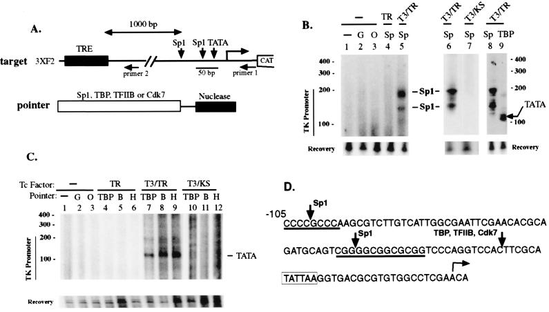
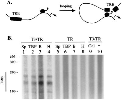
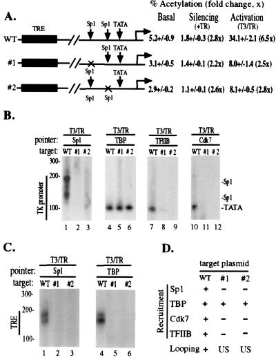
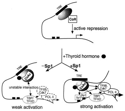
Thyroid hormone receptor (TR) can act as both a transcriptional activator and a silencer. Optimal activation by TR requires synergism with activator(s) bound to the promoter (promoter proximal activator). It is thought that liganded TR either helps to recruit preinitiation complexes (PIC) to the promoter or activates the PIC already recruited. However, the studies analyzing the TR action on the PIC formation were done in vitro and, therefore, it is not clear how relevant they are to the in vivo TR action. For example, in vivo, the TR can act from distances equal to or greater than a kilobase from the promoter, but such distant effect is not reproducible in vitro. In this study, we used the PIN*POINT (ProteIN POsition Identification with Nuclease Tail) assay to define the molecular mechanism of TR action on transcription from the thymidine kinase promoter in the cellular context. We demonstrate that the recruitment of promoter-proximal activator Sp1, and the components of the basal transcription factors such as TBP, TFIIB, and Cdk7, is enhanced with thyroid hormone activation. Our results suggest that DNA forms a loop with TR-mediated activation to accommodate interactions between the liganded TR complex and the complex formed on the promoter. We also show that Sp1 bound to the promoter is essential for the DNA looping and recruitment of basal transcription factors such as TFIIB and Cdk7 but not for recruitment of TBP. On the basis of these findings, we present a model that illustrates the molecular mechanism of TR-mediated activation in vivo.


Figures

Figure 1
Figure 1
Using PIN*POINT to study in vivo transcription factor recruitment during transcriptional silencing and activation of the TRE-regulated reporter gene. (A) Diagram of the target plasmid and pointers. The target DNA, 3XF2 TRE, contains three copies of the high-affinity TRE (30 bp) (ttatTGACCCCAgcTGAGGTCAagttacga, capital letters indicate inverted TREs) from the chicken lysozyme silencer F2 (31) inserted 1-kb 5′ upstream of the herpes simplex virus TK −105/+55 promoter. The TK promoter contains two Sp1 sites at −105/−95 and −56/−45 and the TATA box at −26/−21 position. Horizontal arrows mark the positions of the 3′ antisense primers (primer 1 and primer 2) used for LM-PCR in this study. Primer 1 was derived from the +26/+55 region of the TK promoter and was used to detect cleavage within the TK promoter; primer 2 was derived from a region 51–80 bp downstream from the 3′ end of the 3XF2 TRE and was used to detect cleavage within the TRE region. The pointers were composed of the 25-kDa nuclease domain of FokI restriction endonuclease fused to the carboxyl terminus of Sp1, TBP, TFIIB, or Cdk7 through a flexible glycine linker and were expressed by cotransfecting with the target plasmid. (B) Analysis of recruitment of Sp1 pointer to the TK promoter during the TR-mediated activation and silencing. HeLa cells were cotransfected with target plasmid 3XF2, a TR expression vector (wild type, TR; mutant KS, KS) and one of the pointer expression vectors (G, GAL4 DNA-binding domain; O, Oct-1; Sp, Sp1). Treatment of transfected cells with T3 is indicated. Approximately 24 hr after transfection, the target plasmid was harvested and LM-PCR was performed with primers 1 and MK21 (from the linker). The amplified fragment was detected with Southern blotting followed by hybridization with a radioactively labeled internal probe MK24. The amount of target plasmid was similar in each sample (recovery). The positions of the Sp1 binding sites and the TATA box are indicated. (C) Recruitment of basal transcription factors to the TK promoter during the TR-mediated activation and silencing. Experiments were performed as described for B except that pointers for TBP, TFIIB (B), or TFIIH (H) were used. (D) The cleavage sites for the Sp1 and basal transcription factor pointers as determined against a sequence ladder. Primer extension was performed on the LM-PCR products with radioactively labeled primer 1 and electrophoresed next to a DNA sequence ladder as described previously (29). The cleavage site by each pointer was deduced by subtracting the length of the ligated 29-bp linker (MK21/MK22). By comparing the intensities of the LM-PCR band generated from pointer cleavage with the LM-PCR band generated from the recovered DNA that has been digested with EcoRI, we estimate that approximately 3–4% of the target plasmid was cleaved by the pointers (data not shown).
Figure 2
Figure 2
Detection of DNA looping. (A) Diagram shows how DNA looping will cause the pointers bound to the promoter to cleave within the TRE (see text). (B) To detect cleavage within the TRE region, the DNA samples used in Fig. 1 B and C were analyzed by LM-PCR using primer 2. If cleavage occurs within the 90-bp 3XF2 TRE region, the size of the LM-PCR product with primer 2 is expected to be 110–200 bp.
Figure 3
Figure 3
Role of the Sp1 binding sites in the recruitment of various pointers to the promoter and on DNA looping. (A) The expression levels of the target/reporter plasmids containing a deletion (×) of the distal (#1) or the proximal (#2) Sp1 site were determined by measuring CAT activities (percent acetylation) after transient transfection (32). For these transfections, 5 μg of the reporter gene and 2 μg of the expression vector for TR were used, and cells were treated with or without T3. The extent (fold, in parenthesis) of silencing with TR and activation with T3/TR is in comparison with the basal level of expression (without TR). The data for CAT activities were obtained from three to six independent transfections. (B) Recruitment of Sp1, TBP, TFIIB, and Cdk7 pointers to the target plasmids [wild type (WT), #1, and #2] were analyzed as described in the legend for Fig. 1B. (C) Analysis of DNA looping in target plasmids #1 and #2. Cleavage in the TRE region was analyzed by performing LM-PCR on the samples from B with primer 2. (D) Summary of pointer recruitment and detectable DNA looping for target plasmids WT, #1, and #2. “US” indicates unstable or transient DNA looping.
Figure 4
Figure 4
Model for transcription factor recruitment and DNA looping during the TR-mediated silencing and activation (see text). TR-associated corepressors and coactivators are indicated as CoR and CoAc, respectively. Pol II, RNA polymerase II. To indicate that one protein was essential for the recruitment of another, straight arrows are used. The arrows point to the recruited protein. The transcription initiation site is indicated with a bent arrow.

References

    1. Brent G A, Moore D D, Larsen P R. Annu Rev Physiol. 1991;53:17–35. - PubMed
    1. Glass C K, Holloway J M, Devary O V, Rosenfeld M G. Cell. 1988;54:313–323. - PubMed
    1. Evans R M. Science. 1988;240:889–895. - PMC - PubMed
    1. Samuels H H, Forman B M, Horowitz Z D, Ye Z S. J Clin Invest. 1988;81:957–967. - PMC - PubMed
    1. Weinberger C, Thompson C C, Ong E S, Lebo R, Gruol D J, Evans R M. Nature (London) 1986;324:641–646. - PubMed

MeSH terms

LinkOut - more resources