. 1999 Aug 31;96(18):10194-9.
doi: 10.1073/pnas.96.18.10194.

The hsp70-associating protein Hap46 binds to DNA and stimulates transcription

Affiliations

The hsp70-associating protein Hap46 binds to DNA and stimulates transcription

M Zeiner et al. Proc Natl Acad Sci U S A. .

Abstract

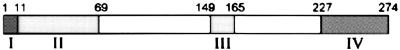
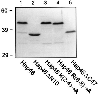
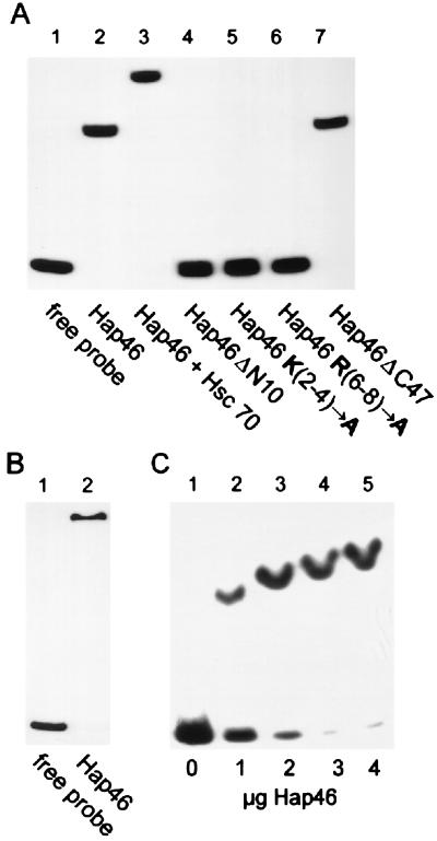
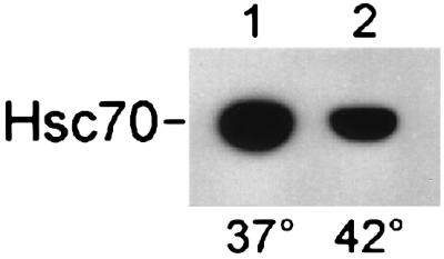
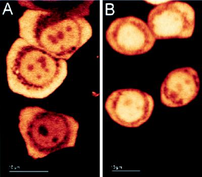
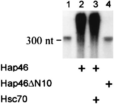
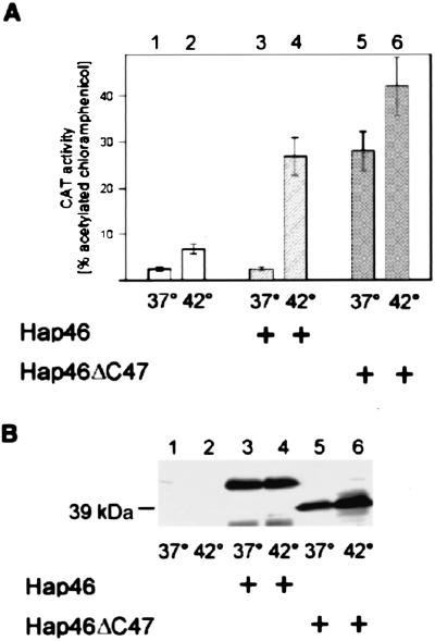
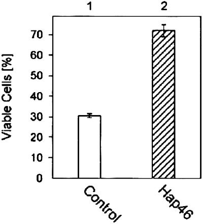
We investigated the ubiquitously expressed hsp70-associating protein Hap46, which is also called RAP46 and is homologous to BAG-1, for activities independent of hsp70 interactions. We observed in vitro binding to various DNA fragments but detected no apparent sequence specificity. Deletion of the amino-terminal decapeptide, which contains two clusters of three basic amino acids each, abolished the DNA-binding ability of Hap46. Similarly, exchange of either of these positively charged clusters for three alanines resulted in loss of DNA binding. Using a fusion of Hap46 and green fluorescent protein, we found preferential accumulation in cell nuclei on heat stress as compared with unstressed cells. The repressive effect of heat shock on overall transcriptional activity in human DU145 carcinoma cells was largely prevented when Hap46 was overexpressed by transfection. Such overproduction of Hap46 also resulted in enhanced expression of specific reporter gene constructs and in increased levels of mRNAs specific for hsp70 and hsp40 after temperature stress. In vitro transcription with nuclear extracts was stimulated greatly by Hap46. Like DNA binding, transcriptional enhancement required amino-terminally located basic amino acid residues but not the carboxyl-terminal portion of Hap46 known to participate in hsp70 interaction. Our results show that Hap46 is a bifunctional protein that can interact with both hsp70s and DNA, employing different portions of the molecule. They also suggest that Hap46 is involved in temperature-sensitive regulation of transcription, acting as a general transcriptional activator.


Figures

Figure 1
Figure 1
The domain structure of Hap46 is shown as a bar diagram. (I) Positively charged sequence MKKKTRRRST required for DNA-binding (residues 1–10). (II) Acidic hexapeptide repeats (residues 11–68). (III) Putative nuclear localization sequence (residues 149–164). (IV) hsp70/hsc70-interacting region (residues 227–274).
Figure 2
Figure 2
Bacterially expressed Hap46. GST fusion proteins of wild-type and mutant Hap46 were expressed in E. coli, purified by affinity chromatography, released by thrombin cleavage, and analyzed by SDS/PAGE. Full-length Hap46 (lane 1), Hap46ΔN10 (lane 2), Hap46 K(2-4)→A (lane 3), Hap46 R(6-8)→A (lane 4), and Hap46ΔC47 (lane 5) were used. Staining was performed with Coomassie blue R250.
Figure 3
Figure 3
Hap46 binding to DNA. (A) A radiolabeled 283-bp fragment of pcDNA3/CAT was either employed as such (lane 1) or used for electrophoretic mobility-shift assays with full-length Hap46 (lane 2), Hap46ΔN10 (lane 4), Hap46 K(2-4)→A (lane 5), Hap46 R(6-8)→A (lane 6), and Hap46ΔC47 (lane 7). In the experiment shown in lane 3, Hap46 was used in combination with hsc70. Analysis was by gel electrophoresis and autoradiography. (B) A 125-bp fragment from phage λ-DNA was subjected to the same assay with full-length Hap46 added (lane 2) or not (lane 1). (C) The 283-bp DNA fragment was used for gel-shift assays with either no protein added (lane 1) or with Hap46 at 1 μg (lane 2), 2 μg (lane 3), 3 μg (lane 4), or 4 μg (lane 5) per 30-μl assay.
Figure 4
Figure 4
Temperature effect on the interaction of Hap46 with hsc70. The GST-Hap46 fusion protein (5 μg) was incubated with hsc70 (1 μg) and GSH-Sepharose for 1 h at 37°C followed by 1 h at either 37° (lane 1) or 42°C (lane 2). Bound protein was analyzed by immunoblotting with hsc70-specific antibody N27F3-4. Quantitation was performed by densitometric scanning and showed that, in the control experiment (lane 1), hsc70 had been retained quantitatively on the matrix.
Figure 5
Figure 5
Cellular distribution of Hap46. HeLa cells were transfected with expression vector encoding the Hap46-GFP fusion protein. Cells were kept overnight at 37°C and analyzed either immediately by confocal laser-scanning microscopy (A) or after exposure to 42°C for 2 h (B). Light areas in photographs denote high fluorescence intensity. Different cells show different levels of transfection. (Bars = 10 μm.)
Figure 6
Figure 6
Hap46 stimulates in vitro transcription. Transcription assays were done with [32P]GTP and nuclear extracts from HeLa cells. Labeled RNA was analyzed by gel electrophoresis and autoradiography. Transcription reactions occurred in the absence (lane 1) or presence of full-length Hap46 (lanes 2 and 3) or mutant Hap46ΔN10 (lane 4) with a CMV promoter carrying DNA template. In the experiment shown in lane 3, Hap46 was used in combination with hsc70. The position of a 300-nt RNA is indicated.
Figure 7
Figure 7
Hap46 stimulates CAT reporter gene expression under heat stress. DU145 cells were transfected with expression vectors encoding CAT alone (lanes 1 and 2), CAT in combination with full-length Hap46 (lanes 3 and 4), or mutant Hap46ΔC47 (lanes 5 and 6). Cells were exposed to heat stress at 42°C for 2 h, as indicated. (A) CAT activity was measured in cell extracts by standard procedures, and turnover is expressed in percentage of acetylated chloramphenicol (18). Data show averages from three independent experiments with error bars indicating maximum deviations. (B) Aliquots of cell extracts, each containing 50 μg of total protein, were analyzed by immunoblotting with Hap46-specific antibody KS-6C8. Endogenous Hap46 contained in untransfected DU145 cells (lanes 1 and 2) did not show up at the short exposure times used here to detect overexpressed wild-type Hap46 and Hap46ΔC47. The position of the 39-kDa marker protein (rabbit muscle aldolase) is indicated.
Figure 8
Figure 8
Hap46 affects cellular mRNA levels. (A) DU145 cells were either transfected with Hap46 cDNA (lanes 3 and 4) or not (lanes 1 and 2) and exposed to heat stress at 42°C for 2 h, as indicated. Metabolic labeling with [33P]phosphoric acid was performed immediately before heat treatment. Poly(A)+ RNA was isolated, run on gels, transferred to filters, and detected by autoradiography on a 16-h exposure. (B) The same filters were submitted to Northern hybridization with 32P-labeled hsp70 cDNA. (C) Northern hybridization with 32P-labeled hsp40 cDNA was performed after stripping the filters (18). RNA was detected by autoradiography. Positions of 28S and 18S rRNAs are shown.
Figure 9
Figure 9
Hap46 protects cells against temperature stress. DU145 cells were transfected with a Hap46 expression vector (column 2) or a control plasmid (column 1). On the day after transfection, cells were exposed to a 2-h heat shock at 42°C and subsequently returned to 37°C. After another 48 h, viable cells were counted. Data show averages from two independent experiments with error bars indicating deviations.

References

    1. Zeiner M, Gehring U. Proc Natl Acad Sci USA. 1995;92:11465–11469. - PMC - PubMed
    1. Takayama S, Sato T, Krajewski S, Kochel K, Irie S, Millan J A, Reed J C. Cell. 1995;80:279–284. - PubMed
    1. Bardelli A, Longati P, Albero D, Goruppi S, Schneider C, Ponzetto C, Comoglio P M. EMBO J. 1996;15:6205–6212. - PMC - PubMed
    1. Takayama S, Bimston D N, Matsuzawa S, Freeman B C, Aime-Sempe C, Xie Z, Morimoto R I, Reed J C. EMBO J. 1997;16:4887–4896. - PMC - PubMed
    1. Zeiner M, Gebauer M, Gehring U. EMBO J. 1997;16:5483–5490. - PMC - PubMed

Publication types

MeSH terms

LinkOut - more resources