Comparative Study
. 1999 Aug 31;96(18):10206-11.
doi: 10.1073/pnas.96.18.10206.

Evidence for a conserved system for iron metabolism in the mitochondria of Saccharomyces cerevisiae

Affiliations
Comparative Study

Evidence for a conserved system for iron metabolism in the mitochondria of Saccharomyces cerevisiae

B Schilke et al. Proc Natl Acad Sci U S A. .

Abstract

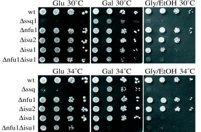
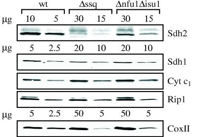
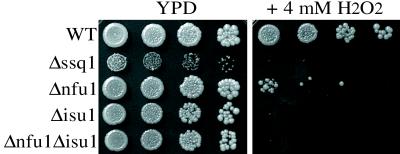
nifU of nitrogen-fixing bacteria is involved in the synthesis of the Fe-S cluster of nitrogenase. In a synthetic lethal screen with the mitochondrial heat shock protein (HSP)70, SSQ1, we identified a gene of Saccharomyces cerevisiae, NFU1, which encodes a protein with sequence identity to the C-terminal domain of NifU. Two other yeast genes were found to encode proteins related to the N-terminal domain of bacterial NifU. They have been designated ISU1 and ISU2. Isu1, Isu2, and Nfu1 are located in the mitochondrial matrix. ISU genes of yeast carry out an essential function, because a Deltaisu1Deltaisu2 strain is inviable. Growth of Deltanfu1Delta isu1 cells is significantly compromised, allowing assessment of the physiological roles of Nfu and Isu proteins. Mitochondria from Deltanfu1Deltaisu1 cells have decreased activity of several respiratory enzymes that contain Fe-S clusters. As a result, Deltanfu1Deltaisu1 cells grow poorly on carbon sources requiring respiration. Deltanfu1Deltaisu1 cells also accumulate abnormally high levels of iron in their mitochondria, similar to Deltassq1 cells, indicating a role for these proteins in iron metabolism. We suggest that NFU1 and ISU1 gene products play a role in iron homeostasis, perhaps in assembly, insertion, and/or repair of mitochondrial Fe-S clusters. The conservation of these protein domains in many organisms suggests that this role has been conserved throughout evolution.


Figures

Figure 1
Figure 1
Tetrad analysis of mutant strains. Growth of spores dissected onto YPD is shown. Plates were replica plated to synthetic media with the indicated amino acids omitted; growth (+) or lack of growth (−). Asci of heterzygous strains Δssq1Δnfu1 at 34°C, (A) Δisu1Δisu2 at 30°C (B), and Δssq1Δisu1 at 34°C (C).
Figure 2
Figure 2
Sequence comparisons of Nfu1, Isu1, and Isu2. (A) Alignment of the amino acid sequence (155–245) of Nfu1 (gp:x69584) from S. cerevisiae (Sc) with that of NifU (pid:g762779) of A. azollae (Aa) (230–311), pid:g2635719 of B. subtilis (Bs) (41–111), pid:g3879150 of C. elegans (Ce), and pid:e1342949 of R. prowazekii (Rp). (B) Alignment of IscU homologs from S. cerevisiae, E. coli (Ec) (gp:AE000339), and Homo sapiens (Hs) (gp:U47101). ∗ indicates conserved cysteine residues. Identical residues between at least two of the sequences are indicated by the black boxes with white letters. Gaps that were inserted during the alignment are denoted by dashes. Alignments were performed by using megalign DNAstar (Madison, WI).
Figure 3
Figure 3
Location of Nfu1, Isu1, or Isu2 in mitochondria. (A) Translocation of preproteins into isolated mitochondria was carried out in the presence of a membrane potential, Δψ, for 5, 15, and 30 min and in the absence of a membrane potential, −Δψ. PK, proteinase K; p, precursor; m, mature. (B) Mitochondria (M) from cells expressing Myc-tagged Nfu1 (Top), Isu1 (Middle), or Isu2 (Bottom) were separated into mitoplast (MP) and intermembrane space (IMS) fractions. An equivalent portion of mitoplasts was disrupted by addition of deoxycholate detergent (MP + DOC). Equivalent amounts of the fractions were either treated (+PK) or not treated (−PK) with proteinase K. Immunoblot analysis was carried out on the fractions with antibodies specific for the MYC epitope, cytochrome b2 (Cyt b2) as a marker for the intermembrane space, or the matrix protein Mge1 after separation by electrophoresis.
Figure 4
Figure 4
Growth of mutant strains. Each of the strains was serially diluted 1:10 and spotted onto complete synthetic media with either glucose (Glu), galactose (Gal), or glycerol/ethanol (Gly/EtOH) as the carbon source. Plates were grown at the indicated temperatures for 4 days.
Figure 5
Figure 5
Levels of mitochondrial proteins. Mitochondrial proteins from each strain were separated by electrophoresis in the amounts indicated, transferred to nitrocellulose, and subjected to immunoblot analyses by using antibodies specific to Sdh1, Sdh2, CoxII, Cyt c1, and Rip1. Densitometry of the autoradiographs was performed by using Ofoto (Light Source Computer Images) and Scan Analysis (Biosoft) software packages.
Figure 6
Figure 6
Sensitivity to H2O2. Cells were serially diluted 1:10 and spotted onto YPD +/− 4 mM H2O2 and incubated at 34°C for 3 days.

References

    1. Tzagoloff A. Mitochondria. New York: Plenum; 1982.
    1. Radisky D C, Kaplan J. J Biol Chem. 1999;274:4481–4484. - PubMed
    1. Radisky D C, Babcock M C, Kaplan J. J Biol Chem. 1999;274:4497–4499. - PubMed
    1. Rötig A, de Lonlay P, Chretien D, Foury F, Koenig M, Sidi D, Munnich A, Rustin P. Nat Genet. 1997;17:215–217. - PubMed
    1. Knight S A B, Sepuri N B V, Pain D, Dancis A. J Biol Chem. 1998;273:18389–18393. - PubMed

Publication types

MeSH terms