Comparative Study
. 1999 Aug 31;96(18):10254-60.
doi: 10.1073/pnas.96.18.10254.

Constructing primate phylogenies from ancient retrovirus sequences

Affiliations
Comparative Study

Constructing primate phylogenies from ancient retrovirus sequences

W E Johnson et al. Proc Natl Acad Sci U S A. .

Abstract

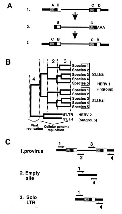
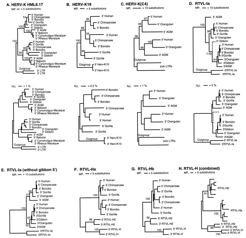
The genomes of modern humans are riddled with thousands of endogenous retroviruses (HERVs), the proviral remnants of ancient viral infections of the primate lineage. Most HERVs are nonfunctional, selectively neutral loci. This fact, coupled with their sheer abundance in primate genomes, makes HERVs ideal for exploitation as phylogenetic markers. Endogenous retroviruses (ERVs) provide phylogenetic information in two ways: (i) by comparison of integration site polymorphism and (ii) by orthologous comparison of evolving, proviral, nucleotide sequence. In this study, trees are constructed with the noncoding long terminal repeats (LTRs) of several ERV loci. Because the two LTRs of an ERV are identical at the time of integration but evolve independently, each ERV locus can provide two estimates of species phylogeny based on molecular evolution of the same ancestral sequence. Moreover, tree topology is highly sensitive to conversion events, allowing for easy detection of sequences involved in recombination as well as correction for such events. Although other animal species are rich in ERV sequences, the specific use of HERVs in this study allows comparison of trees to a well established phylogenetic standard, that of the Old World primates. HERVs, and by extension the ERVs of other species, constitute a unique and plentiful resource for studying the evolutionary history of the Retroviridae and their animal hosts.


Figures

Figure 1
Figure 1
Evolution of ERV LTR sequences. (A) The LTRs of an ERV are identical at the time of integration. Diagram D1, hypothetical provirus with spontaneous LTR mutations at points A, B, C, and D. Diagram D2, a newly transcribed viral RNA genome will contain only the mutations at points B and C. Diagram D3, after reverse transcription and integration, the new provirus has identical LTRs with mutations B and C found in both LTRs. (B) Hypothetical phylogeny based on ERV LTR sequences. The tree contains four distinct types of substitutions. 5′ and 3′ LTRs from the same provirus are expected to cluster separately, because of substitutions that predate all speciation events (“1”). The node joining the two clusters represents the time of integration, when the two LTRs were identical. Substitutions that occur between speciation events (synapomorphies) fall exclusively within one or the other cluster and define the topology of the cluster according to the evolutionary history of the different species (“2”). Assuming that both LTRs have evolved independently and at the same rate, the two clusters will have the same branching pattern, revealing the phylogeny of the input species. Substitutions unique to one species (autoapomorphies) are phylogenetically uninformative (“3”). Branches separating distinct ERV loci, e.g., between the ingroup provirus HERV 1 and the outgroup provirus HERV 2, represent errors accumulated during viral replication (“4”). Thus trees containing sequences from more than one HERV can be rooted at the node joining the two proviruses, because the two loci share a common viral ancestor. Rooting the tree with another ERV is therefore independent of any assumptions about host species phylogeny. (C) PCR amplification of ERV LTRs from genomic DNA. Arrows indicate 5′ → 3′ orientation of PCR primers; thick lines, cellular flanking sequences; thin lines, ERV sequences; boxes, LTR sequences. LTRs and adjacent cellular sequences are amplified by using one flanking sequence-specific primer and one provirus-specific primer (depicted as primers 1 and 2 for the 5′ LTR and primers 3 and 4 for the 3′ LTR). If neither LTR can be detected the presence of the uninterrupted cellular sequence or solo LTR can be determined by using primers 1 and 4. Proviral integration is essentially random, so flanking sequences amplified with each LTR confirm that homologous loci are being compared. Integration also results in a duplication of target sequences (4–6 bp) flanking each provirus, which can be used to confirm that the amplified 5′ LTR and 3′ LTR are from the same provirus.
Figure 2
Figure 2
Phylogenies of seven HERV loci. Maximum parsimony (MP) trees are shown for each locus. Neighbor-joining (NJ) and maximum likelihood (ML) analyses yielded essentially identical results (data not shown). Insertions and deletions were weighted equal to 1 substitution. Tree searches were performed by using the branch-and-bound option [HERV-K(HML6.17), RTVL-Ia, and RTVL-H] or the exhaustive search option [HERV-K18 and HERV-K(C4)]. Final trees were rooted by designating outgroup sequences. Numbers indicate bootstrap values for major nodes (n = 100). AGM, African green monkey. (A) HERV-K(HML6.17) One of three minimum MP trees of length (L) = 258 aligned HERV-K(HML6.17) sequences. Outgroup LTRs are from a provirus related to HERV-K(HML6.17) found in human BAC110P12 (bases 115, 102 to 122, and 217; accession no. U95626) by blast homology search. (B) Single most-parsimonious tree for the HERV-K18 locus (L = 128). The published HERV-K10 LTR sequences were used as an outgroup (31). (C) Single most-parsimonious tree (L = 199) for the HERV-K(C4) sequences. Two HERV-K(C4)-related solo LTRs were included as outgroups. (D) One of eight equally parsimonious trees (L = 393) for the RTVL-Ia locus. Dashed lines indicate the unexpected placement of the gibbon 5′ LTR branch. The outgroup contains RTVL-Ib LTR sequences (14). (E) One of four equally parsimonious trees (L = 372) for the RTVL-Ia locus after excluding the gibbon 5′ LTR. (F) One of seven equally parsimonious trees (L = 145) for RTVL-Ha. (G) One of two equally parsimonious trees (L = 126) for RTVL-Hb. The 5′ LTR from bonobo was not amplified. (H) One of seven equally parsimonious trees containing both the RTVL-Ha and RTVL-Hb loci (L = 200). Published RTVL-H and RTVL-H2 LTRs were designated as outgroups for rooting the final trees in F, G, and H (36, 37).
Figure 3
Figure 3
Identifying conversion of LTR sequences. Open circles represent sites that have retained the ancestral sequence, and filled circles indicate substitutions. Black and white arrows indicate changes discussed in the text. Uninformative sites are not shown. (A) HERV-K18. For clarity, chimpanzee species and humans have been collapsed to a single branch. The boxes highlight 11 sites that cluster the gorilla LTRs separately from their human and chimpanzee counterparts (see Fig. 2B). Although the figure depicts the substitutions as occurring in gorilla, the ancestral sequence (and thus the direction of change) is ambiguous, as indicated by question marks (?) and half-filled circles (◑). (B) RTVL-Ia. Apes are depicted as a single branch. AGM, African green monkey. The pattern of substitutions indicates that a portion of the RTVL-Ia LTR was replaced by conversion with sequences from the 3′ LTR. The pattern between bases 417 and 469 of the gibbon 5′ LTR is identical to the 3′ LTRs (boxed areas).

References

    1. Boeke J D, Stoye J P. In: Retroviruses. Coffin J M, Hughes S H, Varmus H E, editors. Plainview, NY: Cold Spring Harbor Lab. Press; 1997. pp. 343–436. - PubMed
    1. Herniou E, Martin J, Miller K, Cook J, Wilkinson M, Tristem M. J Virol. 1998;72:5955–5966. - PMC - PubMed
    1. Coffin J M. In: RNA Tumor Viruses. Weiss R, Teich N, Varmus H, Coffin J M, editors. Plainview, NY: Cold Spring Harbor Lab. Press; 1982. pp. 1109–1204.
    1. Coffin J M. In: Fundamental Virology. Fields B N, Knipe D M, Howley P M, editors. Philadelphia: Lippincott-Raven; 1996. pp. 763–844.
    1. Benveniste R E, Todaro G J. Nature (London) 1976;261:101–108. - PubMed

Publication types

Associated data

LinkOut - more resources