. 1999 Aug 31;96(18):10349-54.
doi: 10.1073/pnas.96.18.10349.

Correction of the UDP-glucuronosyltransferase gene defect in the gunn rat model of crigler-najjar syndrome type I with a chimeric oligonucleotide

Affiliations

Correction of the UDP-glucuronosyltransferase gene defect in the gunn rat model of crigler-najjar syndrome type I with a chimeric oligonucleotide

B T Kren et al. Proc Natl Acad Sci U S A. .

Abstract

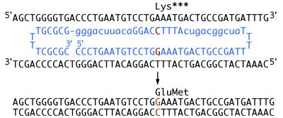
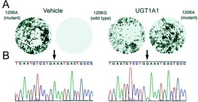
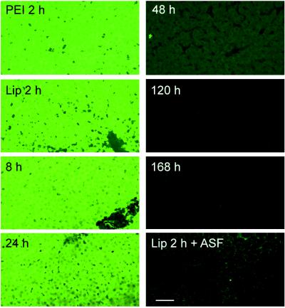
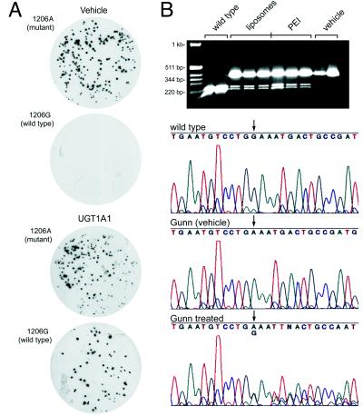
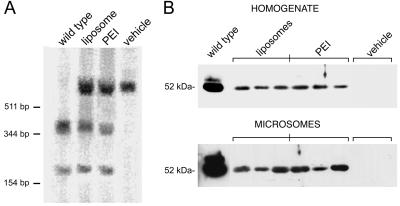
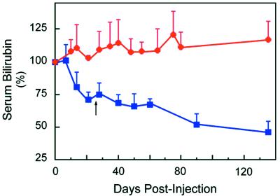
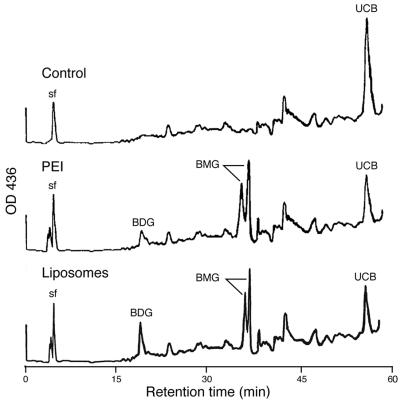
Crigler-Najjar syndrome type I is characterized by unconjugated hyperbilirubinemia resulting from an autosomal recessive inherited deficiency of hepatic UDP-glucuronosyltransferase (UGT) 1A1 activity. The enzyme is essential for glucuronidation and biliary excretion of bilirubin, and its absence can be fatal. The Gunn rat is an excellent animal model of this disease, exhibiting a single guanosine (G) base deletion within the UGT1A1 gene. The defect results in a frameshift and a premature stop codon, absence of enzyme activity, and hyperbilirubinemia. Here, we show permanent correction of the UGT1A1 genetic defect in Gunn rat liver with site-specific replacement of the absent G residue at nucleotide 1206 by using an RNA/DNA oligonucleotide designed to promote endogenous repair of genomic DNA. The chimeric oligonucleotide was either complexed with polyethylenimine or encapsulated in anionic liposomes, administered i.v., and targeted to the hepatocyte via the asialoglycoprotein receptor. G insertion was determined by PCR amplification, colony lift hybridizations, restriction endonuclease digestion, and DNA sequencing, and confirmed by genomic Southern blot analysis. DNA repair was specific, efficient, stable throughout the 6-month observation period, and associated with reduction of serum bilirubin levels. Our results indicate that correction of the UGT1A1 genetic lesion in the Gunn rat restores enzyme expression and bilirubin conjugating activity, with consequent improvement in the metabolic abnormality.


Figures

Figure 1
Figure 1
Targeting strategy to correct the UGT1A1 frameshift mutation in the Gunn rat. The 2′-O-methylated RNA residues of the targeting RNA/DNA ON (blue) are indicated in lowercase and the DNA residues in capital letters. Blocks of 10 modified RNA residues flank both sides of a 9-residue stretch of DNA, which contains the base change required for correction. The ON sequence is complementary to 28 residues of genomic DNA spanning the site of mutation with the exception of a G base (orange) targeted for position 1206. The cell’s endogenous DNA repair process mediates insertion of G at the target site, thereby correcting the frameshift mutation and restoring UGT1A1 activity. The folded double-hairpin structure containing four T residues in each loop, a 5-bp GC clamp, and the modified RNA residues significantly improve resistance to nuclease degradation.
Figure 2
Figure 2
Filter lift hybridizations and sequence analysis of DNA from isolated hepatocytes. (A) Representative hybridization patterns of duplicate filter lifts of the cloned PCR amplicons with either 32P-labeled mutant 1206A or wild-type 1206G 17-mer probes. Hepatocytes were transfected with vehicle (Left) or UGT1A1 ON (Right). (B) The nt sequence of plasmid DNA isolated from clones hybridized with probes to mutant 1206A or wild-type 1206G displaying either A (arrow, Left) or G (arrow, Right), respectively.
Figure 3
Figure 3
In vivo hepatic distribution of fluorescently labeled ONs. Rats received 200 μg of 5′ fluorescein-labeled chimeric ONs encapsulated in anionic liposomes or complexed with PEI by single bolus tail vein injection. At the indicated times, their livers were processed and examined by confocal microscopy. Lip, liposomes. (Bar = 100 μm.)
Figure 4
Figure 4
Filter lift hybridizations, restriction fragment length polymorphism, and sequence analysis of DNA isolated from liver. (A) Hybridization patterns of duplicate filter lifts of the cloned PCR products from liver DNA of Gunn rats 6 months postinjection with vehicle (Upper) or UGT1A1 ONs (Lower). (B) PCR amplicons were subjected to BstNI restriction enzyme digestion and analyzed by agarose gel electrophoresis and ethidium bromide staining (Top). Direct DNA sequencing of the PCR-amplified UGT1A1 gene surrounding the targeted G insertion site at position 1206 (arrow) is shown for wild-type (G, top sequence), vehicle (A, middle), and UGT1A1-treated Gunn rats (A and G, bottom). The size of the DNA standards is indicated at top left.
Figure 5
Figure 5
Southern and Western blot analyses of Gunn rat livers. Liver tissue was harvested for DNA and protein analysis 6 months after in vivo administration of the UGT1A1 ONs as described in Materials and Methods. (A) Southern blot analysis after sequential digestion of genomic DNA with EcoRI and BstNI. (B) Western blot analysis of total liver homogenate and microsomal extracts from the UGT1A1- and vehicle-treated Gunn rats. DNA size markers and protein molecular mass are indicated at left.
Figure 6
Figure 6
Effect of UGT1A1 gene correction on serum bilirubin levels in Gunn rats. Animals were administered UGT1A1 (blue squares) or nonspecific (red circles) ONs complexed to PEI or encapsulated in anionic liposomes as described in Materials and Methods. The dosage was repeated for all groups 30 days after the final injection of the first series (arrow). Each data point is the mean ± SD of 11 animals. There was no significant difference between the PEI and anionic liposome groups. P < 0.001 ≥14 days for UGT1A1 ONs.
Figure 7
Figure 7
HPLC analysis of bile pigments from Gunn rat livers. Bile ducts were cannulated and bile collected for HPLC analysis from both PEI- and liposome-treated Gunn rats as described in Materials and Methods. BMG, bilirubin monoglucuronide; BDG, bilirubin diglucuronide; UCB, unconjugated bilirubin. The HPLC profiles are representative of four animals in each experimental group.

Similar articles

Cited by

References

    1. Roy Chowdhury N, Gross F, Moscioni A D, Kram M, Arias I M, Roy Chowdhury J. J Clin Invest. 1987;79:327–334. - PMC - PubMed
    1. Bosma P J, Seppen J, Goldhoorn B, Bakker C, Oude Elferink R P J, Roy Chowdhury J, Roy Chowdhury N, Jansen P L M. J Biol Chem. 1994;269:17960–17964. - PubMed
    1. Crigler J F, Najjar V A. Pediatrics. 1952;10:169–172. - PubMed
    1. Arias I M. J Clin Invest. 1962;41:2233–2245. - PMC - PubMed
    1. Roy Chowdhury J, Wolkoff A W, Roy Chowdhury N, Arias I M. In: The Metabolic and Molecular Bases of Inherited Disease. Scriver C R, Beaudet A L, Sly W S, Valle D, editors. New York: McGraw-Hill; 1995. pp. 2161–2208.

Publication types