. 1999 Aug 31;96(18):10379-84.
doi: 10.1073/pnas.96.18.10379.

Long-term expression of human coagulation factor VIII and correction of hemophilia A after in vivo retroviral gene transfer in factor VIII-deficient mice

Affiliations

Long-term expression of human coagulation factor VIII and correction of hemophilia A after in vivo retroviral gene transfer in factor VIII-deficient mice

T VandenDriessche et al. Proc Natl Acad Sci U S A. .

Abstract

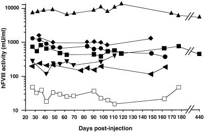
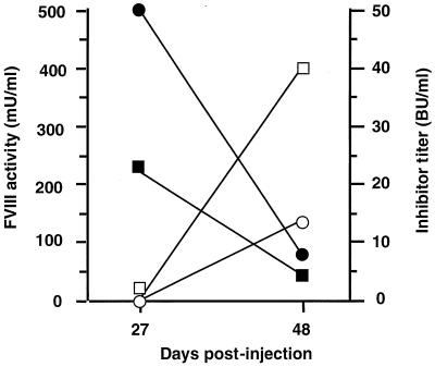
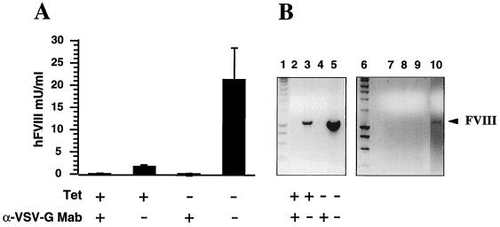
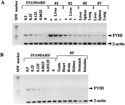
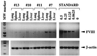
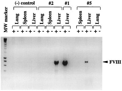
Hemophilia A is caused by a deficiency in coagulation factor VIII (FVIII) and predisposes to spontaneous bleeding that can be life-threatening or lead to chronic disabilities. It is well suited for gene therapy because a moderate increase in plasma FVIII concentration has therapeutic effects. Improved retroviral vectors expressing high levels of human FVIII were pseudotyped with the vesicular stomatitis virus G glycoprotein, were concentrated to high-titers (10(9)-10(10) colony-forming units/ml), and were injected intravenously into newborn, FVIII-deficient mice. High-levels (>/=200 milliunits/ml) of functional human FVIII production could be detected in 6 of the 13 animals, 4 of which expressed physiologic or higher levels (500-12,500 milliunits/ml). Five of the six expressers produced FVIII and survived an otherwise lethal tail-clipping, demonstrating phenotypic correction of the bleeding disorder. FVIII expression was sustained for >14 months. Gene transfer occurred into liver, spleen, and lungs with predominant FVIII mRNA expression in the liver. Six of the seven animals with transient or no detectable human FVIII developed FVIII inhibitors (7-350 Bethesda units/ml). These findings indicate that a genetic disease can be corrected by in vivo gene therapy using retroviral vectors.


Figures

Figure 1
Figure 1
Functional FVIII expression in hemophilia A mice. Neonatal FVIII-deficient littermates were injected with concentrated VSV293-FVIII vector. Shown are mean plasma FVIII activities (n = 3) of recipient mice with prolonged FVIII expression (mice 1–6 in Table 1 are depicted by ▴, ♦, ●, ■, ▾, and ◂, respectively) and negative control mouse injected with PBS (□).The experiment was repeated twice in two independent batches of animals with different vector lots. Mice 1, 4, and 5 (▴, ■ , and ▾) were injected with 1.4 × 10⁸ cfu/ml, and mice 2, 3, and 6, (♦, ●, and ◂) with 0.9 × 10⁸ cfu/ml VSV293-FVIII. Tail-clipping was performed 2 months postinjection except for mice 5 and 6 (3 months). Five of thirteen mice injected with VSV293-FVIII vector (9–13, in Table 1) and all control mice injected with PBS (9 of 9) did not yield detectable FVIII.
Figure 2
Figure 2
Reciprocal correlation between FVIII activity and inhibitor titer in transient FVIII expressers. Functional FVIII expression, (●, ■) and inhibitory antibody titer (○, □) of mice 7 (circles) and 8 (squares) (see Table 1) were shown at different intervals postinjection of VSV293-FVIII.
Figure 3
Figure 3
Modulation of infectivity of VSV293-FVIII. Viral vector producer cells were grown in the presence (+) or absence (−) of tetracycline (Tet) to repress or induce VSV-G expression, respectively, and were incubated with (+) or without (−) neutralizing VSV-G-specific mAb before transduction of COS-7 cells in vitro or injection in FVIII-deficient neonates. FVIII activity was determined in the 24-hr conditioned medium of the transduced COS-7 cells, and the relative transduction efficiencies were determined by FVIII-specific PCR (B, lanes 2–5). PCR also was performed on DNA from liver (lane 7), spleen (lane 8), and lungs (lane 9) of FVIII-deficient recipient mice injected with the same inactivated concentrated vector preparations. Positive control derived from FVIII-containing cells (lane 10) and molecular weight marker corresponding to the Smart Ladder (lanes 1 and 6) (Eurogentec, Liège, Belgium) are included, and FVIII (1.1 kb)-specific fragments are indicated.
Figure 4
Figure 4
Analysis of gene transfer efficiency by quantitative PCR in liver, spleen, and lungs (A) and in testis, heart, brain, kidney, stomach, and intestine (B). The average vector copy number per diploid genomic equivalent was determined by comparison with a serially diluted linear standard (correlation coefficient r2 = 0.98) ranging from 0.5 to 0 (negative control) copies per diploid genomic equivalent. Organs from individual mice were indicated (1, 2, 5, and 7 from Fig. 1 and Table 1). Mice 1 and 2 were not killed, but a liver biopsy was taken instead, and decreasing amounts of target DNA were used as template: 200 ng (a), 100 ng (b), and 50 ng (c). A constant amount of total DNA (200 ng) was maintained in the standard and in the liver samples from mice 1 and 2 by adding spleen DNA from a FVIII-deficient mouse. FVIII (1.1 kb) or control β-actin (0.2 kb)-specific fragments are indicated. The molecular weight (MW) marker corresponds to the Smart Ladder.
Figure 5
Figure 5
Analysis of gene transfer efficiency by quantitative PCR in liver, spleen, and lungs of nonexpressor (10, 11, and 13, from Table 1) and transient expresser (7) mice. Bands corresponding to the amplified FVIII or control β-actin-specific fragments are indicated (1.1 and 0.2 kb, respectively). The same standard was used as described in the legend of Fig. 4. The molecular weight (MW) marker corresponds to the Smart Ladder for the FVIII-PCR and the 1-kb ladder for β-actin.
Figure 6
Figure 6
Expression analysis of FVIII mRNA by RT-PCR in transduced organs derived from either FVIII-expressor mice (1, 2, and 5 from Fig. 1 and Table 1) or a PBS-injected FVIII-deficient mouse as negative (−) control. RNA samples with (+) or without (−) RT as controls were shown to exclude genomic DNA amplification, and FVIII -specific RT-PCR products are indicated (0.7 kb).

Comment in

  • Gene therapy for the hemophilias.
    Kay MA, High K. Kay MA, et al. Proc Natl Acad Sci U S A. 1999 Aug 31;96(18):9973-5. doi: 10.1073/pnas.96.18.9973. Proc Natl Acad Sci U S A. 1999. PMID: 10468539 Free PMC article. Review. No abstract available.

References

    1. Connelly S, Kaleko M. Thromb Haemostasis. 1997;78:31–36. - PubMed
    1. Chuah M K, Collen D, VandenDriessche T. Crit Rev Oncol Hematol. 1998;28:153–171. - PubMed
    1. Koster T, Blann A D, Briet E, Vandenbroucke J P, Roosendael F R. Lancet. 1995;345:152–155. - PubMed
    1. Chuah M K, VandenDriessche T, Morgan R A. Hum Gene Ther. 1995;6:1363–1377. - PubMed
    1. Hoeben R C, van der Jagt R C, Schoute F, van Tilburg N H, Verbeet M P, Briet E, van Ormondt H, van der Eb A J. J Biol Chem. 1990;265:7318–7323. - PubMed

MeSH terms