. 1999 Aug 31;96(18):10412-7.
doi: 10.1073/pnas.96.18.10412.

Bacillus subtilis aconitase is an RNA-binding protein

Affiliations

Bacillus subtilis aconitase is an RNA-binding protein

C Alén et al. Proc Natl Acad Sci U S A. .

Abstract

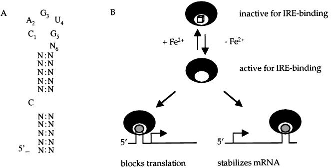
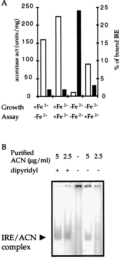
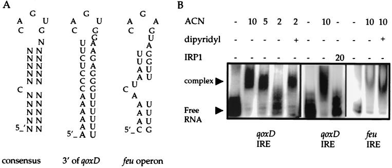
The aconitase protein of Bacillus subtilis was able to bind specifically to sequences resembling the iron response elements (IREs) found in eukaryotic mRNAs. The sequences bound include the rabbit ferritin IRE and IRE-like sequences in the B. subtilis operons that encode the major cytochrome oxidase and an iron uptake system. IRE binding activity was affected by the availability of iron both in vivo and in vitro. In eukaryotic cells, aconitase-like proteins regulate translation and stability of iron metabolism mRNAs in response to iron availability. A mutant strain of B. subtilis that produces an enzymatically inactive aconitase that was still able to bind RNA sporulated 40x more efficiently than did an aconitase null mutant, suggesting that a nonenzymatic activity of aconitase is important for sporulation. The results support the idea that bacterial aconitases, like their eukaryotic homologs, are bifunctional proteins, showing aconitase activity in the presence of iron and RNA binding activity when cells are iron-deprived.


Figures

Figure 1
Figure 1
Iron responsive elements and iron regulatory proteins. (A) Consensus sequence of the eukaryotic IRE (1). (B) IREs are stem-loop structures located in 5′ or 3′ UTRs (shown in gray) of mRNAs encoding proteins involved in iron metabolism. If the IRE is located in the 5′ UTR, binding of IRP (black crescents) inhibits initiation of translation and the level of the protein product decreases. If the IRE is located in the 3′ UTR, binding of IRP protects the mRNA against degradation and the level of the protein increases. In the presence of iron, IRPs are converted to a form that is inactive for RNA binding but may have aconitase enzyme activity. The cube signifies a 4Fe-4S cluster that is essential for aconitase enzyme activity.
Figure 2
Figure 2
Ferritin IRE binding activity of B. subtilis cell extracts. (A) Crude extracts of B. subtilis wild-type and MAB160 (citB∷spc) strains were prepared from cells grown in DS medium without iron supplementation and harvested at the four time points indicated (1–4 hours after the end of exponential growth phase). Aconitase activity of the wild-type crude extracts, after activation with iron, is shown for each time point. The MAB160 extracts had no detectable aconitase activity. (B) A rabbit ferritin IRE-containing, radiolabeled 90-nt RNA (≈20 cps per reaction) was mixed with extracts of wild-type or MAB160 (50 μg of total protein per reaction), was incubated at room temperature for 15 min, and then was subjected to polyacrylamide gel electrophoresis.
Figure 3
Figure 3
Ferritin IRE binding activity of purified aconitase. (A) Binding reactions were performed as described in Fig. 2 using successive 2-fold dilutions of purified B. subtilis aconitase (see Materials and Methods) starting from 5 μg/ml (lanes 2–5). Lanes 7 and 8 show similar binding reactions using wild-type crude extracts. Lanes 9 and 10 show the complex formed by rabbit IRP1 (5 and 2.5 μg/ml respectively) with the same RNA probe. Unbound RNA was run in lanes 1 and 6. (B) The sequence complementary to the ferritin IRE (asIRE) was synthesized in vitro and was used as a target for binding (lanes 1 and 2). In lanes 3 and 4, the IRE probe was tested with or without B. subtilis ACN (5 μg/ml). (C) Ferritin IRE binding activity of purified ACNC517A. Binding reactions were set up with radiolabeled ferritin IRE and either wild-type or ACNC517A protein preparations (see Materials and Methods). The respective protein concentrations are indicated.
Figure 4
Figure 4
Iron-responsiveness of ferritin IRE binding activity. (A) A wild-type strain was grown either in DS medium or in the same medium without addition of FeSO4. Cells were harvested at the start of the stationary phase, and crude extracts were prepared. All samples were assayed for aconitase activity (open bars) and IRE binding activity (filled bars) before and after activation in vitro with Fe2+ and DTT (see Materials and Methods). The resulting binding gels were scanned, and both free and bound IRE were quantified by using the program image quant 1.2 (Molecular Dynamics). (B) Binding reactions of purified aconitase in binding buffer with or without addition of dipyridyl (0.5 mM). In this experiment, the unbound probe ran off the bottom of the gel.
Figure 5
Figure 5
B. subtilis IRE-like structures. (A) Sequences of the consensus eukaryotic IRE and putative IREs located 3′ of the qoxD coding sequence and in-between the feuA and feuB genes of B. subtilis. (B) The sequences shown in A were cloned and transcribed in vitro and were tested for binding to purified B. subtilis aconitase preparations. Similar binding reactions were performed by using a purified preparation of rabbit IRP1. Dipyridyl, to a final concentration of 0.5 mM, was added to the binding reactions as indicated.

References

    1. Hentze M W, Kuhn L C. Proc Natl Acad Sci USA. 1996;93:8175–8182. - PMC - PubMed
    1. Rouault T A, Klausner R D. Trends Biochem Sci. 1996;21:174–178. - PubMed
    1. Aziz N, Munro H N. Proc Natl Acad Sci USA. 1987;84:8478–8482. - PMC - PubMed
    1. Casey J L, Di Jeso B, Rao K, Klausner R D, Harford J B. Proc Natl Acad Sci USA. 1988;85:1787–1791. - PMC - PubMed
    1. Owen D, Kühn L C. EMBO J. 1987;6:1287–1293. - PMC - PubMed

Publication types

MeSH terms

LinkOut - more resources