. 1999 Sep;117(3):540-9.
doi: 10.1046/j.1365-2249.1999.00986.x.

Colony-stimulating factor-1 (CSF-1) expression in the uteroplacental unit of mice with spontaneous and induced pregnancy loss

Affiliations

Colony-stimulating factor-1 (CSF-1) expression in the uteroplacental unit of mice with spontaneous and induced pregnancy loss

M Gorivodsky et al. Clin Exp Immunol. 1999 Sep.

Abstract

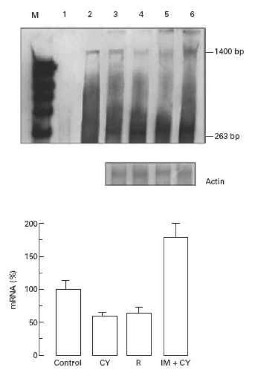
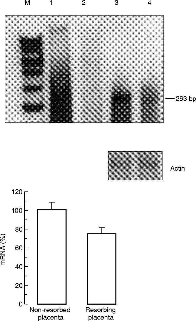
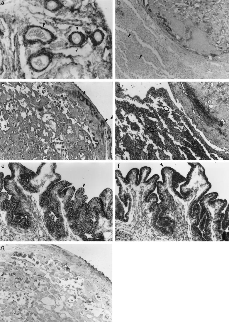
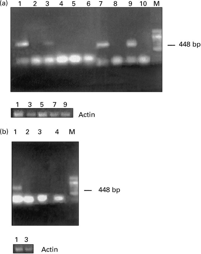
CSF-1 plays an important role in female reproduction and normal embryo development. To understand further CSF-1 function in normal and, especially, in compromised pregnancy, we studied the pattern of its mRNA expression as well as expression of its receptor (c-fms) in the uteroplacental units of mice with induced (cyclophosphamide (CY)-treated) and spontaneous (CBA/J x DBA/2J mating combination) pregnancy loss. RNase protection analysis demonstrated the presence of two forms of CSF-1 mRNA in the uteroplacental unit corresponding to 1400- and 263-bp protective fragments. Densitometric analysis demonstrated that the level of 1400-bp mRNA form was decreased by 40% in the uteroplacental units of mice with CY-induced pregnancy loss compared with the control mice. About 20% decrease in 263-bp protective fragment was registered in resorbing versus non-resorbed placenta of CBA/J females mated to DBA/2J males. As judged by in situ hybridization assay, CSF-1 mRNA transcripts were localized in the uterine epithelium and stroma, while c-fms mRNA was found mainly in the trophoblast. The number of metrial gland cells as well as the number of uterine leucocytes expressing CSF-1 and c-fms mRNAs was substantially lower in the uteroplacental unit of mice with pregnancy loss than in control animals. Maternal immunostimulation, while significantly decreasing the resorption rate in mice with CY-induced pregnancy loss, also strengthened CSF-1 mRNA expression at the fetomaternal interface and resulted in reconstitution in the number of CSF-1+ uterine leucocytes and metrial gland cells. These data suggest a role for uterine CSF-1 in the physiology of normal and compromised pregnancy and demonstrate a possible involvement of CSF-1-associated signalling in mechanisms of placenta and endometrium repair following immunopotentiation.


Figures

Fig. 1
Fig. 1
RNase protection analysis of CSF-1 mRNA in the uteroplacental unit of cyclophosphamide (CY)-treated ICR mice. Top: hybridization with CSF-1-specific anti-sense RNA probe. Lane 1, undigested CSF-1 riboprobe; lane 2, yeast tRNA used as negative control; lane 3, uteroplacental unit from control mice; lane 4, non-resorbed uteroplacental unit from CY-treated mice; lane 5, resorbing uteroplacental unit from CY-treated mice; lane 6, non-resorbed uteroplacental unit from immunopotentiated CY-treated mice. Middle: hybridization with β-actin-specific riboprobe (250 bp protected fragment). Bottom: densitometry analysis of protected fragment corresponding to 1400 bp. CY, Non-resorbed uteroplacental unit from mice treated with CY; R, resorbing uteroplacental unit from mice treated with CY; IM + CY, non-resorbed uteroplacental unit from immunopotentiated CY-treated mice. To evaluate results of RNase protection assay statistically, four samples obtained from different litters were analysed.
Fig. 2
Fig. 2
RNase protection analysis of CSF-1 mRNA in the uteroplacental unit of CBA/J mice. Top: hybridization with CSF-1-specific RNA probe. Lane 1, undigested CSF-1 riboprobe; lane 2, yeast tRNA negative control; lane 3, non-resorbed uteroplacental unit from CBA/J × DBA/2J mouse combination; lane 4, resorbing uteroplacental unit from CBA/J × DBA/2J mouse combination. Middle: hybridization with β-actin-specific riboprobe (250 bp protected fragment). Bottom: densitometry analysis of RNA blots. Five samples were analysed to evaluate results of RNase protection assay statistically.
Fig. 3
Fig. 3
Distribution of CSF-1 and c-fms mRNAs in placentas and uteri of cyclophosphamide (CY)-treated ICR mice. (a) Expression of CSF-1 mRNA in the uterus of CY-treated mice The positive signal is detected in stromal cells and muscle cells of uterine vasculature (b) Leucocytes in necrotic zone in the resorbing uteroplacental unit of CY-treated mice demonstrate no expression of CSF-1 mRNA. (c) c-fms mRNA expression in giant cells in the placenta of CY-treated mice. (d) Strong positive signal for c-fms mRNA in leucocytes infiltrating necrotic zone of the resorbing uteroplacental unit of CY-treated mice. (e,f) CSF-1 mRNA expression in the uterine epithelium of non-immunopotentiated (e) and immunopotentiated (f) CY-treated mice. (g) Negative control—a placental section of CY-treated mice hybridized with non-homologous RNA (× 100).
Fig. 4
Fig. 4
Reverse transcriptase-polymerase chain reaction (RT-PCR) analysis of c-fms mRNA. Top: (a) Lanes 1 and 2, uteroplacental unit of control mice. Cyclophosphamide (CY)-treated mice: lanes 3 and 4, non-resorbed placenta; lanes 5 and 6, resorbing placenta; lanes 7 and 8, placenta of immunopotentiated control mice; lanes 9 and 10, placenta of immunopotentiated and CY-treated mice. Lanes 2, 4, 6, 8 and 10 demonstrate negative controls for RT-PCR in which reverse transcriptase was omitted. (b) CBA/J × DBA/2J mating combination. Lanes 1 and 2, non-resorbed placenta; lanes 3 and 4, resorbing placenta. Lanes 2 and 4 represent negative controls. Bottom: (a,b) RT-PCR with β-actin-specific primers.

References

    1. Wegmann TG. The cytokine basis for cross-talk between the maternal immune and reproductive systems. Curr Opin Immunol. 1990;2:765–9. - PubMed
    1. Toder V, Shomer B. The role of lymphokines in pregnancy. Immunol Allerg Clin N Am. 1990;10:65–69.
    1. Stanley ER, Guilbert LJ, Tushinsky RJ, Bartelmez SH. CSF-1—a mononuclear phagocyte lineage-specific hemopoietic growth factor. J Cell Biochem. 1983;21:151–9. - PubMed
    1. De M, Sanford T, Wood GW. Relationship between macrophage colony-stimulating factor production by uterine epithelial cells and accumulation and distribution of macrophages in the uterus of pregnant mice. J Leukoc Biol. 1993;53:240–8. - PubMed
    1. Bartocci A, Pollard JW, Stanley ER. Regulation of colony stimulating factor 1 during pregnancy. J Exp Med. 1986;164:956–61. - PMC - PubMed

Publication types

Substances