. 1999 Sep;114(3):393-403.
doi: 10.1085/jgp.114.3.393.

Role of calcium permeation in dihydropyridine receptor function. Insights into channel gating and excitation-contraction coupling

Affiliations

Role of calcium permeation in dihydropyridine receptor function. Insights into channel gating and excitation-contraction coupling

R T Dirksen et al. J Gen Physiol. 1999 Sep.

Abstract

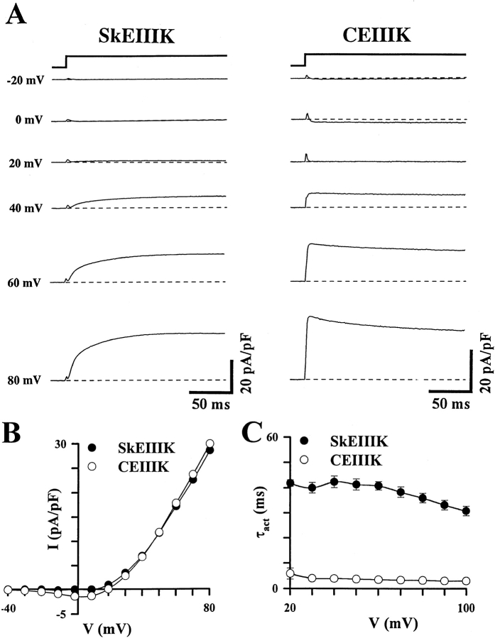
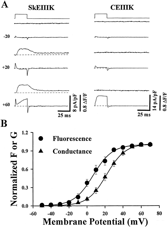
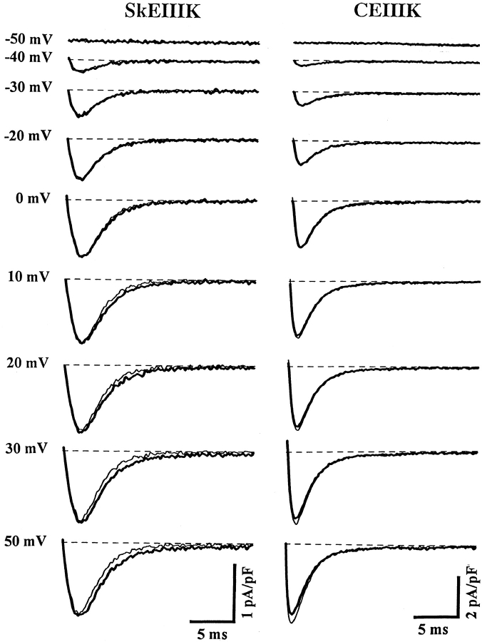
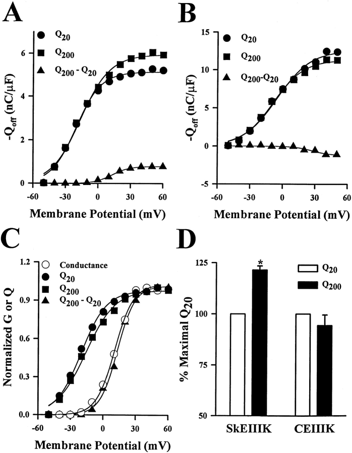
The skeletal and cardiac muscle dihydropyridine receptors (DHPRs) differ with respect to their rates of channel activation and in the means by which they control Ca2+ release from the sarcoplasmic reticulum (Adams, B.A., and K.G. Beam. 1990. FASEB J. 4:2809-2816). We have examined the functional properties of skeletal (SkEIIIK) and cardiac (CEIIIK) DHPRs in which a highly conserved glutamate residue in the pore region of repeat III was mutated to a positively charged lysine residue. Using expression in dysgenic myotubes, we have characterized macroscopic ionic currents, intramembrane gating currents, and intracellular Ca2+ transients attributable to these two mutant DHPRs. CEIIIK supported very small inward Ca2+ currents at a few potentials (from -20 to +20 mV) and large outward cesium currents at potentials greater than +20 mV. SkEIIIK failed to support inward Ca2+ flux at any potential. However, large, slowly activating outward cesium currents were observed at all potentials greater than + 20 mV. The difference in skeletal and cardiac Ca2+ channel activation kinetics was conserved for outward currents through CEIIIK and SkEIIIK, even at very depolarized potentials (at +100 mV; SkEIIIK: tau(act) = 30.7 +/- 1.9 ms, n = 11; CEIIIK: tau(act) = 2.9 +/- 0.5 ms, n = 7). Expression of SkEIIIK in dysgenic myotubes restored both evoked contractions and depolarization-dependent intracellular Ca(2+) transients with parameters of voltage dependence (V(0.5) = 6.5 +/- 3.2 mV and k = 9.3 +/- 0.7 mV, n = 5) similar to those for the wild-type DHPR (Garcia, J., T. Tanabe, and K.G. Beam. 1994. J. Gen. Physiol. 103:125-147). However, CEIIIK-expressing myotubes never contracted and failed to exhibit depolarization-dependent intracellular Ca2+ transients at any potential. Thus, high Ca2+ permeation is required for cardiac-type excitation-contraction coupling reconstituted in dysgenic myotubes, but not skeletal-type. The strong rectification of the EIIIK channels made it possible to obtain measurements of gating currents upon repolarization to -50 mV (Qoff) following either brief (20 ms) or long (200 ms) depolarizing pulses to various test potentials. For SkEIIIK, and not CEIIK, Qoff was significantly (P < 0.001) larger after longer depolarizations to +60 mV (121.4 +/- 2.0%, n = 6). The increase in Qoff for long depolarizations exhibited a voltage dependence similar to that of channel activation. Thus, the increase in Q(off) may reflect a voltage sensor movement required for activation of L-type Ca2+ current and suggests that most DHPRs in skeletal muscle undergo this voltage-dependent transition.


Figures

Scheme S1
Scheme S1
Figure 1
Figure 1
A glutamate-to-lysine mutation in the pore region of repeat III of the skeletal (SkEIIIK) and cardiac (CEIIIK) DHPRs alters channel permeation, but not the rate of channel activation. (A) Representative whole-cell ionic currents obtained from dysgenic myotubes expressing either SkEIIIK (left) or CEIIIK (right). Currents were elicited by 200-ms depolarizations to the indicated potentials after a prepulse protocol (see methods) used to inactivate T-type Ca2+ currents (Adams et al. 1990). Inward currents were not observed at any potential for the SkEIIIK-expressing myotube and minimal inward currents were observed at a few potentials for the CEIIIK-expressing myotube. The dashed lines represent zero current levels. (B) Superimposed current–voltage relationships for the two experiments shown in A. Compared with their wild-type counterparts (Tanabe et al. 1988, 1990), the potential at which SkEIIIK and CEIIIK permit outward current flux is shifted by ∼60 mV in the hyperpolarized direction. (C) The difference between the rate of skeletal and cardiac L-current activation persists in the SkEIIIK and CEIIIK mutants (data from 11 SkEIIIK- and 7 CEIIIK-expressing myotubes). Note that the rate of skeletal L-current activation is nearly independent of voltage and remains slow even at +100 mV.
Figure 2
Figure 2
Skeletal-type EC coupling is unaffected, and cardiac-type EC coupling is abolished, by the repeat III glutamate-to-lysine mutations in the skeletal and cardiac DHPRs, respectively. (A) Simultaneously recorded intracellular Ca2+ transients (top) and ionic currents (bottom) elicited by test pulses to the indicated potentials. The dysgenic myotube expressing SkEIIIK (left) exhibited depolarization-induced intracellular Ca2+ transients at potentials greater than −10 mV, whereas the CEIIIK-expressing myotube (right) failed to exhibit Ca2+ transients at any potential. The dashed lines represent basal fluorescence (top) and zero current (bottom) levels (B) Comparison of the voltage dependence of the normalized peak intracellular Ca2+ transient (•, n = 5) and peak conductance (▴, n = 16) obtained from dysgenic myotubes expressing SkEIIIK L-channels. The smooth solid curves through the data were obtained by Boltzmann fitting of the averaged data points. These fits yielded the following values: F V; V0.5 = +6.9 mV and k = 10.7 mV and GV; V0.5 = +21.8 mV and k = 10.5.
Figure 3
Figure 3
Long depolarizations recruit additional OFF gating current for dysgenic myotubes expressing SkEIIIK, but not CEIIIK. Representative Qoff recordings obtained from dysgenic myotubes expressing either SkEIIIK (left) or CEIIIK (right). Data were obtained in the presence of 2.0 mM Cd2+ and 0.2 mM La3+. Expression of constructs was confirmed in these experiments by the presence of large outward ionic currents (>40 pA/pF at +100 mV) under control conditions (i.e., in the absence of Cd2+/La3+). Qoff gating currents were elicited by repolarization to −50 mV after either brief (20 ms, light traces) or long (200 ms, dark traces) depolarizing pulses to the indicated potentials. The 20-ms pulse was sufficient to fully activate current through CEIIIK channels, but too brief to significantly activate current through SkEIIIK L-channels (see Fig. 1). The 200-ms pulse fully activated both SkEIIIK and CEIIIK channels. The dashed lines represent zero current levels. A significant increase in Qoff was only observed at potentials >0 mV for dysgenic myotubes expressing the SkEIIIK L-channel. A prepulse protocol (see methods) preceded all test depolarizations in order to immobilize gating currents due to sodium and T-type Ca2+ channels.
Figure 4
Figure 4
Long depolarizations increase Qoff in dysgenic expressing SkEIIIK, and not CEIIIK, with a voltage dependence that mirrors that of channel conductance. (A) Dependence of the absolute magnitude of Qoff for SkEIIIK on voltage during test pulses of 20- (Q20, •) or 200-ms (Q200, ▪) duration. The voltage dependence of the additional Qoff gating current recruited by the 200-ms depolarization pulses was determined by subtracting the value of Q20 from the value of Q200 for each voltage (Q200–Q20, ▪). (B) Dependence of the absolute magnitude of Qoff for CEIIIK on test potential of either 20- (Q20, •) or 200-ms (Q200, ▪) duration, together with the voltage dependence of Q200–Q20 (▪). Data in A and B are taken from the same myotubes as those shown in Fig. 3. (C) Comparisons of the voltage dependence of the normalized values for Q20, Q200, Q200–Q20, and channel conductance (G) for SkEIIIK-expressing myotubes. Each data set was fitted by a Boltzmann distribution which yielded values of Q20, V0.5 = −20.3 mV and k = 11.6 mV; Q200: V0.5 = −16.0 mV and k = 13.4 mV; Q200–Q20: V0.5 = +13.0 mV and k = 8.0 mV; and G: V0.5 = +10.6 mV and k = 8.5 mV. Similar results were obtained from a total of six different SkEIIIK-expressing myotubes. (D) Effect of depolarization duration on maximal Qoff recorded from dysgenic myotubes expressing either SkEIIIK (left, n = 6) or CEIIIK (right, n = 3). A significant (P < 0.001) increase in maximal Qoff was observed only for dysgenic myotubes expressing SkEIIIK.
Figure 5
Figure 5
Lack of inward ionic tail currents under the recording conditions used to measure Qoff. Representative data collected from a SkEIIIK-expressing dysgenic myotube obtained under both ionic (10 mM Ca2+, ○) and gating current recording conditions (8 mM Ca2+, 2.0 mM Cd2+, and 0.2 mM La3+, •). Cells were depolarized for 200 ms to +60 mV (to fully activate SkEIIIK channels) and subsequently repolarized to the potentials indicated on the abscissa. Currents during the repolarization period (20 ms) were subsequently integrated and plotted against the repolarization potential. Outward ionic currents through SkEIIIK (seen as positive integrals at potentials >0 mV) were significantly attenuated by Cd2+ and La3+. At potentials hyperpolarized to −20 mV (inset), negative integrals were not largely affected by either the Cd2+ and La3+ solution or by the increase in driving force, indicating a lack of inward ionic currents through SkEIIIK channels. Similar results were obtained from four separate experiments.

References

    1. Adams B.A., Tanabe T., Mikami A., Numa S., Beam K.G. Intramembrane charge movement restored in dysgenic skeletal muscle by injection of dihydropyridine receptor cDNAs. Nature. 1990;346:569–572. - PubMed
    1. Almers W., McCleskey E.W. Non-selective conductance in calcium channels of frog musclecalcium selectivity in a single-file pore. J. Physiol. 1984;353:585–608. - PMC - PubMed
    1. Armstrong C.M., Bezanilla F.M., Horowicz P. Twitches in the presence of ethylene glycol bis(-aminoethyl ether)-N,N′-tetraacetic acid. Biochim. Biophys. Acta. 1972;267:605–608. - PubMed
    1. Beam K.G., Franzini-Armstrong C. Functional and structural approaches to the study of excitation–contraction coupling. Methods Cell Biol. 1997;52:283–306. - PubMed
    1. Beam K.G., Knudson C.M. Calcium currents in embryonic and neonatal mammalian skeletal muscle. J. Gen. Physiol. 1988;91:781–798. - PMC - PubMed

Publication types

MeSH terms