

Prevalence and anatomic characteristics of infarct-like lesions on MR images of middle-aged adults: the atherosclerosis risk in communities study
- PMID: 10472985
- PMCID: PMC7055978
Prevalence and anatomic characteristics of infarct-like lesions on MR images of middle-aged adults: the atherosclerosis risk in communities study
Abstract
Background and purpose: MR imaging has revealed putative evidence of subclinical cerebrovascular disease (CVD) as reflected by white matter signal changes and infarct-like lesions (ILLs). Nonetheless, the prevalence of this condition in the general population has been defined only to a limited extent. We herein report the prevalence and anatomic characteristics of ILLs seen on cranial MR images obtained as part of a population-based study of cardiovascular disease in middle-aged adults. These results are contrasted to those of previous similar studies, particularly those of an elderly population in the Cardiovascular Health Study (CHS).
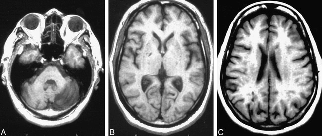
Methods: This Atherosclerosis Risk in Communities (ARIC) cohort consists of a probability sample of community-living persons who were 55 to 72 years old at the time of MR examination. MR imaging of 1890 participants was performed at two ARIC field centers, based on a common protocol. MR studies were evaluated by trained readers at the MR Reading Center using original digital data displayed on a high-resolution workstation. The measures of lesion size, anatomic location, and signal intensity were collected. The definition for an ILL was a non-mass, hyperintense region with an arterial vascular distribution on spin-density and T2-weighted images.
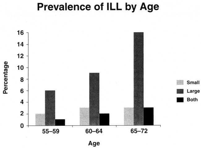
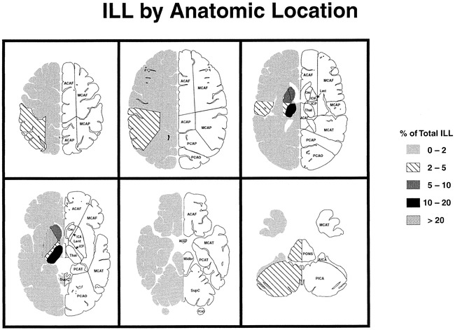
Results: Two hundred ninety participants had ILLs, for an overall prevalence of 15.3%. Eighty-two percent of participants with ILLs had lesions that were 3 mm or larger in maximal dimension, although 87% of these lesions were 20 mm or smaller in maximal dimension. The prevalence of ILLs increased with age, from 7.9% in the 55- to 59-year-old age group to 22.9% in the 65- to 72-year-old age group (P < .001). Lesion prevalence was greater in black (20.7%) than in white persons (10.2% [P < .0001]), but did not differ significantly between male and female participants. The basal ganglia and thalamic region was the most commonly affected anatomic site, accounting for 78.9% of the lesions.
Conclusion: Considering that the prevalence of self-reported stroke or transient ischemic attack in ARIC participants is 1.5%, these results suggest that there is significantly more subclinical than clinical CVD in the general population. Furthermore, the prevalence of this subclinical disease increases with age, and is greater in black persons. ILLs are dominated by "lacunae" in the basal ganglia and thalamus. These results are, in general, similar to those of a comparable study of elderly participants in the CHS, except for a 60% lower prevalence of ILLs in this younger population.
Figures
References
-
- National Center for Health Statistics. Current Estimates from the National Health Interview Survey, 1991.. Hyattsville, MD: National Center for Health Statistics, 1992 - PubMed
-
- Mittelmark MB, Psaty BM, Rautaharju PM, et al. Prevalence of cardiovascular diseases among older adults. The Cardiovascular Health Study. Am J Epidemiol 1993;137(3):311-317 - PubMed
-
- Laffey PA, Peyster RG, Nathan R, Haskin ME, McGinley JA. Computed tomography and aging. Results in a normal elderly population. Neuroradiology 1984;26:273-278 - PubMed
-
- Lindgren A, Roijer A, Rudling O, et al. Cerebral lesions on magnetic resonance imaging, heart disease, and vascular risk factors in subjects without stroke. A population based study. Stroke 1994;25:929-934 - PubMed
Publication types
MeSH terms
Grants and funding
LinkOut - more resources
Full Text Sources
Medical
Molecular Biology Databases