

Review
Anomalous origin of the right vertebral artery: review of the literature and case report of right vertebral artery origin distal to the left subclavian artery
Affiliations
- PMID: 10472992
- PMCID: PMC7055987
Item in Clipboard
Review
Anomalous origin of the right vertebral artery: review of the literature and case report of right vertebral artery origin distal to the left subclavian artery
AJNR Am J Neuroradiol.
1999 Aug.
Abstract
We present the case of a 57-year-old patient who was admitted to the hospital for preoperative cerebral angiography because of an intraspinal mass at the level of C1 and C2. Angiographic examination revealed an abnormal origin of the right vertebral artery, which normally originates from the right subclavian artery. Thus, the right vertebral artery was the last branch of the supraaortic vessels. We also review herein the incidence of the various anomalous origins of the right vertebral artery in the literature and discuss their potential embryologic development and clinical significance.
Figures
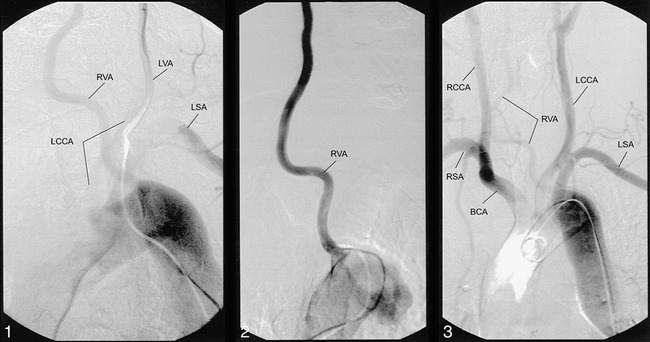
During the selective contrast medium injection into the left vertebral artery, the catheter was pulled back to the aortic arch. A filling of the left common carotid artery and the anomalous right vertebral artery origin is shown. Note.—RVA, right vertebral artery; LVA, left vertebral artery; LSA, left subclavian artery; LCCA, left common carotid artery. fig 2. The selective position of the catheter within the right vertebral artery shows the direct origin of this artery from aortic arch. fig 3. Angiography of the aortic arch confirms the anomalous origin and connection to other supraaortic arteries. Note.—RCCA, right common carotid artery; RSA, right subclavian artery; BCA, brachiocephalic artery.
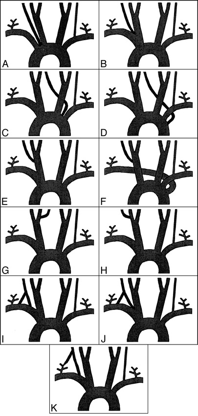
Overview of the different origin variants of the right vertebral artery. A, Right vertebral artery from the aorta between the right subclavian and common carotid artery in cases of missing brachiocephalic arteries. One case has been described in the literature (Lippert H [1985]). B, Right vertebral artery from the aorta on the left between the left common carotid artery and left subclavian artery. One case has been described in the literature (Wasserman BA [1992]). C, Right vertebral artery from the aorta on the left, distal to the left subclavian artery. Seven cases have been described in the literature (Lie TA [1968], Newton TH [1974], Argenson C [1980], Sakamoto H [1980], Schwarzacher SW [1989], Takagi T [1992]), and in one case this was combined with coactation (Stoesslein F [1982]). D, Right vertebral artery directly from the brachiocephalic artery. Three cases have been described in the literature (Daseler EH [1959], Argenson C [1980], Lippert H [1985]). E, Right vertebral artery from the right common carotid artery without A. lusoria. Six cases have been reported in the literature (Daseler EH [1959], Koo K [1966], Lippert H [1985]). F, Right vertebral artery from the right common carotid artery with A. lusoria. Ten cases have been reported in the literature (Iyer AA [1927], Windel WF [1928] Newton TH [1974], Bernard L [1975], Palmer FJ [1977] Tan WS [1979] Wackenheim A [1979]). G. Right vertebral artery from the right internal carotid artery via the hypoglossal artery. One case has been reported in the literature (Keller HL [1973]). H, Right vertebral artery from the right external carotid artery. There have been no known cases of this vertebral artery origin. I, Right vertebral artery with double origin from the right subclavian artery. Eleven cases have been reported (Lie TA [1968], Babin E [1974], Argenson C [1980], Rath G [1984], Harada J [1987], Hashimoto [1987], Nishijima M [1989], Cavdar S [1989], Nogueira TE [1997], Takasato Y [1992]). J, Right vertebral artery with double origin from the right subclavian artery and the brachiocephalic artery. One case has been reported in the literature (Kiss J [1968]). K, Right vertebral artery with double origin from the right subclavian artery and the right thyreocervical trunk. One case has been reported in the literature (Lippert H [1985]).
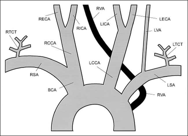
Drawing of the case presented herein
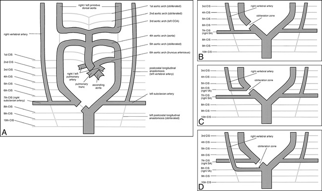
Drawing of the embryologic development of the aorta and brachiocephalic vessels (14-mm embryonic stage). A, Normal development. B, Development of lusorian artery; origin of right vertebral artery also moves to the left. C, Development of lusorian artery, origin of right vertebral artery remains on the right (common carotid artery). D, Development of the right vertebral artery originating from the left and right subclavian artery arising from the right.
References
-
- Adachi B. Das Arteriensystem der Japaner. Kyoto: Kenkyu-Sha; 1928
-
- Newton TH, Mani RL. The vertebral artery. . In: Newton TH, Potts DG, eds. Radiology of the Skull and Brain. St. Louis: Mosby; 1974;1659-1672
-
- Stoesslein F, Porstmann W, Schueler F, Schoepke W. Aberrant vertebral artery originating from the descending aorta: a new congenital steal syndrome in coarctation. . Eur J Radiol 1982;2:157-159 - PubMed
-
- Moore KL. The Developing Human. . 3rd ed. Philadelphia: Saunders; 1982
Publication types
MeSH terms
LinkOut - more resources
Full Text Sources
Miscellaneous