. 1999 Sep;65(9):4099-107.
doi: 10.1128/AEM.65.9.4099-4107.1999.

Molecular and biochemical characterization of two xylanase-encoding genes from Cellulomonas pachnodae

Affiliations

Molecular and biochemical characterization of two xylanase-encoding genes from Cellulomonas pachnodae

A E Cazemier et al. Appl Environ Microbiol. 1999 Sep.

Abstract

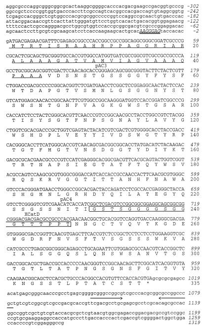
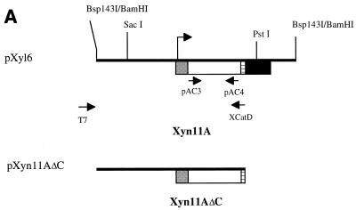
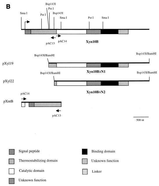
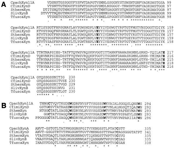
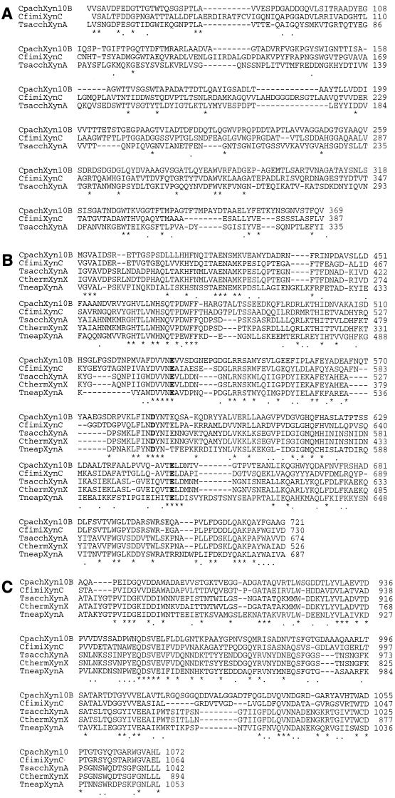
Two xylanase-encoding genes, named xyn11A and xyn10B, were isolated from a genomic library of Cellulomonas pachnodae by expression in Escherichia coli. The deduced polypeptide, Xyn11A, consists of 335 amino acids with a calculated molecular mass of 34,383 Da. Different domains could be identified in the Xyn11A protein on the basis of homology searches. Xyn11A contains a catalytic domain belonging to family 11 glycosyl hydrolases and a C-terminal xylan binding domain, which are separated from the catalytic domain by a typical linker sequence. Binding studies with native Xyn11A and a truncated derivative of Xyn11A, lacking the putative binding domain, confirmed the function of the two domains. The second xylanase, designated Xyn10B, consists of 1,183 amino acids with a calculated molecular mass of 124,136 Da. Xyn10B also appears to be a modular protein, but typical linker sequences that separate the different domains were not identified. It comprises a N-terminal signal peptide followed by a stretch of amino acids that shows homology to thermostabilizing domains. Downstream of the latter domain, a catalytic domain specific for family 10 glycosyl hydrolases was identified. A truncated derivative of Xyn10B bound tightly to Avicel, which was in accordance with the identified cellulose binding domain at the C terminus of Xyn10B on the basis of homology. C. pachnodae, a (hemi)cellulolytic bacterium that was isolated from the hindgut of herbivorous Pachnoda marginata larvae, secretes at least two xylanases in the culture fluid. Although both Xyn11A and Xyn10B had the highest homology to xylanases from Cellulomonas fimi, distinct differences in the molecular organizations of the xylanases from the two Cellulomonas species were identified.


Figures

FIG. 1
FIG. 1
Nucleotide sequence of xyn11A and its flanking regions and deduced amino acid sequence. The putative Shine-Dalgarno-type ribosome binding site is indicated in capital italics and is double underlined. The positions of the primers pAC3, pAC4, and XCatD are indicated. The amino acids underlined at the N terminus are the deduced signal peptide. The linker sequence between the family 11 catalytic domain and the XBD is boxed. The translational stop codon is indicated by an asterisk (∗). A palindrome is indicated by arrows.
FIG. 2
FIG. 2
(A) Schematic representation of the Bsp143I fragment containing Xyn11A, which was ligated into the BamHI site of the multiple cloning site of the pBK-CMV phagemid vector. The translational start codon is indicated by an arrow. (B) Schematic representation of Xyn10B and its derivatives Xyn10BΔN1 and Xyn10BΔN2 encoded by Bsp143I fragments in pXyl19 and pXyl22, respectively. The translational start codon is indicated by an arrow. The N-terminal fragment of xyn10B was obtained by cloning of an inverse PCR product, yielding pXntB.
FIG. 2
FIG. 2
(A) Schematic representation of the Bsp143I fragment containing Xyn11A, which was ligated into the BamHI site of the multiple cloning site of the pBK-CMV phagemid vector. The translational start codon is indicated by an arrow. (B) Schematic representation of Xyn10B and its derivatives Xyn10BΔN1 and Xyn10BΔN2 encoded by Bsp143I fragments in pXyl19 and pXyl22, respectively. The translational start codon is indicated by an arrow. The N-terminal fragment of xyn10B was obtained by cloning of an inverse PCR product, yielding pXntB.
FIG. 3
FIG. 3
Alignment of the amino acid sequences of family 11 catalytic domains (A) and XBDs (B) of Xyn11A of C. pachnodae (this study), XynD of C. fimi (accession no. P54865), a xylanase of S. thermoviolaceus (accession no. D85897), XynB of Streptomyces lividans (accession no. P26515), and a xylanase of Thermomonospora fusca (accession no. U01242). Numbering of the amino acids starts at the N termini of the proteins. Conserved and identical amino acids are indicated by asterisks (∗) and points (.), respectively. Highly conserved amino acid residues are shown in boldface letters. Gaps are indicated by dashes.
FIG. 4
FIG. 4
Alignment of the amino acid sequences of thermostabilizing domains (A), family 10 catalytic domains (B), and CBDs (C) of Xyn10B of C. pachnodae (this study), XynC of C. fimi (accession no. Z50866), XynA of T. saccharolyticum (accession no. P36917), XynX of Clostridium thermocellum (accession no. P38535), and a xylanase of Thermotoga neapolitana (accession no. Q60042). Numbering starts at the N termini of the proteins. Conserved and identical amino acids are indicated by asterisks (∗) and points (.), respectively. Highly conserved glutamic acid residues are shown in boldface letters. Gaps are indicated by dashes.
FIG. 5
FIG. 5
Influence of the pH (A) and temperature (B) on the activity of partially purified recombinant Xyn11A. For the pH profile, the enzyme activity was measured at 50°C in 100 mM phosphate-citrate buffer adjusted to the correct pH. For the temperature profile, enzyme activity was measured in 100 mM phosphate-citrate buffer (pH 6.0) at different temperatures. Values are the means of results of duplicate experiments; separate values do not differ more than 10%.
FIG. 6
FIG. 6
Zymogram of xylanases found in C. pachnodae culture fluids and of recombinant Xyn11A. Lanes: 1, supernatant xylan culture; 2, supernatant beech litter culture; 3, CFE of E. coli harboring pXyl6. Samples contained 0.5 to 1 mU of xylanase activity. Position of molecular mass markers are indicated by horizontal lines and are expressed in kilodaltons.

Similar articles

Cited by

References

    1. Bayon C, Mathelin J. Carbohydrate fermentation and by-product adsorption studied with labelled cellulose in Oryctes nasicornis larvae (Coleoptera: Scrarabaeidae) J Insect Physiol. 1980;26:833–840.
    1. Bhalerao J, Patki A H, Bhave M, Khurana I, Deobagkar D N. Molecular cloning and expression of a xylanase gene from Cellulomonas sp. into Escherichia coli. Appl Microbiol Biotechnol. 1990;34:71–76.
    1. Black G W, Hazlewood G P, Millward-Sadler S J, Laurie J I, Gilbert H J. A modular xylanase containing a novel non-catalytic xylan-specific binding domain. Biochem J. 1995;307:191–195. - PMC - PubMed
    1. Cazemier A E, Op den Camp H J M, Hackstein J H P, Vogels G D. Fibre digestion in arthropods. Comp Biochem Physiol. 1997;118A:101–109.
    1. Cazemier, A. E., H. J. M. Op den Camp, J. C. Verdoes, J. H. P. Hackstein, and G. D. Vogels. Cellulomonas pachnodae sp. nov., a member of the (hemi)cellulolytic hindgut flora of larvae of the scarab beetle Pachnoda marginata. Submitted for publication. - PubMed

Publication types

Associated data

LinkOut - more resources