. 1999 Sep;65(9):4126-33.
doi: 10.1128/AEM.65.9.4126-4133.1999.

Identification of nitrite-oxidizing bacteria with monoclonal antibodies recognizing the nitrite oxidoreductase

Affiliations

Identification of nitrite-oxidizing bacteria with monoclonal antibodies recognizing the nitrite oxidoreductase

S Bartosch et al. Appl Environ Microbiol. 1999 Sep.

Abstract

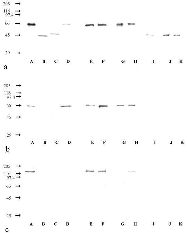
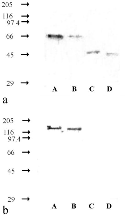
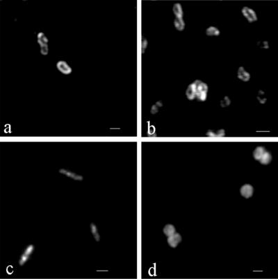
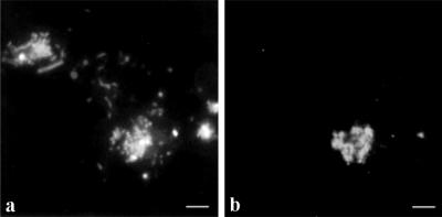
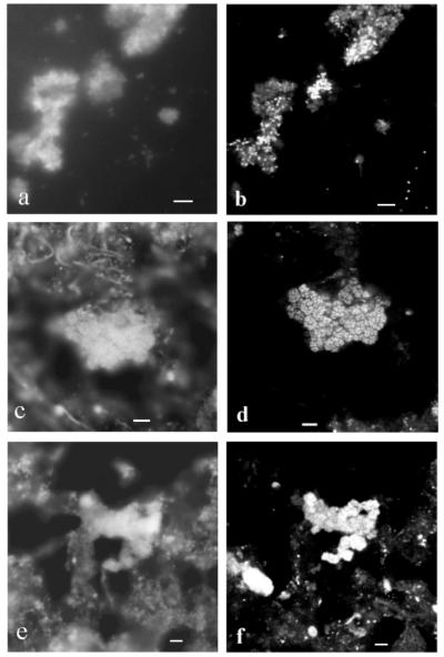
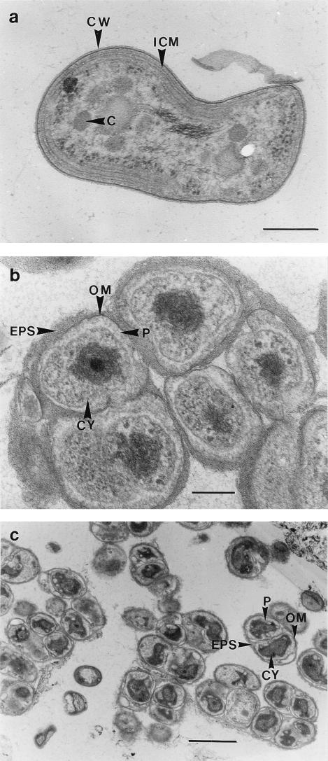
Immunoblot analyses performed with three monoclonal antibodies (MAbs) that recognized the nitrite oxidoreductase (NOR) of the genus Nitrobacter were used for taxonomic investigations of nitrite oxidizers. We found that these MAbs were able to detect the nitrite-oxidizing systems (NOS) of the genera Nitrospira, Nitrococcus, and Nitrospina. The MAb designated Hyb 153-2, which recognized the alpha subunit of the NOR (alpha-NOR), was specific for species belonging to the genus Nitrobacter. In contrast, Hyb 153-3, which recognized the beta-NOR, reacted with nitrite oxidizers of the four genera. Hyb 153-1, which also recognized the beta-NOR, bound to members of the genera Nitrobacter and Nitrococcus. The molecular masses of the beta-NOR of the genus Nitrobacter and the beta subunit of the NOS (beta-NOS) of the genus Nitrococcus were identical (65 kDa). In contrast, the molecular masses of the beta-NOS of the genera Nitrospina and Nitrospira were different (48 and 46 kDa). When the genus-specific reactions of the MAbs were correlated with 16S rRNA sequences, they reflected the phylogenetic relationships among the nitrite oxidizers. The specific reactions of the MAbs allowed us to classify novel isolates and nitrite oxidizers in enrichment cultures at the genus level. In ecological studies the immunoblot analyses demonstrated that Nitrobacter or Nitrospira cells could be enriched from activated sludge by using various substrate concentrations. Fluorescence in situ hybridization and electron microscopic analyses confirmed these results. Permeated cells of pure cultures of members of the four genera were suitable for immunofluorescence labeling; these cells exhibited fluorescence signals that were consistent with the location of the NOS.


Figures

FIG. 1
FIG. 1
Immunoblots of different nitrite-oxidizing bacteria. (a) Hyb 153-3, which recognized the β-NOR. (b) Hyb 153-1, which recognized the β-NOR. (c) Hyb 153-2, which recognized the α-NOR. The values on the left are molecular masses (in kilodaltons). Lane A, Nitrobacter hamburgensis X14; lane B, Nitrospira moscoviensis M-1; lane C, Nitrospina gracilis 3/211; lane D, Nitrococcus mobilis 231; lane E, Nitrobacter alkalicus AN 1; lane F, Nitrobacter alkalicus AN 4; lane G, strain BS 5/6; lane H, strain BS 5/13; lane I, Nitrospira marina 295; lane J, strain Ns (42°C); lane K, strain Ns (47°C). All cells were disrupted by sonication and were added to the gel at protein concentrations of 0.25 to 1 mg ml−1.
FIG. 2
FIG. 2
Immunoblots of enrichment cultures from activated sludge from the sewage treatment plant in Dradenau. (a) Hyb 153-3, which recognized the β-NOR. (b) Hyb 153-2, which recognized the α-NOR. The values on the left are molecular masses (in kilodaltons). Lane A, Nitrobacter hamburgensis X14; lane B, enrichment culture containing 2 g of NaNO2 liter−1; lane C, enrichment culture containing 0.2 g of NaNO2 liter−1; lane D, Nitrospira moscoviensis M-1. All cells were disrupted by sonication and were added to the gel at protein concentrations of 0.25 to 2.5 mg ml−1.
FIG. 3
FIG. 3
IF labeling with Hyb 153-3. (a) Nitrobacter vulgaris K55 (zoom step 7.4). (b) Nitrospira moscoviensis M-1 (zoom step 9.9). (c) Nitrospina gracilis 3/211 (zoom step 7.3). (d) Nitrococcus mobilis 231 (zoom step 7.3). Bars = 1 μm. The objective used was a Neoflutar objective (100×/1.4oil). The images were obtained with a CLSM by using different zoom steps (model TCS 4D microscope; Leica); excitation was provided by an argon krypton laser (568 exc.; LP 590 em.).
FIG. 4
FIG. 4
Epifluorescence micrographs of the enrichment culture from activated sludge from the sewage treatment plant in Dradenau containing 2 g of NaNO2 liter−1. (a) DAPI staining. (b) IF labeling with the Nitrobacter-specific Hyb 153-2. Bars = 5 μm. The objective used was a Neoflutar objective (100×/1.4oil). DAPI was visualized with Leica filter set A (BP 340-380 exc.; RKP 400; LP 425 em.), and IF labeling was visualized with Leica filter set I3 (BP 450-490 exc.; RKP 510; LP 515 em.).
FIG. 5
FIG. 5
FISH analyses of activated sludge from the sewage treatment plant in Dradenau and subsequent enrichment of nitrite oxidizers. (a) Epifluorescence micrograph of DAPI-stained enrichment culture containing 2 g of NaNO2 liter−1. (b) CLSM image after FISH of panel a with oligonucleotide probe NIT3, which recognizes Nitrobacter species (36). (c) Epifluorescence micrograph of DAPI-stained enrichment culture containing 0.2 g of NaNO2 liter−1. (d) CLSM image after FISH of panel c with oligonucleotide probe S-∗-Ntspa-1026-a-A-18, which is specific for Nitrospira moscoviensis (17). (e) Epifluorescence micrograph of DAPI-stained activated sludge from the sewage treatment plant in Dradenau. (f) CLSM image after FISH of panel e with oligonucleotide probe S-∗-Ntspa-1026-a-A-18, which is specific for Nitrospira moscoviensis (17). Bars = 5 μm. The objective used was a Neoflutar objective (100×/1.4oil). DAPI was visualized with Leica filter set A (BP 340-380 exc.; RKP 400; LP 425 em.), and FISH was visualized with a CLSM (Leica model TCS 4D); excitation was provided by an argon krypton laser (568 exc.; LP 590 em.)].
FIG. 6
FIG. 6
Electron micrographs of ultrathin sections of activated sludge and enrichment cultures containing nitrite oxidizers. (a) Pleomorphic short rods were the dominant cells in the nitrite-oxidizing enrichment culture containing 2 g of NaNO2 liter−1. The cells each had an asymmetric cell wall and cytomembranes with an electron-dense layer on the inner side, a polar cap consisting of intracytoplasmic membranes, and carboxysomes like Nitrobacter. Bar = 0.25 μm. (b) Nitrospira-like cells were the dominant cells in the nitrite-oxidizing enrichment culture containing 0.2 g of NaNO2 liter−1. The cells had no intracytoplasmic membranes and carboxysomes but had enlarged periplasmic spaces. Bar = 0.25 μm. (c) Activated sludge from the sewage treatment plant in Dradenau contained microcolonies of Nitrospira-like cells. Bar = 1 μm. C, carboxysome; CW, cell wall; CY, cytoplasm; ICM, intracytoplasmic membranes; OM, outer membrane; P, periplasm.

References

    1. Aamand J, Ahl T, Spieck E. Monoclonal antibodies recognizing nitrite oxidoreductase of Nitrobacter hamburgensis, N. winogradskyi, and N. vulgaris. Appl Environ Microbiol. 1996;62:2352–2355. - PMC - PubMed
    1. Beimfohr C, Krause A, Amann R, Ludwig W, Schleifer K-H. In situ identification of lactococci, enterococci and streptococci. Syst Appl Microbiol. 1993;16:450–456.
    1. Belser L W. Population ecology of nitrifying bacteria. Annu Rev Microbiol. 1979;33:309–333. - PubMed
    1. Belser L W, Schmidt E L. Serological diversity within a terrestrial ammonia-oxidizing population. Appl Environ Microbiol. 1978;36:589–593. - PMC - PubMed
    1. Bock E, Koops H-P. The genus Nitrobacter and related genera. In: Balows A, Trüper H G, Dworkin M, Harder W, Schleifer K-H, editors. The prokaryotes. New York, N.Y: Springer-Verlag; 1992. pp. 2302–2309.

MeSH terms

LinkOut - more resources