. 1999 Sep;10(9):2817-28.
doi: 10.1091/mbc.10.9.2817.

Division of labor among the alpha6beta4 integrin, beta1 integrins, and an E3 laminin receptor to signal morphogenesis and beta-casein expression in mammary epithelial cells

Affiliations
Free PMC article

Division of labor among the alpha6beta4 integrin, beta1 integrins, and an E3 laminin receptor to signal morphogenesis and beta-casein expression in mammary epithelial cells

J Muschler et al. Mol Biol Cell. 1999 Sep.
Free PMC article

Abstract

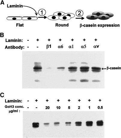
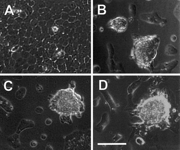
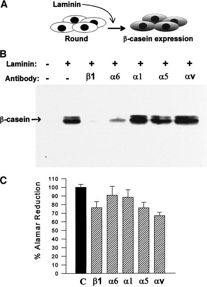
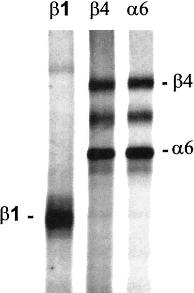
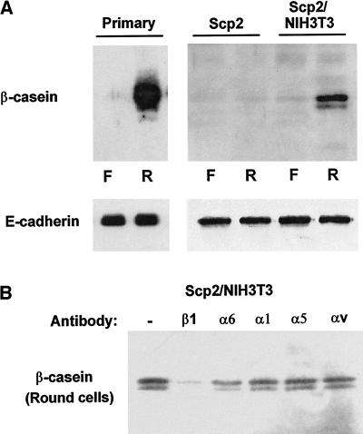
Contact of cultured mammary epithelial cells with the basement membrane protein laminin induces multiple responses, including cell shape changes, growth arrest, and, in the presence of prolactin, transcription of the milk protein beta-casein. We sought to identify the specific laminin receptor(s) mediating the multiple cell responses to laminin. Using assays with clonal mammary epithelial cells, we reveal distinct functions for the alpha6beta4 integrin, beta1 integrins, and an E3 laminin receptor. Signals from laminin for beta-casein expression were inhibited in the presence of function-blocking antibodies against both the alpha6 and beta1 integrin subunits and by the laminin E3 fragment. The alpha6-blocking antibody perturbed signals mediated by the alpha6beta4 integrin, and the beta1-blocking antibody perturbed signals mediated by another integrin, the alpha subunit(s) of which remains to be determined. Neither alpha6- nor beta1-blocking antibodies perturbed the cell shape changes resulting from cell exposure to laminin. However, the E3 laminin fragment and heparin both inhibited cell shape changes induced by laminin, thereby implicating an E3 laminin receptor in this function. These results elucidate the multiplicity of cell-extracellular matrix interactions required to integrate cell structure and signaling and ultimately permit normal cell function.


Figures

Figure 1
Figure 1
Inhibition of β-casein expression by function-blocking integrin antibodies. Assays for the induction of β-casein expression in SCp2 mammary epithelial cells were performed on cells initially spread on plastic and subsequently exposed to medium containing the lactogenic hormone prolactin, plus or minus laminin, and function-blocking antibodies against the β1, α6, α1, α5, and αv integrin subunits. (A) Schematic representation of the assay. A two-step signaling process leads to β-casein expression after cell contact with laminin, beginning with a prerequisite cell shape change (rounding), followed by subsequent biochemical signaling events. (B) β-Casein expression was assayed by immunoblots of cell extracts and appears as a doublet migrating at ∼34 kDa. Laminin-induced β-casein expression was inhibited in the presence of β1 and α6 integrin-blocking antibodies. (C) Titration of the α6-blocking (GoH3) antibody shows maximal inhibition in the range of 2–5 μg/ml.
Figure 2
Figure 2
Morphogenic changes induced by laminin are unaltered by the presence of α6- and β1-blocking antibodies. Assays for the induction of β-casein expression in SCp2 mammary epithelial cells were performed on cells initially spread on plastic and subsequently exposed to medium containing the lactogenic hormone prolactin, plus or minus laminin, and function-blocking antibodies against the β1 and α6 integrin subunits. In the absence of added laminin, cells remained attached and spread on the plastic and continued to grow to confluence (A). In contrast, cells exposed to laminin underwent cell rounding, and those in contact with other cells clustered into multicellular aggregates, leaving much of the plastic culture dish exposed (B). Cells exposed to laminin in the presence of function-blocking antibodies against the α6 (C) and β1 (D) integrin subunits continued to undergo the cell shape changes induced by laminin even though these same antibody treatments perturbed signals for β-casein expression.
Figure 3
Figure 3
β-Casein expression and cell survival in the presence of function-blocking antibodies against β1 and α6 integrins in prerounded cells. SCp2 cells cultured in suspension were treated with prolactin, plus or minus laminin, and function-blocking antibodies against the β1, α6, α1, α5, and αv integrin subunits. (A) Assays of β-casein expression in suspension cultures measure signaling events subsequent to, and independent of, the required cell shape changes. (B) Immunoblots of cell extracts after 3 d of laminin exposure show that laminin-induced β-casein expression in prerounded cells was still inhibited in the presence of β1 and α6 integrin-blocking antibodies. (C) Cell viability was assayed by Alamar Blue dye reduction in duplicate wells under β-casein assay conditions identical to those described for B, but culture was for 4 d (1 d longer) to measure any cell death that may have been initiated during the course of the β-casein assay.
Figure 4
Figure 4
Immunoprecipitation of α6 integrins from the SCp2 cells. The β1, β4, and α6 integrins were immunoprecipitated from [35S]methionine-labeled cell extracts and separated on a 7% SDS-polyacrylamide gel. Bands for both the β4 and α6 subunits are evident in the immunoprecipitations using the β4 and α6 subunit antibodies, demonstrating the presence of the α6β4 heterodimer. The β1 subunit was not detectable in precipitations of the α6 integrins, demonstrating the absence of the α6β1 heterodimer in the SCp2 cells. The exclusive dimerization of α6 with the β4 subunit is further supported by the fact that the yield of the α6 and β4 subunits is equal between the α6 and β4 immunoprecipitations, indicating that most or all of the α6 subunit is dimerized with the β4 subunit in these cells.
Figure 5
Figure 5
Morphogenic changes and β-casein expression are blocked by the laminin E3 fragment and by heparin. (A) Assays for the cell shape changes induced by laminin in SCp2 mammary epithelial cells were performed on cells initially spread on plastic and subsequently exposed to medium containing laminin plus or minus the elastase-generated laminin fragments E3 and E8. In the absence of added laminin (a), cells remain attached and spread on the plastic and continue to grow to confluence. Cells exposed to laminin (b) undergo cell rounding, and cells sharing cell-cell contacts cluster into multicellular aggregates. Cells continue to undergo the cell shape changes when exposed to laminin in the presence of BSA at 100 μg/ml (c), the laminin E8 fragment at 100 μg/ml (d), the laminin E3 fragment at 50 μg/ml (e), heparansulfate at 400 μg/ml (g), or heparin at 100 μg/ml (h). However, cells are strongly inhibited from undergoing the cell rounding and clustering when exposed to laminin in the presence of the laminin E3 fragment at 100 μg/ml (f) or heparin at 400 μg/ml (i). (B) The E3 laminin fragment was tested as a competitive inhibitor in assays of β-casein expression in prerounded cells. The E3 fragment continues to inhibit β-casein expression even in assays of prerounded cells, with partial inhibition at concentrations as low as 20 μg/ml. (C) Heparin and heparan sulfate were tested as competitive inhibitors in assays of β-casein expression in prerounded cells. Heparin inhibits β-casein expression in assays of prerounded cells at 400 μg/ml, whereas heparan sulfate has no effect at the same concentration.
Figure 6
Figure 6
Assays of β-casein expression, in the absence of added laminin, in primary cell cultures, clonal epithelial cells, and cocultures of clonal epithelial cells and fibroblasts. (A) Primary murine mammary epithelial cell cultures, SCp2 clonal epithelial cells, and cocultures of SCp2 cells and NIH3T3 fibroblasts (10:1) were assayed for β-casein expression in both flat cells (F) and rounded cells (R) (suspension culture) exposed to prolactin in the absence of added laminin. Cell rounding in suspension cultures permitted the induction of β-casein in primary cultures but not in the clonal SCp2 cell line. Coculture of SCp2 cells with a mesenchymal component (NIH3T3 fibroblasts) permitted the induction of β-casein. The same immunoblot filters were also probed for E-cadherin to demonstrate normalization for equal cell number. (B) SCp2 cells and NIH3T3 fibroblasts were cocultured in suspension (prerounded) and exposed to prolactin in the presence of function-blocking antibodies against the β1, α6, α1, α5, and αv integrin subunits, without the addition of laminin. β-Casein expression induced by endogenous basement membrane formation was inhibited by the β1 integrin-blocking antibody but was not inhibited effectively by the α6 integrin-blocking antibody GoH3.

Similar articles

Cited by

References

    1. Bao ZZ, Lakonishok M, Kaufman S, Horwitz AF. Alpha 7 beta 1 integrin is a component of the myotendinous junction on skeletal muscle. J Cell Sci. 1993;106:579–589. - PubMed
    1. Barcellos-Hoff MH, Aggeler J, Ram TG, Bissell MJ. Functional differentiation and alveolar morphogenesis of primary mammary cultures on reconstituted basement membrane. Development. 1989;105:223–235. - PMC - PubMed
    1. Boudreau N, Sympson CJ, Werb Z, Bissell MJ. Suppression of ICE and apoptosis in mammary epithelial cells by extracellular matrix. Science. 1995;267:891–893. - PMC - PubMed
    1. Boudreau N, Werb Z, Bissell MJ. Suppression of apoptosis by basement membrane requires three-dimensional tissue organization and withdrawal from the cell cycle. Proc Natl Acad Sci USA. 1996;93:3509–3513. - PMC - PubMed
    1. Burgeson RE, Christiano AM. The dermal-epidermal junction. Curr Opin Cell Biol. 1997;9:651–658. - PubMed

Publication types

MeSH terms