. 1999 Sep;10(9):2861-78.
doi: 10.1091/mbc.10.9.2861.

Stimulation of beta1-integrin function by epidermal growth factor and heregulin-beta has distinct requirements for erbB2 but a similar dependence on phosphoinositide 3-OH kinase

Affiliations
Free PMC article

Stimulation of beta1-integrin function by epidermal growth factor and heregulin-beta has distinct requirements for erbB2 but a similar dependence on phosphoinositide 3-OH kinase

M A Adelsman et al. Mol Biol Cell. 1999 Sep.
Free PMC article

Abstract

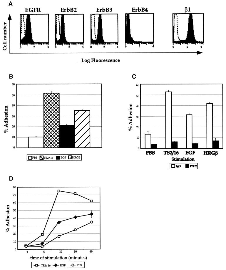
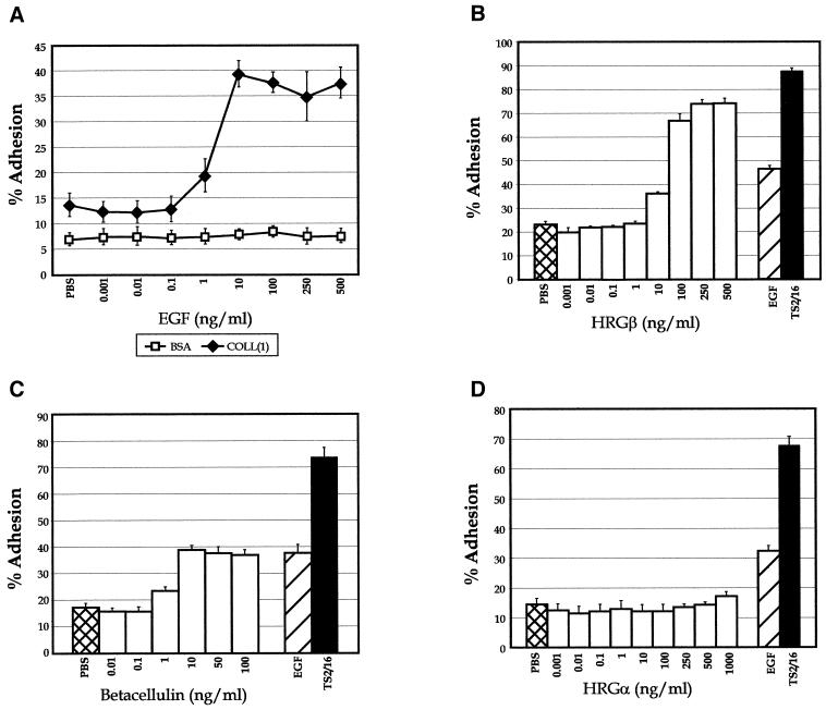
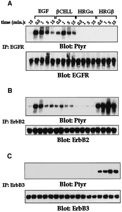
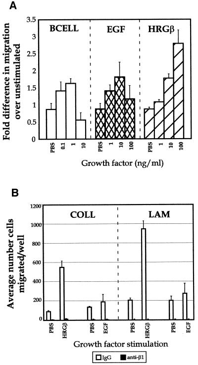
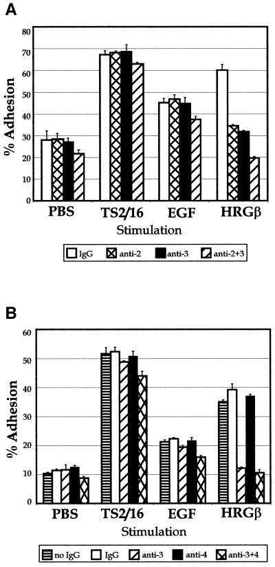
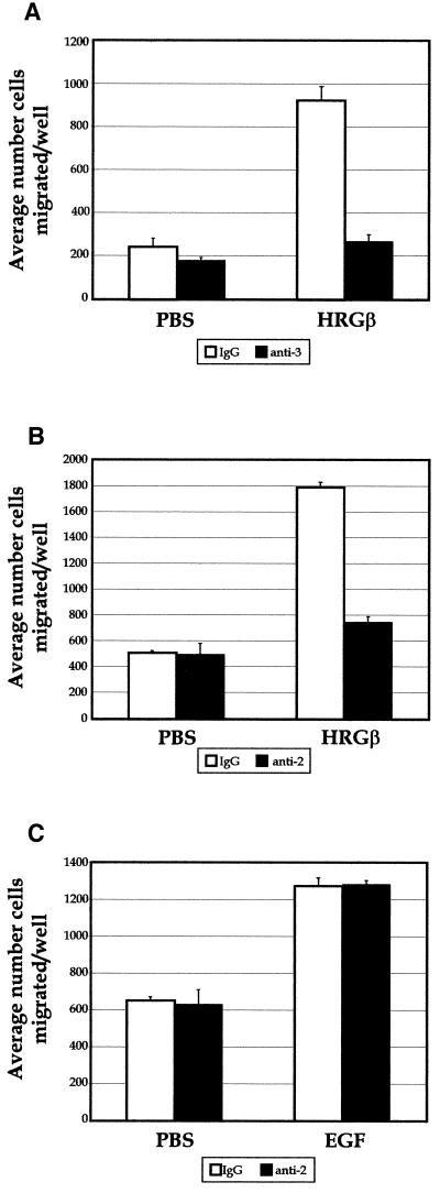
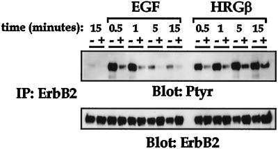
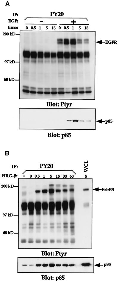
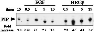
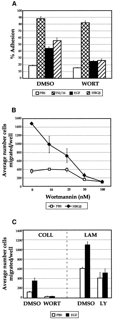
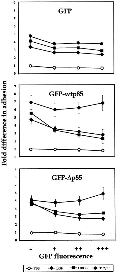
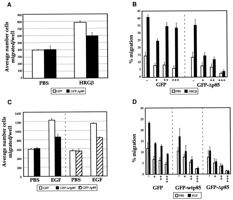
Integrins and growth factor receptors are important participants in cellular adhesion and migration. The EGF receptor (EGFR) family of tyrosine kinases and the beta1-integrin adhesion receptors are of particular interest, given the implication for their involvement in the initiation and progression of tumorigenesis. We used adhesion and chemotaxis assays to further elucidate the relationship between these two families of transmembrane signaling molecules. Specifically, we examined integrin-mediated adhesive and migratory characteristics of the metastatic breast carcinoma cell line MDA-MB-435 in response to stimulation with growth factors that bind to and activate the EGFR or erbB3 in these cells. Although ligand engagement of the EGFR stimulated modest beta1-dependent increases in cell adhesion and motility, heregulin-beta (HRGbeta) binding to the erbB3 receptor initiated rapid and potent induction of breast carcinoma cell adhesion and migration and required dimerization of erbB3 with erbB2. Pharmacologic inhibitors of phosphoinositide 3-OH kinase (PI 3-K) or transient expression of dominant negative forms of PI 3-K inhibited both EGF- and HRGbeta-mediated adhesion and potently blocked HRGbeta- and EGF-induced cell motility. Our results illustrate the critical role of PI 3-K activity in signaling pathways initiated by the EGFR or erbB3 to up-regulate beta1-integrin function.


Figures

Figure 1
Figure 1
EGF and HRGβ modulate breast carcinoma cell adhesion to ECM ligands. (A) Surface expression of the EGFR, erbB2, erbB3, erbB4, or β1-integrin on MDA-MB-435 cells was determined by flow cytometry as described in MATERIALS AND METHODS. Representative histogram profiles are shown, with open histograms representing negatively stained control cells. (B) Cells were analyzed for their ability to respond to EGF or HRGβ treatment in cell adhesion assays performed on human type IV COLL. Cells that had been serum starved for 12–24 h were harvested by release from tissue culture flasks in 1 mM EDTA. Suspended cells were than washed in serum-free media to remove excess EDTA before quantitation and labeling with Calcein AM as previously described (Zell et al., 1996). Adhesion assays were performed in 96-well plates that were precoated overnight at 4°C with COLL at 1 μg/well. Cells were then added to wells containing assay media alone (open bar) or stimulators: the activating β1-integrin-specific monoclonal antibody TS2/16 (cross-hatched bar), EGF at 100 ng/ml final concentration (shaded bar), or HRGβ at 100 ng/ml (hatched bar). (C) For blocking of β1-integrins, cells were preincubated at 4°C with control mouse IgG (open bars) or with the inhibitory β1-integrin-specific antibody P5D2 (shaded bars; a kind gift from T. LeBien) at 1 μg/1 × 106 cells for 10 min before addition to plates coated with 1 μg/well COLL. The time course of adhesion to COLL (D) was determined in the presence of 1 μg/well TS2/16 (open squares) or 100 ng/ml EGF (solid diamonds) in comparison with unstimulated adhesion (PBS; open circles). Cells were allowed to settle briefly in wells before analyzing preadherent fluorescence on a fluorescence plate reader. Cells were then stimulated at 37°C for 10 min or for designated times before removing nonadherent cells by hand washing with a syringe–manifold system. Comparison of adhesion with human type IV collagen or mouse EHS-collagen did not show significant differences (our unpublished data). Adhesion data are representative of at least three independent experiments and reflect the average of triplicate samples per condition.
Figure 2
Figure 2
MDA-MB-435 cell adhesion is stimulated by multiple growth factors that bind to and activate members of the EGF receptor family. For analysis of dose response to growth factor, increasing amounts of EGF ranging from 1 pg/ml to 500 ng/ml were added to COLL-coated wells before addition of cells and stimulation at 37°C (A; solid diamonds). Adhesion to BSA-coated wells (open squares) was performed as a control. Dose responses for adhesion to COLL were determined in the presence of increasing amounts of HRGβ (B), the EGF-like growth factor betacellulin (C), or HRGα (D). Unstimulated (PBS; B–D, cross-hatched bars), EGF-stimulated (B–D, hatched bars), or TS2/16-stimulated (B–D, solid bars) adhesion was analyzed for comparison. Cells plated on BSA alone as a control for nonspecific adhesion generally showed <10% adhesion (A; our unpublished data). Data are representative of at least three separate assays.
Figure 3
Figure 3
Multiple growth factors induce tyrosine phosphorylation of the EGFR, erbB2, and erbB3 in MDA-MB-435 cells. Receptor activation was assessed after stimulation of MDA-MB-435 cells (10 × 106 cells per sample) for increasing periods at 37°C with PBS alone (first lane in each panel), EGF at 100 ng/ml, betacellulin (βCELL) at 10 ng/ml, HRGα at 100 ng/ml, or HRGβ at 100 ng/ml. Lysis buffer (2×) was added to each sample after the indicated stimulation time, and cleared lysates were immunoprecipitated for EGFR (A), erbB2 (B), or erbB3 (C). Western blotting was performed on samples after separation by SDS-PAGE and transfer to polyvinylidene difluoride membranes. Total phosphotyrosine content in each sample was assessed by probing with anti-phosphotyrosine Ab 4G10 (upper panels). Equivalent receptor loading was confirmed by stripping and reprobing each blot for the presence of the indicated receptor (lower panels). Blots were also stripped and reprobed for the presence of p85 (our unpublished data). Similar results were observed in at least three independent experiments.
Figure 4
Figure 4
Betacelluin, EGF, and HRGβ induce β1-integrin-dependent MDA-MB-435 cell migration on LAM. (A) Cells were grown to ∼75% confluency and placed in serum-free media for 16 h before harvesting as for adhesion assays. Polycarbonate filters (8 μm) were coated overnight at 4°C in PBS containing mouse EHS-LAM or EHS-COLL (our unpublished data) at 20 μg/ml. Forty-eight-well chemotaxis chambers were assembled with assay media alone or containing increasing amounts of growth factors in the lower chambers, as indicated. LAM-coated filters were placed over the lower wells and, ∼23,000 MDA-MB-435 cells were placed in the upper wells and allowed to migrate at 37°C for 4–6 h. Migrated cells in each well were quantitated on fixed and stained filters. The sum of four microscopic fields was taken for each well, and three wells were averaged for each stimulation condition. (B) Antibody blocking studies were carried out by preincubating cells with control IgG (open bars) or the inhibitory β1-integrin-specific mAb P5D2 (solid bars) at 1 μg/1 × 106 cells on ice for 10 min before adding the cells to the upper wells of chambers containing EGF or HRGβ and containing filters coated with LAM or COLL. Some variability was observed with the levels of basal migration in the data shown in A, because each growth factor titration was carried out in a separate chemotaxis chamber. However, the level of stimulation over that of basal migration for each growth factor was reproducible over at least three separate assays.
Figure 5
Figure 5
HRGβ–mediated adhesion of MDA-MB-435 cells to COLL requires heterodimerization with erbB2. Adhesion assays were performed as described for Figure 1. (A) To examine contributions by erbB2 and erbB3, control IgG (open bars) or blocking mAbs specific for erbB2 (cross-hatched bars), erbB3 (shaded bars), or a combination of anti-erbB2 and anti-erbB3 mAbs (hatched bars) were incubated with cells before addition to plates containing TS2/16, EGF, or HRGβ. (B) To examine contributions by erbB3 and erbB4, cells were incubated with no IgG (horizontally striped bars), control IgG (open bars), or blocking mAbs specific for erbB3 (hatched bars), erbB4 (solid bars), or a combination of anti-erbB3 and anti-erbB4 mAbs (cross-hatched bars) before addition to plates containing TS2/16, EGF, or HRGβ. The data reflect an average of three wells per condition and are representative of at least three separate assays.
Figure 6
Figure 6
HRGβ-mediated migration of MDA-MB-435 cells on LAM requires heterodimerization with erbB2. Migration assays were carried out as described for Figure 4. To examine contributions by erbB3 (A) or erbB2 (B and C), control IgG (open bars) or blocking mAbs specific for erbB3 (A, solid bars) or erbB2 (B and C, solid bars) were incubated with cells before addition to chemotaxis chambers containing control media, EGF, or HRGβ. Cell migration was quantitated by taking the sum of four microscopic fields per well and the average of at least three wells per condition. Experiments were performed a minimum of three times.
Figure 7
Figure 7
Tyrosine phosphorylation of erbB2 after EGF or HRGβ stimulation is inhibited by the anti-erbB2 blocking Ab-16. MDA-MB-435 cells (5 × 10⁶ per sample) were coated for 15 min on ice with either control mouse IgG (− lanes) or erbB2 Ab-16 (+ lanes) at 0.5 μg/1 × 106 cells. Cells were then stimulated for various times with 100 ng/ml EGF or HRGβ at 37°C followed by lysis in an equal volume of 2× lysis buffer. Immunoprecipitation for erbB2 was performed on cleared lysates, and samples were separated by SDS-PAGE. Immunoblotting for phosphotyrosine using 4G10 (upper panel) was performed after Western transfer. The membrane was then stripped and reprobed for erbB2 (lower panel) to confirm receptor levels. The figure shown is representative of a minimum of three independent assays.
Figure 8
Figure 8
EGF and HRGβ stimulation of MDA-MB-435 cells induces recruitment of the p85 subunit of PI 3-K to the phosphotyrosine cellular fraction. Cells were serum starved for 24 h and harvested as previously described. Cells (7.5 × 10⁶ per sample) were incubated in the absence or presence of 100 ng/ml EGF (A) or 100 ng/ml HRGβ (B) for the indicated periods at 37°C. Cells were then lysed, and phosphotyrosine-containing proteins were immunoprecipitated with the anti-phosphotyrosine mAb PY20 coupled to protein A-Sepharose beads. Washed immunocomplexes were separated by SDS-PAGE and transferred to polyvinylidene difluoride membranes. Western blotting was performed on membranes with the anti-phosphotyrosine mAb 4G10 to detect phosphotyrosine-containing proteins (upper panels). Blots were stripped and reprobed for EGFR or erbB3 (A and B, respectively; our unpublished data) and for p85 (lower panels). Similar data were obtained in at least two additional assays.
Figure 9
Figure 9
EGF and HRGβ activate PI 3-K enzymatic activity in vitro. Cells were left unstimulated or stimulated in the presence of EGF or HRGβ (each at 100 ng/ml) for various times at 37°C. PY20 immunoprecipitates were prepared for a PI 3-K in vitro kinase assay as previously described (Chan et al., 1997). Kinase activity was detected by the in vitro phosphorylation of sonicated phosphatidylinositol, the products of which were visualized and quantitated after TLC separation, autoradiography, and phosphorimage analysis.
Figure 10
Figure 10
PI 3-K inhibitors block EGF- or HRGβ-mediated adhesion and migration of MDA-MB-435 cells. (A) Adhesion assays were carried out in the presence of 100 nM wortmannin (WORT) with no stimulation (open bars) or after stimulation for 10 min at 37°C with TS2/16 (cross-hatched bars), EGF (shaded bars), or HRGβ(hatched bars). (B) Migration analysis was carried out in the presence of increasing amounts of wortmannin in the presence of media only (open squares) or HRGβ (solid triangles). Similar effects were observed when dose–response analysis was carried out on COLL or when assays were performed in the presence of 25 μM LY294002 (our unpublished data). (C) Migration on COLL or LAM was performed in the presence of control DMSO, 100 nM wortmannin (WORT), or 25 μM LY294002 (LY) with no stimulation (open bars) or stimulation with 10 ng/ml EGF (solid bars). HRGβ stimulation was slightly lower than normal in the adhesion assay shown in A in comparison with EGF or TS2/16 stimulation, but the data shown are otherwise representative of at least three separate experiments.
Figure 11
Figure 11
Overexpression of the wild-type or dominant negative p85 subunit of PI 3-K inhibits EGF- or HRGβ-mediated increases in MDA-MB-435 adhesion to COLL. Control vector expressing GFP (top panel) or constructs expressing a GFP-wt p85 (middle panel) or GFP-Δp85 (bottom panel) fusion protein were transiently transfected into MDA-MB-435 cells as described in MATERIALS AND METHODS. Transfected cells were allowed to recover for 24 h and then placed in serum-free media overnight. Cells were harvested as for standard adhesion assays, except that no Calcein AM labeling was performed, and cells (∼300,000 cells per well) were added to 24-well plates coated with 6 μg/well COLL. Adhesion was analyzed in the presence of PBS alone (open circles) or containing 1 μg/well TS2/16 (solid circles), 100 ng/ml EGF (solid diamonds), or 100 ng/ml HRGβ (solid squares) for 10 min at 37°C. Nonadherent cells were washed away, and adherent cells were collected fromwells using a 1:1 trypsin:1 mM EDTA solution. Collected cells were then analyzed by flow cytometry using aliquots of preadherent cell populations to confirm cell numbers added to wells for each transfectant and to determine the percent expression of GFP in the starting cell populations. Percent adhesion was determined by gating GFP-negative (−), GFP-low (+), -middle- (++), and -high (+++)-positive cells and comparing preadherent and adherent cell numbers from each population. The data shown reflect fold differences in adhesion when compared with the GFP-negative, unstimulated cell subpopulation from each transfectant. Average percent adhesion was determined from samples examined in triplicate for each stimulation condition, and results were similar for a minimum of three independent assays.
Figure 12
Figure 12
Overexpression of the dominant negative p85 subunit of PI 3-K inhibits HRGβ- and EGF-mediated increases in MDA-MB-435 migration on LAM. Control vector expressing GFP or a construct expressing a GFP-Δp85 fusion protein were transiently transfected into MDA-MB-435 cells. Transfected cells were allowed to recover for 24 h and then placed in serum-free media for 24 h. Cells were harvested and quantitated as previously described. (A) To monitor the efficiency of stimulated migration on bulk transfected populations, standard chemotaxis analysis was performed as described in MATERIALS AND METHODS with aliquots of cells at the same concentration as used for migration in transwells (below). Total cell populations expressing GFP (open bars) or GFP-Δp85 (solid bars) were allowed to migrate in Boyden chambers in the presence or absence of HRGβ in the lower wells on LAM-coated filters overnight, and migrated cells were fixed, stained, and quantitated as described. (B) The migration of GFP-positive versus GFP-negative cells in each bulk transfectant cells was assessed specifically by allowing cells to migrate overnight in transwell chambers that had been precoated with LAM in the absence (open bars) or presence of 100 ng/ml HRGβ (solid bars). After ∼16 h, migrated cells were collected from the lower surface of LAM-coated filters with trypsin/EDTA and analyzed by flow cytometry as described for transient adhesion assays in Figure 11. Data represent the average of three wells per condition and show the percent migration of each GFP-negative or GFP-positive cell population. (C) Cells expressing GFP (open bars), GFP-wtp85 (solid bars), or GFP-Δp85 (hatched bars) were placed in the upper wells of Boyden chambers and allowed to migrate in the presence or absence of 10 ng/ml EGF on LAM-coated filters overnight, and migrated cells were fixed, stained, and quantitated as described. (D) The migration of GFP-positive transfected cells was assessed specifically by allowing cells to migrate overnight in transwell chambers that had been precoated with LAM in the absence (open bars) or presence of 10 ng/ml EGF (solid bars). After ∼16 h, migrated cells were collected from the lower surface of LAM-coated filters with trypsin/EDTA and analyzed by flow cytometry as described for transient adhesion assays in Figure 11. Data represent the average of three wells per condition and show the percent migration of each GFP-negative or GFP-positive cell population. Migration observed under standard Boyden chamber conditions (A and C) reflected increases in migration in response to stimuli comparable with those seen in the transwell analysis. Modest effects of GFP-wtp85 or GFP-Δp85 on migration were observed in the mixed cell populations in A and C but are more apparent upon single-cell analysis as shown in B and D. Comparison of assays carried out for 4 h vs. 16 h gave similar results, and the experimental data are representative of at least three independent assays.

References

    1. Adam L, Vadlamudi R, Kondapaka SB, Chernoff J, Mendelsohn J, Kumar R. Heregulin regulates cytoskeletal reorganization and cell migration through the p21-activated kinase-1 via phosphatidylinositol-3 kinase. J Biol Chem. 1998;273:28238–28246. - PubMed
    1. Akiyama SK, Larjava H, Yamada KM. Differences in the biosynthesis and localization of the fibronectin receptor in normal and transformed cultured human cells. Cancer Res. 1990;50:1601–1607. - PubMed
    1. Albelda SM. Role of integrins and other cell adhesion molecules in tumor progression and metastasis. Lab Invest. 1993;68:4–17. - PubMed
    1. Alroy I, Yarden Y. The ErbB signaling network in embryogenesis and oncogenesis: signal diversification through combinatorial ligand-receptor interactions. FEBS Lett. 1997;410:83–86. - PubMed
    1. Aplin AE, Howe A, Alahari SK, Juliani RL. Signal transduction and signal modulation by cell adhesion receptors: the role of integrins, cadherins, immunoglobulin-cell adhesion molecules, and selectins. Pharmacol Rev. 1998;50:197–263. - PubMed

Publication types

MeSH terms