. 1999 Sep;10(9):2955-69.
doi: 10.1091/mbc.10.9.2955.

Biparental inheritance of gamma-tubulin during human fertilization: molecular reconstitution of functional zygotic centrosomes in inseminated human oocytes and in cell-free extracts nucleated by human sperm

Affiliations
Free PMC article

Biparental inheritance of gamma-tubulin during human fertilization: molecular reconstitution of functional zygotic centrosomes in inseminated human oocytes and in cell-free extracts nucleated by human sperm

C Simerly et al. Mol Biol Cell. 1999 Sep.
Free PMC article

Abstract

Human sperm centrosome reconstitution and the parental contributions to the zygotic centrosome are examined in mammalian zygotes and after exposure of spermatozoa to Xenopus laevis cell-free extracts. The presence and inheritance of the conserved centrosomal constituents gamma-tubulin, centrin, and MPM-2 (which detects phosphorylated epitopes) are traced, as is the sperm microtubule-nucleating capability on reconstituted centrosomes. gamma-Tubulin is biparentally inherited in humans (maternal >> than paternal): Western blots detect the presence of paternal gamma-tubulin. Recruitment of maternal gamma-tubulin to the sperm centrosome occurs after sperm incorporation in vivo or exposure to cell-free extract, especially after sperm "priming" induced by disulfide bond reduction. Centrin is found in the proximal sperm centrosomal region, demonstrates expected calcium sensitivity, but appears absent from the zygotic centrosome after sperm incorporation or exposure to extracts. Sperm centrosome phosphorylation is detected after exposure of primed sperm to egg extracts as well as during the early stages of sperm incorporation after fertilization. Finally, centrosome reconstitution in cell-free extracts permits sperm aster microtubule assembly in vitro. Collectively, these results support a model of a blended zygotic centrosome composed of maternal constituents attracted to an introduced paternal template after insemination.


Figures
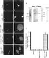
Figure 1
Figure 1
γ-Tubulin in human and bovine sperm centrosomes. More than 98% of human sperm do not immunostain with the XG-1-4 γ-tubulin antibody (A; arrow points to the sperm centrosomal region) after lysolecithin permeabilization and methanol fixation (B, DNA). Likewise, no significant increase in the detection of γ-tubulin at the human sperm centrosome is observed after 5 mM DTT priming treatment (C; arrows point to the sperm centrosomal region), although some DNA decondensation occurs in vitro (D). Very similar observations have been observed in bovine spermatozoa treated in exactly the same manner. However, both human (E and F) and bovine (I and J) spermatozoa treated with 5 mM DTT followed by CSF-arrested cell-free extract exposure demonstrated extensive DNA decondensation after 1 h (F and J) and XG-1-4 γ-tubulin immunolocalization at the sperm centrosomal regions (E and I; arrows point to the sperm centrosomal region).Immunodepletion of γ-tubulin from the CSF-arrested extracts by the XG-1-4 γ-tubulin abolishes detection of γ-tubulin at the base of permeabilized, DTT-treated human spermatozoa, demonstrating that the vast majority of γ-tubulin is maternally derived (G and H). (K) Western blot analysis of Xenopus, human, and bovine sperm demonstrates prominent bands at ∼55 kDa with the XG-1-4 antibody, indicating the presence of paternal γ-tubulin in these sperm. Lane 1, Xenopus sperm, 1.25 × 10⁶ sperm per lane; lane 2, bovine sperm subjected to Percoll density centrifugation, at ∼2.6 × 10⁶ sperm per lane; lane 3, washed bovine sperm without Percoll separation, at ∼2.6 × 10⁶ sperm per lane; lane 4, human sperm subjected to Percoll density separation and labeled with XG-1-4 γ-tubulin antibody, ∼2.5 × 10⁶ sperm per lane; lane 5, 0.5 μg of purified α- and β-tubulin, demonstrating no cross-reactivity of these tubulin superfamily members with the XG-1-4 γ-tubulin antibody. (L) Graphic representation of permeabilized human spermatozoa immunostained with γ-tubulin XG-1-4 antibody after permeabilization, DTT priming, and CSF-arrested cell-free extract. By immunofluorescence, very little paternal γ-tubulin is observed in permeabilized human sperm (left bar) or permeabilized human sperm primed by exposure to 5 mM DTT (middle bar). However, a significant increase in the detection of γ-tubulin is observed when permeabilized and primed sperm are treated with CSF-arrested cell-free extract (right bar). All images were double labeled for γ-tubulin and Hoechst DNA. (Arrows) Sperm centrosomal region as observed with phase or differential interference contrast optics. Bar in J, 10 μm.
Figure 2
Figure 2
Centrin in human sperm centrosomes. Centrin is localized exclusively as either a pair of punctate sources or a single spot (depending on orientation) at the centrosome in permeabilized human sperm (A, centrin antibody 20H5; B, DNA) or permeabilized spermatozoa exposed to 2 mM CaCl2 (C and D). This staining is not dependent on calcium exposure. Sperm exposed to 5 mM DTT (E and F) for 40 min show no change in the centrin-staining pattern from controls. However, spermatozoa exposed to DTT and 2 mM CaCl2 show a dissipation of centrin staining (G and H). Arrows indicate the point of tail attachment to the sperm head as observed with phase or differential interference contrast optics. Identical results were found with bovine sperm. (I) Western blots of bovine and human sperm, demonstrating a single band at ∼20 kDa that comigrates with bacterially expressed centrin and the calcium sensitivity of sperm centrosomal centrin. Lane 1, DTT-primed bovine sperm (60 μg of total protein per lane); lane 2, DTT-primed bovine sperm treated for 30 min with 2 mM CaCl2, showing the loss in 20H5 centrin detection after high external calcium treatment (43 μg of total protein per lane); lane 3, human sperm, immunoprecipitated with 20H5 anti-centrin and immunostained with anti-centrin serum 24/14-1; lane 4, purified bacterially expressed centrin protein immunostained with anti-centrin serum 24/14-1. (J). Graphic representation of human spermatozoa immunostained with 20H5 centrin after permeabilization, DTT priming, and elevated external calcium exposure. Analysis reveals that DTT-primed spermatozoa treated with either high external calcium (fourth bar) or CSF-arrested extract (fifth bar) demonstrate a significant reduction in the detection of centrin at the sperm centrosome. Bar in H, 10 μm.
Figure 3
Figure 3
Ultrastructural detection of centrin in the bovine mature sperm centrosome and its sensitivity to CSF-arrested cell-free extract. (A and B) Immunogold labeling of mature bovine spermatozoa with anti-β-tubulin antibody, demonstrating extensive immunolabeling of the sperm proximal centriole (asterisks) and outer microtubule doublets of the sperm axoneme (A, arrowheads). (C and D) Immunogold labeling of mature bovine sperm with anti-centrin antibody 20H5. In C, a longitudinal section of proximal centriole in the sperm tail connecting piece is observed, with centrin localized to its capitulum-attached end (arrows). (D) Oblique sections of the proximal centriole demonstrating centrin detection in an area of the centriole adjacent to the striated columns of the connecting piece. (E and F) Control bovine spermatozoa immunolabeled with colloidal gold–conjugated secondary antibody only. No labeling is observed in the connecting piece structures (E, longitudinal section of the centriole) or in the proximal centriole (F, cross-section). (G and H) Human permeabilized spermatozoa treated sequentially with 5 mM DTT and CSF-arrested cell-free extract and then immunostained with the 20H5 centrin antibody. The primed sperm has begun to decondense in the presence of egg extract (G), but 20H5 centrin is no longer detected at the base of the sperm head (H, arrow). if, implantation fossa; c, capitulum; sc, striated columns; odf, outer dense fibers; asterisks, proximal centriole. Bars in A, B, D, E, and F, 0.2 μm; bar in C, 0.5 μm; bar in H, 1 μm.
Figure 4
Figure 4
MPM-2 detection in human sperm and early bovine sperm penetration. X. laevis sperm become phosphorylated after incubation in X. laevis CSF-arrested cell-free extract (A, MPM-2; B, DNA). The mature human sperm centrosome does not immunostain with MPM-2 antibody after methanol fixation (C, MPM-2; D, DNA), although punctate staining is occasionally observed in the head, midpiece, and principal sperm tailpiece (not shown). Human spermatozoa permeabilized in lysolecithin and exposed to CSF-arrested cell-free extract demonstrate MPM-2 immunostaining at the sperm centrosomal region (E, MPM-2; F, DNA). Sperm priming with 5 mM DTT does not significantly increase the detection of centrosome phosphorylation (G, MPM-2; H, DNA) until after exposure to CSF-arrested cell-free extract (I, MPM-2; J, DNA). Arrows depict sperm tail attachment to the sperm head as observed with phase or differential interference contrast optics. Similar results were observed in mature bovine sperm exposed to CSF-arrested extracts. (K and L) Dispermic penetration in a bovine oocyte after in vitro fertilization (8 h after insemination). MPM-2 phosphorylation of the sperm centrosomes (L, arrows) is observed, suggesting that the positive MPM-2 staining of the assembling zygotic centrosome observed after exposure to frog extracts is mimicked in vivo. (M) Graphic representation of human spermatozoa immunostained with MPM-2 antibody after permeabilization, DTT treatment, and exposure to cell-free extract. The analysis demonstrates that significant MPM-2 immunostaining at the sperm centrosome is observed only when permeabilized or DTT-primed spermatozoa are exposed to CSF-arrested cell-free extract (second and fourth bars). Bars in B, J, and L, 10 μm.
Figure 5
Figure 5
Microtubule assembly in vitro nucleated by X. laevis, human, and bovine sperm. (A) Lysolecithin-permeabilized Xenopus sperm incubated in CSF-arrested cell-free extract containing 0.08 mg/ml rhodamine-conjugated bovine brain tubulin. Microtubule assembly (red) is radially symmetric and tightly focused at the sperm centrosome (blue, DNA). Bull (B) and human (C) sperm (blue), exposed to 5 μM ionomycin and primed with 5 mM DTT, also demonstrate assembly of microtubules in vitro (red) from the centrosomal region after 40–60 min of incubation in CSF-arrested cell-free extract. No free asters or assembled microtubules are present in the background, suggesting that microtubule nucleation, as opposed to microtubule capture, has occurred. Primed human sperm (blue) that was not exposed to CSF extract did not nucleate microtubules when exposed to rhodamine-conjugated bovine brain tubulin in Pipes buffer alone (D, red; arrow points to sperm axoneme). Bar in A, 30 μm; bars in B, C, and D, 1 μm.
Figure 6
Figure 6
γ-Tubulin, centrin, and MPM-2 detection in human and bovine zygotes. (A) Fertilized bovine oocyte fixed 12 h after insemination and immunostained with γ-tubulin antibody (red), XG-1-4 γ-tubulin antibody (green), and Hoechst DNA (blue). The sperm aster is a radially symmetrical array of microtubules emanating from the base of the sperm head (M). γ-Tubulin is detectable as a bright dot at the focal point of the sperm aster (arrow). (B) A dispermic human zygote fixed 26.5 h after insemination and triple labeled for microtubules (red, costained with β-tubulin and acetylated α-tubulin antibodies), XG-1-4 γ-tubulin antibody (green), and Hoechst DNA (blue). Each pole of the bipolar mitotic spindle has a sperm axoneme (red, arrows). γ-Tubulin is detected as four bright dots at the spindle poles (green), two of which are associated with the incorporated sperm axonemes (red, arrows). (C) An arrested, monospermically inseminated human oocyte fixed 48 h after insemination and immunostained with antibodies to microtubules (red, glutamate α-tubulin antibody), 20H5 mAb to centrin (inset), and Hoechst DNA (blue). A replicated and split centrosome, indicated by the two small microtubule asters (red, arrows) around the adjacent male (M) and female (F) pronuclei, is shown. The incorporated sperm axoneme (arrowhead) is associated with one of the two microtubule asters, but no centrin is detected at the centrosome (inset). (D) A bovine oocyte fertilized in vitro and fixed 10 h after insemination demonstrating sperm aster formation (red) and a pair of MPM-2 immunoreactive foci within the assembled astral microtubules (green). After activation of bovine oocytes, cortical microtubule assembly increases (red) and other cytoplasmic MPM-2 reactive foci (green) can be seen within this polymerizing array. (E) A 55-kDa band is prominently detected in 50 mature bovine oocytes after Western blotting with the rabbit poly-clonal XG-1-4 antibody, indicating the presence of abundant maternal γ-tubulin protein. All images were triple labeled for microtubules (red), γ-tubulin, centrin, or MPM-2 (green), and DNA (blue). Bars, 10 μm.

Similar articles

Cited by

References

    1. Baron AT, Greenwood TM, Bazinet CW, Salisbury JL. Centrin is a component of the pericentriolar lattice. Biol Cell. 1992;76:383–388. - PubMed
    1. Baron AT, Suman VJ, Nemeth E, Salisbury JL. The pericentriolar lattice of PtK2 cells exhibits temperature and calcium-modulated behavior. J Cell Sci. 1994;107:2993–3003. - PubMed
    1. Baum P, Furlong C, Byers B. Yeast gene required for spindle pole body duplication: homology of its product with Ca2+-binding proteins. Proc Natl Acad Sci USA. 1986;83:5512–5516. - PMC - PubMed
    1. Bellvé AR, Zheng W, Martinova YS. Recovery, capacitation, acrosome reaction, and fractionation of sperm. Methods Enzymol. 1993;225:113–136. - PubMed
    1. Biggins S, Rose MD. Direct interaction between yeast spindle pole body components: Kar1p is required for Cdc31p localization to the spindle pole body. J Cell Biol. 1994;125:843–852. - PMC - PubMed

Publication types

MeSH terms

LinkOut - more resources