. 1999 Sep;10(9):2971-86.
doi: 10.1091/mbc.10.9.2971.

A p90(rsk) mutant constitutively interacting with MAP kinase uncouples MAP kinase from p34(cdc2)/cyclin B activation in Xenopus oocytes

Affiliations
Free PMC article

A p90(rsk) mutant constitutively interacting with MAP kinase uncouples MAP kinase from p34(cdc2)/cyclin B activation in Xenopus oocytes

A C Gavin et al. Mol Biol Cell. 1999 Sep.
Free PMC article

Abstract

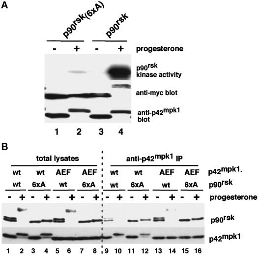
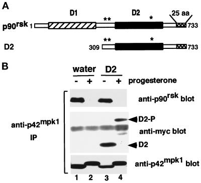
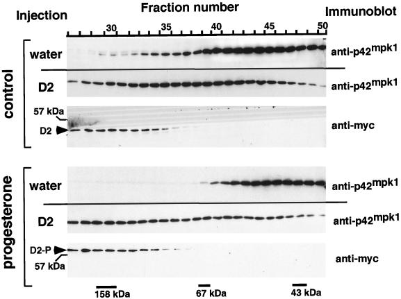
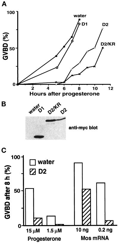
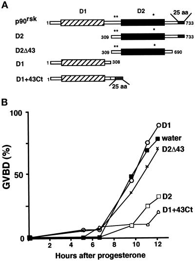
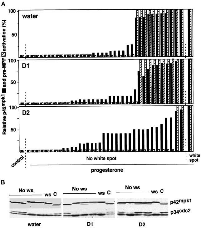
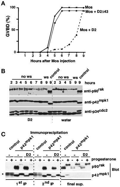
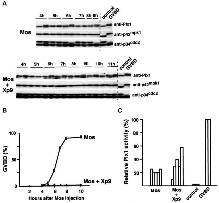
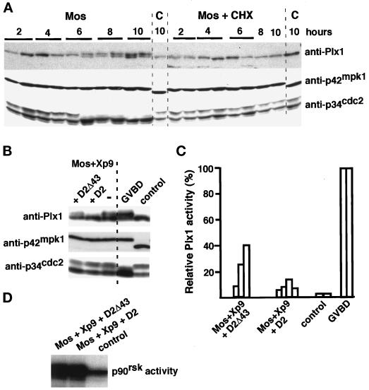
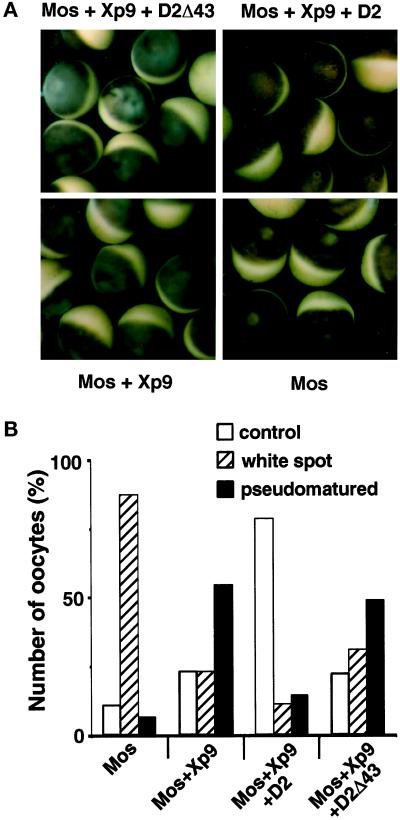
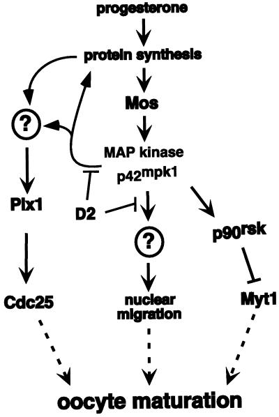
The efficient activation of p90(rsk) by MAP kinase requires their interaction through a docking site located at the C-terminal end of p90(rsk). The MAP kinase p42(mpk1) can associate with p90(rsk) in G(2)-arrested but not in mature Xenopus oocytes. In contrast, an N-terminally truncated p90(rsk) mutant named D2 constitutively interacts with p42(mpk1). In this report we show that expression of D2 inhibits Xenopus oocyte maturation. The inhibition requires the p42(mpk1) docking site. D2 expression uncouples the activation of p42(mpk1) and p34(cdc2)/cyclin B in response to progesterone but does not prevent signaling through p90(rsk). Instead, D2 interferes with a p42(mpk1)-triggered pathway, which regulates the phosphorylation and activation of Plx1, a potential activator of the Cdc25 phosphatase. This new pathway that links the activation of p42(mpk1) and Plx1 during oocyte maturation is independent of p34(cdc2)/cyclin B activity but requires protein synthesis. Using D2, we also provide evidence that the sustained activation of p42(mpk1) can trigger nuclear migration in oocytes. Our results indicate that D2 is a useful tool to study MAP kinase function(s) during oocyte maturation. Truncated substrates such as D2, which constitutively interact with MAP kinases, may also be helpful to study signal transduction by MAP kinases in other cellular processes.


Figures

Figure 1
Figure 1
Mutation to alanine of six phosphorylatable residues in p90rsk impairs its activation by progesterone and its ability to dissociate from p42mpk1. (A) Lysates were prepared from untreated or progesterone-treated oocytes expressing either myc-tagged p90rsk or p90rsk (6xA) and were analyzed by immunoblotting using anti-myc and anti-p42mpk1, as indicated. Anti-myc immunoprecipitates were prepared from the same oocyte lysates, and their associated kinase activity was assayed using GST-Myt1 as an in vitro substrate. (B) Lysates were prepared from either untreated or progesterone-treated oocytes expressing the indicated p90rsk and p42mpk1 proteins and were immunoprecipitated with anti-p42mpk1 antibodies. The total lysates (lanes 1–8) and the anti-p42mpk1 immunoprecipitates (lanes 9–16) were analyzed by immunoblotting with anti-p90rsk and anti-myc antibodies.
Figure 2
Figure 2
The C-terminal D2 domain of p90rsk constitutively interacts with endogenous p42mpk1 in oocytes. (A) Schematic representation of full-length p90rsk indicating the two kinase domains, D1 and D2, the p42mpk1 docking site located in the last 25 amino acids (25 aa) and the three p42mpk1 phosphorylation sites (asterisks). The N-terminally truncated p90rsk protein D2 is also shown. (B) Oocytes injected with water (lanes 1 and 2) or expressing myc-tagged D2 (lanes 3 and 4) were induced to mature by progesterone or not as indicated. Anti-p42mpk1 immunoprecipitates prepared from the oocyte lysates were analyzed by immunoblotting with anti-p90rsk, anti-myc and anti-p42mpk1 antibodies. The unphosphorylated and hyperphosphorylated D2 are indicated as D2 and D2-P, respectively.
Figure 3
Figure 3
D2 expressed in oocytes comigrates with endogenous p42mpk1 in a high-molecular-weight complex upon gel filtration chromatography. Oocytes injected with water or expressing myc-tagged D2 were induced to mature by progesterone or not as indicated. The oocyte lysates were separated by gel filtration chromatography on Superose 12, and the fractions were analyzed by immunoblot using anti-myc and anti-p42mpk1 antibodies. The elution of the molecular weight markers is indicated at the bottom. The unphosphorylated and hyperphosphorylated D2 are indicated as D2 and D2-P, respectively.
Figure 4
Figure 4
Expression of D2 inhibits oocyte maturation. (A) Groups of 25–30 oocytes were injected with water or mRNAs encoding the indicated proteins and after overnight incubation were treated with progesterone and scored for GVBD. D2 was found to inhibit or delay progesterone-induced maturation in 27 of 34 experiments. (B) Lysates were prepared from the same oocytes as in A after overnight incubation and analyzed by immunoblotting using anti-myc antibodies. (C) Groups of 25–30 oocytes were injected with water or D2 mRNA, incubated overnight, and then either injected with Mos mRNA or incubated with progesterone. GVBD was scored 8 h later.
Figure 5
Figure 5
Expression of the p42mpk1 docking site is necessary and sufficient to inhibit oocyte maturation. (A) Schematic representation of the recombinant proteins expressed in oocytes. (B) Groups of 25–30 oocytes were injected with water or mRNAs encoding the indicated proteins and after overnight incubation were treated with progesterone and scored for GVBD.
Figure 6
Figure 6
Expression of D2 uncouples the activation of p42mpk1 and pre-MPF in response to progesterone. (A) Groups of 30 oocytes were injected with water or mRNAs encoding either D1 or D2 and after overnight incubation were treated with progesterone. Every hour, starting 3 h after the addition of progesterone, two or three progesterone-treated oocytes without a white spot were transferred to dry ice. Untreated oocytes (control) and progesterone-treated oocytes that had a white spot were also taken for comparison. Oocyte lysates were prepared from single oocytes and analyzed by immunoblotting with anti-p42mpk1 and anti-p34cdc2 antibodies. Each bar represents a single oocyte. A total of 30 oocytes were analyzed in three independent experiments. The activation of p42mpk1 was scored by the phosphorylation of the protein, which causes its upward mobility shift in the blots. The activation of pre-MPF was scored for the disappearance in the blots of the slow migrating p34cdc2 form, which corresponds to Tyr-phosphorylated cyclin B-bound p34cdc2. (B) Representative examples of the p42mpk1 and p34cdc2 immunoblots from single oocytes described in A.
Figure 7
Figure 7
Expression of D2 does not affect the Mos-induced activation of p42mpk1 and p90rsk. (A) Groups of 25–30 oocytes were injected with water or mRNAs encoding either D2 or D2Δ43 and after overnight incubation were injected with malE-mos protein and scored for GVBD. (B) Lysates were prepared from the same oocytes as in A and analyzed by immunoblotting with anti-p90rsk, anti-p42mpk1, and anti-p34cdc2 antibodies. (C) Groups of 100 oocytes were injected with either water or D2 mRNA, incubated overnight, and then treated or not treated with progesterone for 12 h. Oocyte lysates were subjected to two sequential rounds of immunoprecipitation using either anti-p42mpk1 or control rabbit IgG. The immunoprecipitates and the final supernatant (after the second round of immunoprecipitation) were analyzed by immunoblotting with anti-p90rsk and anti-p42mpk1 antibodies. The asterisk indicates a nonspecific band that probably corresponds to control IgG leaking from the beads.
Figure 8
Figure 8
Mos induces partial phosphorylation and activation of Plx1 independently of pre-MPF activation. (A) Groups of 35 oocytes were injected with either Xp9 mRNA or water and after overnight incubation were injected again with malE-mos protein. At the indicated times injected oocytes that did not have a normal white spot were transferred to dry ice. Uninjected oocytes (control) and malE-mos matured oocytes (GVBD) were also taken for comparison. Lysates were prepared from single oocytes, and half of this lysate was analyzed by immunoblotting with anti-Plx1, anti-p42mpk1, and anti-p34cdc2 antibodies. (B) Kinetics of maturation of the same oocytes as in A. (C) Lysates corresponding to half an oocyte (the second half of the lysates prepared from single oocytes that in A were used for immunblotting) were immunoprecipitated with anti-Plx1 antibodies, and their associated kinase activity was assayed using casein as an in vitro substrate. Each bar represents single oocytes that were uninjected (control), malE-mos injected and matured (GVBD), or injected with malE-mos either alone or plus Xp9 but had no GVBD (these corresponded to the oocytes that in A showed partial Plx1 phosphorylation but had no preMPF activation). The Plx1 activity taken as 100% in mature oocytes (GVBD) was usually 30- to 40-fold higher than in control oocytes.
Figure 9
Figure 9
Activation of Plx1 by Mos requires protein synthesis and is inhibited by expression of D2. (A) Groups of 30 oocytes that had been either preincubated for 45 min or not with cycloheximide (CHX, 50 μg/ml) were injected with malE-mos protein. At the indicated times oocytes were individually transferred to dry ice. Uninjected oocytes (C) were also taken for comparison. Oocyte lysates were prepared from single oocytes and analyzed by immunoblotting with anti-Plx1, anti-p42mpk1, and anti-p34cdc2 antibodies. (B) Groups of 25 oocytes were injected first with mRNAs encoding either D2 or D2Δ43 and 2 h later with Xp9 mRNA. After overnight incubation the oocytes were injected again with malE-mos protein and incubated for 12 h. Lysates prepared from three oocytes were analyzed by immunoblotting with anti-Plx1, anti-p42mpk1, and anti-p34cdc2 antibodies. Uninjected oocytes (control) and Mos-matured oocytes (GVBD) were also analyzed in parallel. (C) Groups of 35 oocytes were injected first with mRNAs encoding either D2 or D2Δ43 and 2 h later with Xp9 mRNA. After overnight incubation the oocytes were injected again with malE-mos protein, and 4 h later oocytes that did not have a normal white spot were individually taken every hour and stored on dry ice. Lysates were prepared from single oocytes and immunoprecipitated with anti-Plx1 antibodies to assay their associated kinase activity using casein as an in vitro substrate. Each bar represents single oocytes that were uninjected (control), malE-mos injected and matured (GVBD), or injected with malE-mos plus Xp9 and either D2 or D2Δ43. The Plx1 activity taken as 100% in mature oocytes (GVBD) was usually 30- to 40-fold higher than in control oocytes. (D) Groups of 25 oocytes were injected first with mRNAs encoding Xp9 and either D2 or D2Δ43 and later with malE-mos protein as in A. After 12 h, lysates were prepared from three oocytes, immunoprecipitated with anti-p90rsk antibodies, and assayed for kinase activity using GST-Myt1 Ct as an in vitro substrate. Uninjected oocytes (control) were also analyzed in parallel.
Figure 10
Figure 10
The sustained activation of p42mpk1 in the absence of pre-MPF activation produces morphological changes in the oocytes. (A) Oocytes were injected with Xp9 mRNA alone or together with either D2 or D2Δ43 mRNAs, incubated overnight, and then injected again with malE-mos protein. Pictures were taken 10 h after malE-mos injection. (B) Oocytes were injected as in A and 8–24 h later were fixed by boiling for 90 s in PBS. After dissection, the oocytes were monitored for the presence of the nucleus (control), GVBD (white spot), or nuclear migration (pseudomatured). The results represent four independent experiments with ∼40 oocytes being dissected for each treatment.
Figure 11
Figure 11
A summary of possible p42mpk1 functions during Xenopus oocyte maturation. Progesterone stimulates the translation of maternal mRNAs and triggers the synthesis of the protein kinase Mos, which activates p42mpk1 via MAP kinase kinase. p42mpk1 activates p90rsk, which in turn down-regulates the p34cdc2 inhibitory kinase Myt1. Our results indicate that D2 interferes with p42mpk1 signaling in oocytes but does not affect the activation of p42mpk1 and p90rsk. Instead, D2 interferes with the phosphorylation and activation of Plx1. This pathway, which may lead to Cdc25 up-regulation, is triggered by p42mpk1 independently of pre-MPF activation but requires protein synthesis. Expression of D2 also inhibits the nuclear migration and pseudomaturation, which appears to be triggered by the sustained activation of p42mpk1 in the absence of MPF activation.

Similar articles

Cited by

References

    1. Abrieu A, Brassac T, Galas S, Fisher D, Labbe JC, Doree M. The polo-like kinase plx1 is a component of the MPF amplification loop at the G2/M-phase transition of the cell cycle in Xenopus eggs. J Cell Sci. 1998;111:1751–1757. - PubMed
    1. Adams PD, Sellers WR, Sharma SK, Wu AD, Nalin CM, Kaeling WG. Identification of a cyclin-cdk2 recognition motif present in substrates and p21-like cyclin-dependent kinase inhibitors. Mol Cell Biol. 1996;16:6623–6633. - PMC - PubMed
    1. Atherton-Fessler S, Liu F, Gabrielli B, Lee MS, Peng CY, Piwnica-Worms H. Cell cycle regulation of the p34cdc2 inhibitory kinases. Mol Biol Cell. 1994;5:989–1001. - PMC - PubMed
    1. Barkoff A, Ballantyne S, Wickens M. Meiotic maturation in Xenopus requires polyadenylation of multiple mRNAs. EMBO J. 1998;17:3168–3175. - PMC - PubMed
    1. Birchmeier C, Broek D, Wigler M. RAS proteins can induce meiosis in Xenopus oocytes. Cell. 1985;43:615–621. - PubMed

Publication types

MeSH terms