. 1999 Sep;10(9):3015-33.
doi: 10.1091/mbc.10.9.3015.

A role for the GSG domain in localizing Sam68 to novel nuclear structures in cancer cell lines

Affiliations
Free PMC article

A role for the GSG domain in localizing Sam68 to novel nuclear structures in cancer cell lines

T Chen et al. Mol Biol Cell. 1999 Sep.
Free PMC article

Abstract

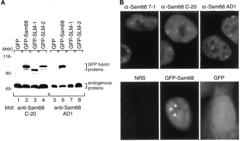
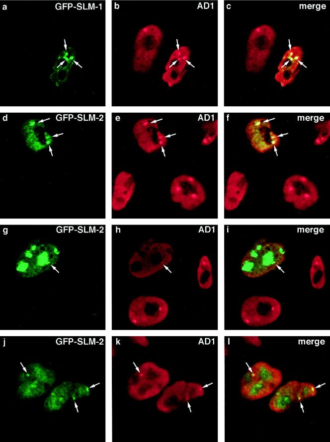
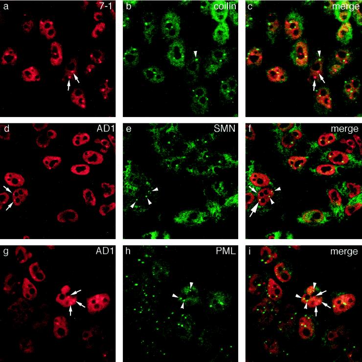
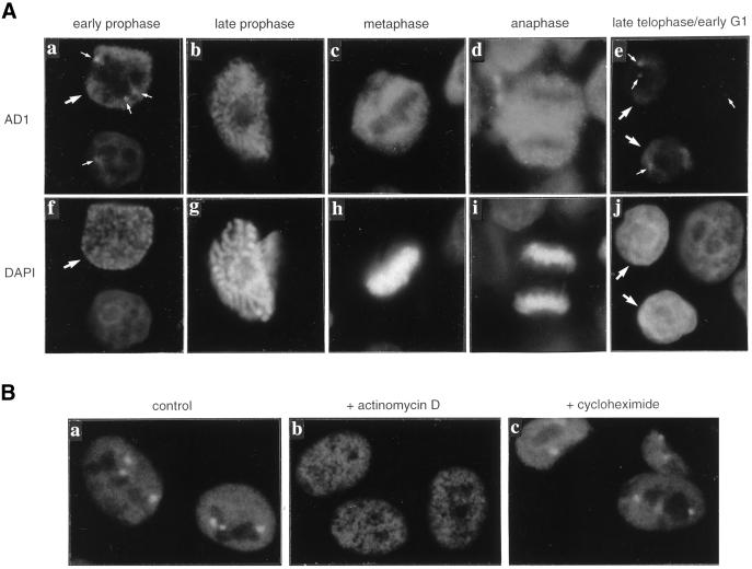
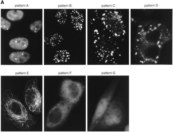
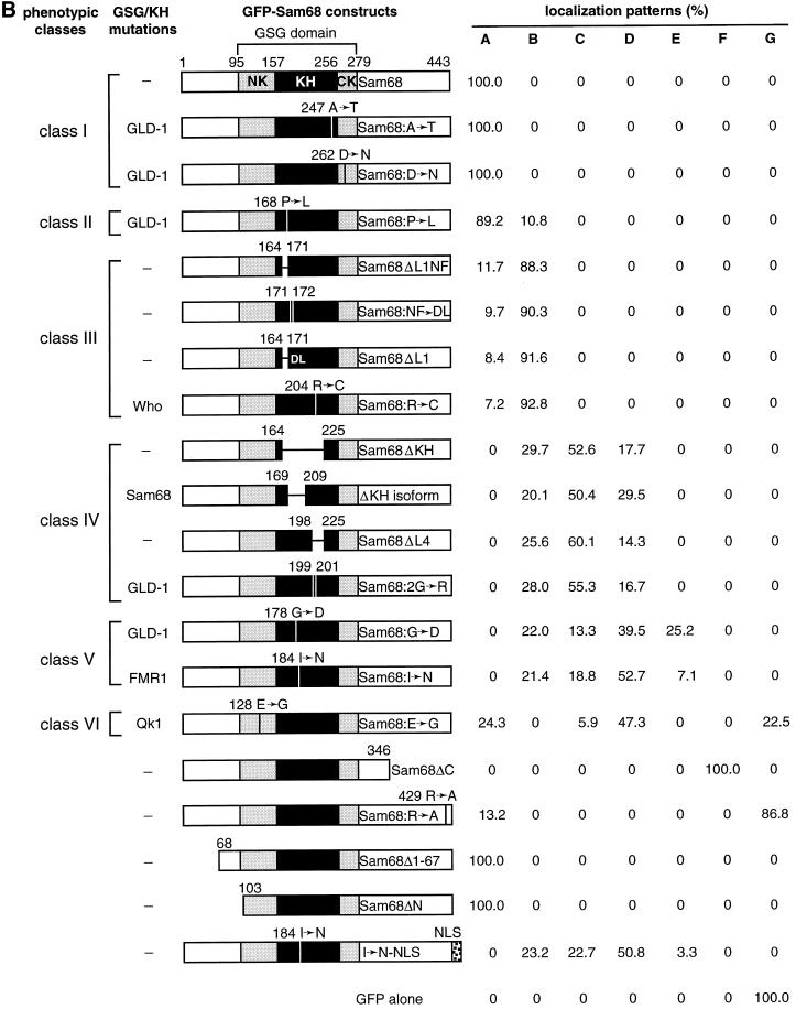
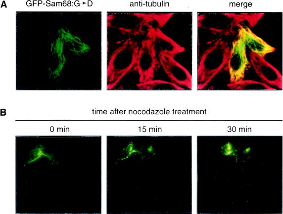
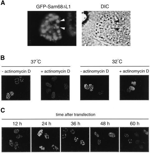
The GSG (GRP33, Sam68, GLD-1) domain is a protein module found in an expanding family of RNA-binding proteins. The numerous missense mutations identified genetically in the GSG domain support its physiological role. Although the exact function of the GSG domain is not known, it has been shown to be required for RNA binding and oligomerization. Here it is shown that the Sam68 GSG domain plays a role in protein localization. We show that Sam68 concentrates into novel nuclear structures that are predominantly found in transformed cells. These Sam68 nuclear bodies (SNBs) are distinct from coiled bodies, gems, and promyelocytic nuclear bodies. Electron microscopic studies show that SNBs are distinct structures that are enriched in phosphorus and nitrogen, indicating the presence of nucleic acids. A GFP-Sam68 fusion protein had a similar localization as endogenous Sam68 in HeLa cells, diffusely nuclear with two to five SNBs. Two other GSG proteins, the Sam68-like mammalian proteins SLM-1 and SLM-2, colocalized with endogenous Sam68 in SNBs. Different GSG domain missense mutations were investigated for Sam68 protein localization. Six separate classes of cellular patterns were obtained, including exclusive SNB localization and association with microtubules. These findings demonstrate that the GSG domain is involved in protein localization and define a new compartment for Sam68, SLM-1, and SLM-2 in cancer cell lines.


Figures

Figure 1
Figure 1
Sam68 localizes in nuclear bodies. (A) Anti-Sam68 AD1 recognizes Sam68 but not SLM-1 and SLM-2. HeLa cells transfected with GFP alone, GFP-Sam68, GFP-SLM-1, or GFP-SLM-2 were lysed and the cell lysates were divided equally, resolved by SDS-PAGE, transferred to nitrocellulose membranes, and immunoblotted with anti-Sam68 C-20 (lanes 1–4) or AD1 (lanes 5–8) antibodies. The migration of endogenous and GFP-Sam68/SLM proteins is indicated on the right, and the positions of molecular mass markers (in kilodaltons) are shown on the left. (B) HeLa cells were fixed, permeabilized, and immunostained with anti-Sam68 mAb 7-1, rabbit polyclonal Sam68/SLM C-20 antibody, rabbit polyclonal antibody AD1, or normal rabbit serum (NRS), followed by a rhodamine-conjugated goat anti-mouse or anti-rabbit secondary antibody, and visualized by fluorescence microscopy. Expression vectors encoding GFP-Sam68 or GFP alone were transfected into HeLa cells, and 12 h after transfection, live cells were visualized by fluorescence microscopy.
Figure 2
Figure 2
GFP-SLM-1 and GFP-SLM-2 colocalize with Sam68 in nuclear bodies. GFP-SLM-1 (a–c) and GFP-SLM-2 (d–l) were transfected individually in HeLa cells, and the cells were fixed, permeabilized, and immunostained with anti-Sam68 AD1 antibody followed by a rhodamine-conjugated goat anti-rabbit secondary antibody (b, e, h, and k). Colocalization was determined by confocal microscopy, and the merged images are shown on the right (c, f, i, and l). The arrows point to nuclear bodies that colocalize. Three major localization patterns were observed with GFP-SLM-2: extranucleolar staining (d), accumulation in nucleoli (g), and diffuse staining in both nucleoplasm and nucleoli (j).
Figure 3
Figure 3
Sam68 nuclear bodies are novel nuclear structures. HeLa cells were fixed, permeabilized, and double immunostained with anti-Sam68 7-1 and anti-coilin (a–c), anti-Sam68 AD1 and anti-SMN (d–f), or anti-Sam68 AD1 and anti-PML (g–i). Anti-Sam68 antibodies were followed by rhodamine-conjugated secondary antibodies (left panels), and the other antibodies were followed by FITC-conjugated secondary antibodies (middle panels). Colocalization was determined by merging the confocal images of the left and middle panels using confocal microscopy (right panels). Sam68 nuclear bodies are indicated by arrows, and coiled bodies, gems, and PML nuclear bodies are indicated by arrowheads. The large arrow in panel f indicates partial overlap between a Sam68 nuclear body and a gem.
Figure 4
Figure 4
Sam68 nuclear bodies are dynamic structures. (A) HeLa cells were fixed, permeabilized, and immunostained with anti-Sam68 AD1 antibody followed by rhodamine-conjugated goat anti-rabbit secondary antibody, and the cell nuclei were stained with DAPI. Fluorescence of representative cells in interphase (a and f, lower cells; e and j, right cells), early prophase (a and f, large arrow), late prophase (b and g), metaphase (c and h), anaphase (d and i), and late telophase/early G1 phase (e and j, large arrows) are shown. Sam68 nuclear bodies are indicated by small arrows. (B) HeLa cells were mock-treated or treated with 5 μg/ml actinomycin D (b) or 20 μg/ml cycloheximide (c) for 3 h, then fixed, permeabilized, and immunostained with anti-Sam68 AD1 antibody.
Figure 5
Figure 5
Amino acid substitutions or deletions in the Sam68 GSG domain alter Sam68 localization. (A) GFP-Sam68 constructs were transfected into HeLa cells, and 12 h after transfection the protein expression patterns were analyzed by fluorescence microscopy. The GFP fusion partner was located at the N terminus. The typical localization patterns observed are shown: diffuse nuclear staining (excluding nucleolus) with several SNBs (pattern A); exclusive SNB accumulation (pattern B); punctate nuclear and cytoplasmic staining (pattern C); punctate cytoplasmic staining (pattern D); fibrous structure (pattern E); diffuse cytoplasmic staining (pattern F); and diffuse nuclear and cytoplasmic staining (pattern G). (B) (facing page) Schematic diagram of GFP-Sam68 constructs and the quantitation of different localization patterns. In the diagrams of the constructs, the GSG domain is depicted with a bracket. The GSG domain contains an extended KH domain (denoted by the black box). The regions in the GSG domain at the N terminus of the KH domain and at the C terminus of the KH domain were called the NK and CK regions, respectively. The genes in which the genetic missense mutations were identified that alter amino acids in the GSG or KH domain are shown on the left. Δ indicates a deletion; → represents an amino acid substitution; and a horizontal or vertical thin line denotes the position of a deletion or point mutation. The NLS represents the SV40 large T antigen nuclear localization sequence (PKKKRKV). For each construct, an average of 250–300 transfected (green) cells from three separate experiments were counted, and the localization patterns were expressed as percentages. The Sam68 proteins harboring amino acid substitutions or deletions in their GSG mutations were grouped into six phenotypic classes based on their localization patterns.
Figure 5
Figure 5
Amino acid substitutions or deletions in the Sam68 GSG domain alter Sam68 localization. (A) GFP-Sam68 constructs were transfected into HeLa cells, and 12 h after transfection the protein expression patterns were analyzed by fluorescence microscopy. The GFP fusion partner was located at the N terminus. The typical localization patterns observed are shown: diffuse nuclear staining (excluding nucleolus) with several SNBs (pattern A); exclusive SNB accumulation (pattern B); punctate nuclear and cytoplasmic staining (pattern C); punctate cytoplasmic staining (pattern D); fibrous structure (pattern E); diffuse cytoplasmic staining (pattern F); and diffuse nuclear and cytoplasmic staining (pattern G). (B) (facing page) Schematic diagram of GFP-Sam68 constructs and the quantitation of different localization patterns. In the diagrams of the constructs, the GSG domain is depicted with a bracket. The GSG domain contains an extended KH domain (denoted by the black box). The regions in the GSG domain at the N terminus of the KH domain and at the C terminus of the KH domain were called the NK and CK regions, respectively. The genes in which the genetic missense mutations were identified that alter amino acids in the GSG or KH domain are shown on the left. Δ indicates a deletion; → represents an amino acid substitution; and a horizontal or vertical thin line denotes the position of a deletion or point mutation. The NLS represents the SV40 large T antigen nuclear localization sequence (PKKKRKV). For each construct, an average of 250–300 transfected (green) cells from three separate experiments were counted, and the localization patterns were expressed as percentages. The Sam68 proteins harboring amino acid substitutions or deletions in their GSG mutations were grouped into six phenotypic classes based on their localization patterns.
Figure 6
Figure 6
Sam68:G→D and Sam68:I→N associate with microtubules. (A) HeLa cells transfected with GFP-Sam68:G→D were fixed, permeabilized, and immunostained with anti-tubulin antibody followed by a rhodamine-conjugated goat anti-mouse secondary antibody and then analyzed by confocal microscopy. Colocalization of Sam68 fibers (green) with microtubule fibers (red) resulted in yellow color when the confocal images of GFP-Sam68:G→D and anti-tubulin immunostaining were merged. (B) HeLa cells transfected with GFP-Sam68:G→D were incubated with 40 ng/ml nocodazole, and cells with fibrous phenotype were photographed live before (0 min) and 15 or 30 min after the addition of nocodazole.
Figure 7
Figure 7
Characterization of GFP-Sam68ΔL1 nuclear bodies. (A) HeLa cells transfected with GFP-Sam68ΔL1 were fixed and analyzed with fluorescence and difference interference contrast (DIC) microscopy. (B) GFP-Sam68ΔL1 nuclear bodies are dynamic structures. HeLa cells were transfected with GFP-Sam68ΔL1 and then incubated at 37 or 32°C for 24 h; the last 3 h of incubation was performed with or without 5 μg/ml actinomycin D. The cells were fixed and visualized by fluorescence microscopy. (C) HeLa cells transfected with GFP-Sam68ΔL1 were fixed and observed 12, 24, 36, 48, or 60 h after transfection.
Figure 8
Figure 8
Sam68 nuclear bodies do not contain nascent RNAs and snRNPs. (A) HeLa cells transfected with GFP-Sam68 (a–c), GFP-Sam68ΔL1 (d–f), or GFP-PTB (g–i) were permeabilized with 50 μg/ml saponin for 5 min at 4°C and then incubated with a transcription cocktail containing Br-UTP for 20 min at 33°C. The cells were fixed, and the sites of Br-UTP incorporation were detected by immunolabeling using anti-BrdU antibody (which also recognizes Br-UTP) followed by a rhodamine-conjugated secondary antibody (b, e, and h). Colocalization was determined by confocal microscopy (c, f, and i). The arrows in panel i indicate colocalization of PNC with sites of Br-UTP incorporation. (B) HeLa cells (a–c) or HeLa cells transfected with GFP-Sam68ΔL1 (d–f) were fixed, permeabilized, and immunostained with anti-SC35 antibody followed by a secondary antibody conjugated to rhodamine (b and e). Endogenous Sam68 nuclear bodies were detected by immunolabeling using anti-Sam68 AD1 antibody followed by FITC-conjugated goat anti-rabbit antibody (a). Colocalization of SNBs and SC35 was analyzed by confocal microscopy (c and f). The arrows in panel f indicate partial overlap of Sam68ΔL1 SNBs with SC35, most likely random overlapping.
Figure 9
Figure 9
Correlative microscopy of Sam68 nuclear bodies. An ultrathin section (30 nm) of HeLa cells, previously labeled with anti-Sam68 AD1 antibody and embedded for electron microscopy, was examined under an immunofluorescence microscope (A) and then an electron microscope (B). The respective images were resized and rotationally aligned before being merged (C). It was then possible to identify the locations of structures labeled by immunofluorescence and to characterize them, by electron spectroscopic imaging, for phosphorus content (D) and nitrogen content (E). (Magnification: B and C, ×3,000; D and E, ×12,000).

References

    1. Andrade LEC, Tan EM, Chan EKL. Immunocytochemical analysis of the coiled body in the cell cycle and during cell proliferation. Proc Natl Acad Sci USA. 1993;90:1947–1951. - PMC - PubMed
    1. Arning S, Gruter P, Bilbe G, Kramer A. Mammalian splicing factor SF1 is encoded by variant cDNAs and binds to RNA. RNA. 1996;2:794–810. - PMC - PubMed
    1. Baehrecke EH. who encodes a KH RNA binding protein that functions in muscle development. Development. 1997;124:1323–1332. - PubMed
    1. Barlat I, Maurier F, Duchesne M, Guitard E, Tocque B, Schweighoffer F. A role for Sam68 in cell cycle progression antagonized by a spliced variant within the KH domain. J Biol Chem. 1997;272:3129–3132. - PubMed
    1. Bazett-Jones DP, Hendzel MJ. Electron spectroscopic imaging of chromatin. Methods. 1999;17:188–200. - PubMed

Publication types

MeSH terms

LinkOut - more resources