. 1999 Sep;10(9):3045-59.
doi: 10.1091/mbc.10.9.3045.

The conserved core of a human SIR2 homologue functions in yeast silencing

Affiliations
Free PMC article

The conserved core of a human SIR2 homologue functions in yeast silencing

J M Sherman et al. Mol Biol Cell. 1999 Sep.
Free PMC article

Abstract

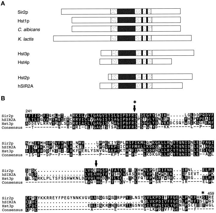
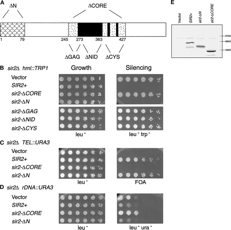
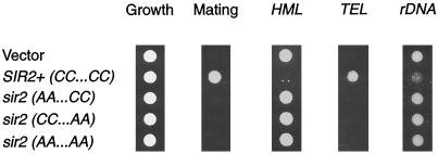
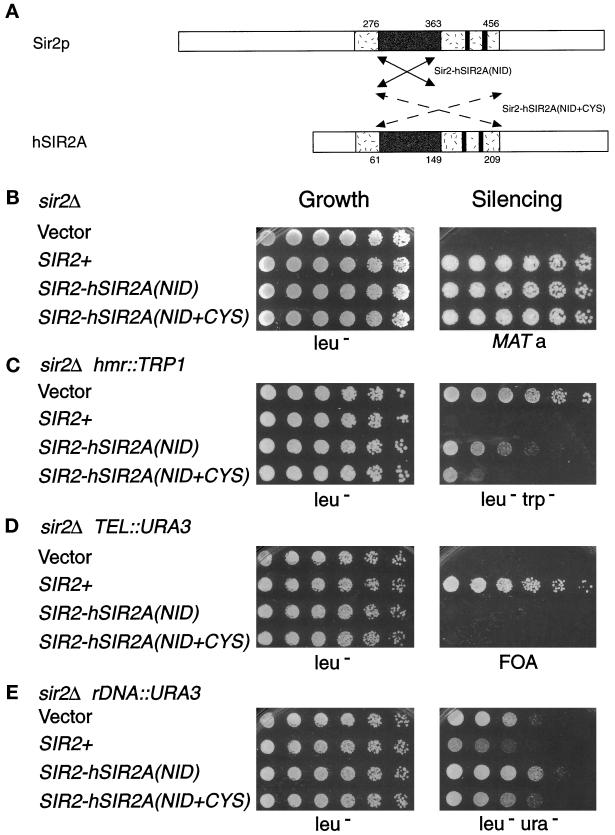
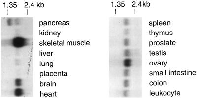
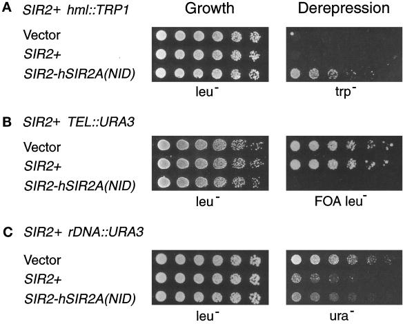
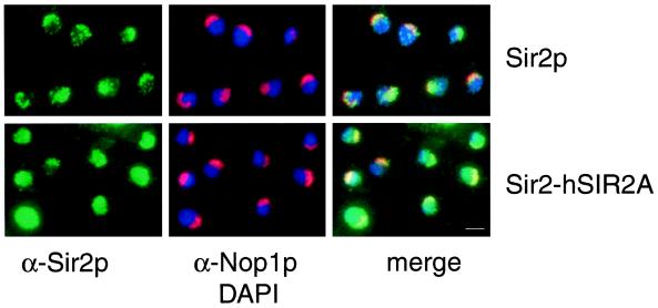
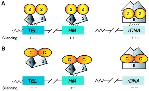
Silencing is a universal form of transcriptional regulation in which regions of the genome are reversibly inactivated by changes in chromatin structure. Sir2 (Silent Information Regulator) protein is unique among the silencing factors in Saccharomyces cerevisiae because it silences the rDNA as well as the silent mating-type loci and telomeres. Discovery of a gene family of Homologues of Sir Two (HSTs) in organisms from bacteria to humans suggests that SIR2's silencing mechanism might be conserved. The Sir2 and Hst proteins share a core domain, which includes two diagnostic sequence motifs of unknown function as well as four cysteines of a putative zinc finger. We demonstrate by mutational analyses that the conserved core and each of its motifs are essential for Sir2p silencing. Chimeras between Sir2p and a human Sir2 homologue (hSir2Ap) indicate that this human protein's core can substitute for that of Sir2p, implicating the core as a silencing domain. Immunofluorescence studies reveal partially disrupted localization, accounting for the yeast-human chimeras' ability to function at only a subset of Sir2p's target loci. Together, these results support a model for the involvement of distinct Sir2p-containing complexes in HM/telomeric and rDNA silencing and that HST family members, including the widely expressed hSir2A, may perform evolutionarily conserved functions.


Figures

Figure 1
Figure 1
Alignment of yeast Sir2p and the Hst proteins. (A) The Hst proteins from bacteria to humans are ∼30–65% identical to Sir2p overall. Sir2p and the Hst proteins all contain a characteristic core domain (shaded) that includes four cysteines of a putative zinc finger (paired lines). In the most highly conserved region of this domain (dark), bounded by the GAG and NID motifs, which are of unknown function, identity reaches 84%. The Hst proteins can be grouped into three subfamilies based on the length and sequence of their relatively distinct N and C termini. Members of the first two of these subfamilies have previously been implicated in silencing (Brachmann et al., 1995). (B) Core domain sequences of members of each of the three subfamilies (Sir2p, hSir2Ap, and Hst3p) are aligned. The consensus sequence highlights the four conserved cysteines of the putative zinc finger (at positions 372, 374, 396, and 398 in Sir2p), as well as the two conserved motifs of unknown function, which generally consist of the sequences GAG(I/V)Sxxx G(I/V)PDFRS, and (Y/I)TQNID. Some additional variation at the positions in brackets is observed for published sequences and at other positions for incomplete sequences in the databases. These motifs, although somewhat degenerate, are diagnostic of members of this gene family. The arrows and stars indicate the boundaries of the region swapped in the Sir2-hSir2A/Hst3(NID) chimeras and the Sir2-hSir2A(NID+CYS) chimera, respectively. See Brachmann et al. (1995) for additional flanking sequences.
Figure 2
Figure 2
The conserved core and its motifs are essential for Sir2p silencing. (A) The N- and C-terminal end points of the sir2 mutants are indicated diagramatically for the sir2-ΔN (ΔN, Δ2–79) and sir2-ΔCORE (ΔCORE, Δ245–427) constructs, as well as the smaller motif deletions [sir2-ΔGAG (ΔGAG, Δ245–273), sir2-ΔNID (ΔNID, Δ277–363), and sir2-ΔCYS (ΔCYS, Δ364–427)]. (B–D) Complementation of the sir2Δ silencing defects by the core and N-terminal deletion mutants. LPY1403 (MATα sir2Δ hml:: TRP1) (B), LPY1953 (MATa sir2Δ TEL:: URA3) (C), and LPY2447 (MATα sir2Δ rDNA:: URA3) (D) were transformed with vector alone (YEp351) or containing SIR2+ (pLP349), sir2-ΔCORE (pLP387), or sir2-ΔN (pLP411) constructs. LPY1403 was also transformed with plasmids carrying the motif deletion constructs [sir2-ΔGAG (pLP385), sir2-ΔNID (pLP656), and sir2-ΔCYS (pLP386)]. The transformants were assayed for growth by plating serial dilutions on leu plates (left panel) and for function by plating on leutrp (B), 5-FOA (C), and leuura (D) plates (right panel). The rDNA silencing assays were performed on leuura plates to distinguish between silencing and recombination resulting in loss of the rDNA reporter. In LPY2447, such recombination occurs at high frequency on FOA-containing medium because of selection against URA3 expression and the enhanced rDNA recombination caused by a sir2Δ mutation. (E) Immunoblot analysis was performed using an antiserum directed against a C-terminal peptide derived from Sir2p (Smith et al., 1998) on whole-cell lysates of LPY1953 transformants.
Figure 3
Figure 3
Mutation of the conserved cysteines in Sir2p causes a complete loss of silencing function. sir2Δ strains were transformed with vector alone (YEp351) or containing wild-type SIR2 (pLP349) or the cysteine point mutants (pLP531 - - AA . . CC, pLP555 - -CC . . AA, and pLP570 - - AA . . AA). Equal cell densities (1 A600) for each transformant were stamped onto leu plates (growth control) and onto selective plates to assay silencing function. Shown is a representative growth control panel and LPY1953 (MATa sir2Δ TEL::URA3) transformants stamped on a lawn of α his4 (LPY78) mating tester on minimal plates (only those that form diploids grow under these conditions) and 5-FOA to assay telomeric silencing. Also shown are LPY1403 transformants on leutrp plates to assay HM silencing and LPY2447 transformants on leuura plates to assay rDNA silencing. Quantitation indicated that the point mutants are completely nonfunctional in silencing (our unpublished results).
Figure 4
Figure 4
The Sir2-hSir2A chimeras function in transcriptional silencing. (A) The arrows denote the region of the human Sir2A core exchanged for that of the Sir2p core in the Sir2-hSir2A(NID) chimera (solid lines indicate aa 276–363 of Sir2p were replaced with aa 61–149 of hSir2Ap) and in the Sir2-hSir2A(NID+CYS) chimera (dashed lines indicate aa 276–456 of Sir2p were replaced with aa 61–209 of hSir2Ap). (See Figure 1B for the sequences exchanged in the chimeras.) (B–E) Results of dilution assays to test the SIR2-hSIR2A(NID) (pLP999) and SIR2-hSIR2A(NID+CYS) (pLP905) chimeras’ complementation of the sir2Δ mating defect in LPY1403 (MATα sir2Δ hml:: TRP1) (B), hmr::TRP1 silencing defect in LPY3923 (MATa sir2Δ hmrΔ::TRP1) (C), telomeric silencing defect in LPY1953 (MATa sir2Δ TEL::URA3) (D), and rDNA silencing defect in LPY2447 (MATα sir2Δ rDNA::URA3) (E). The left panel depicts growth of serial dilutions on leu plates as a control, and the right panel depicts growth on a his4 (LPY143) mating tester on minimal plates (B), leutrp (C), 5-FOA (D), and leuura (E).
Figure 5
Figure 5
hSIR2A mRNA is widely distributed. A multiple-tissue RNA blot was probed with an hSIR2A cDNA probe. The blot was hybridized separately with an actin cDNA probe and shown to be evenly loaded except that the skeletal muscle sample was overloaded by aproximately twofold.
Figure 6
Figure 6
The SIR2-hSIR2A(NID) chimera exhibits dominant derepression. Dilution assays were performed on LPY253 (MATa SIR2+ hml::TRP1), LPY1683 (MATa SIR2+ TEL::URA3 TEL::ADE2), and LPY2446 (MATα SIR2+ rDNA::URA3) transformed with high-copy vector alone (YEp351) or containing SIR2+ (pLP349) or the SIR2-hSIR2A(NID) chimera (pLP999). Similar results were obtained with low-copy versions of these constructs as well as with the SIR2-hSIR2A(NID+CYS) chimera on high- and low-copy plasmids (our unpublished results). Serial dilutions were plated on leu plates as a control for growth (left panel) and onto trp, 5-FOA leu, and ura to assay dominance (right panel).
Figure 7
Figure 7
The Sir2-hSir2A-(NID+CYS) chimera fails to localize properly within the nucleus. SIR2+ and SIR2-hSIR2A-(NID+CYS) plasmids (pLP907, top row, and pLP888, bottom row, respectively) were introduced into the homozygous sir2/sir2 diploid strain LPY3380. Immunofluorescence was performed by staining cells with anti-Sir2p antibodies (to localize Sir2p and the chimeras, in green), anti-Nop1p antibodies (to identify the nucleolus, in red), and DAPI (to identify DNA, in blue). At right are merged images of the left and center panels. Bar, 2 μm.
Figure 8
Figure 8
Models for Sir2p silencing function and the involvement of distinct Sir2p-containing complexes in HM/telomeric and rDNA silencing. (A) Wild-type Sir2p function. Sir2p interacts with itself and Sir3p/Sir4p in a complex (Moazed and Johnson, 1996; Holmes et al., 1997; Moazed et al., 1997; Strahl-Bolsinger et al., 1997) and localizes to and silences the HM loci and telomeres (Hecht et al., 1996; Gotta et al., 1997; Strahl-Bolsinger et al., 1997). Because Sir3p and Sir4p are not required for rDNA silencing (Smith and Boeke, 1997), a second Sir2p-containing complex involving one or more unidentified factors (I and II) localizes to and silences the rDNA. Single silencing complexes are shown for simplicity, although it is believed that numerous complexes, perhaps in multimeric forms, localize to and silence each of the loci. (B) Sir2-hSir2A chimera function. The chimeras continue to dimerize or oligomerize and interact with other silencing factors. They localize to the HM loci to silence them but fail to localize properly to the telomeres and rDNA, leading to a loss of silencing. In the presence of wild-type Sir2p, Sir2p-chimera heteromers form. These heteromeric forms continue to interact with HM and telomeric silencing factor(s), e.g., Sir3p and Sir4p, and titrate them, leading to a loss of silencing at these loci. On the other hand, the Sir2p-chimera heteromers interact correctly with rDNA silencing factors (I and II) and localize subnucleolarly, leading to rDNA silencing function. Although Sir2p and the Sir2p-hSir2A chimeras are diagrammed as dimers based on wild-type Sir2p’s demonstrated ability to interact with itself (Moazed et al., 1997), the functional form of Sir2p and its variants has yet to be determined. Thus, they may function as higher-order multimers, dimers, or even monomers. Furthermore, the functional form of the Sir2-hSir2A chimeras may differ from that of wild-type Sir2p.

Similar articles

Cited by

References

    1. Aparicio OM, Billington BL, Gottschling DE. Modifiers of position effect are shared between telomeric and silent mating-type loci in S. cerevisiae. Cell. 1991;66:1279–1287. - PubMed
    1. Aris JP, Blobel G. Identification and characterization of a yeast nucleolar protein that is similar to a rat liver nucleolar protein. J Cell Biol. 1988;107:17–31. - PMC - PubMed
    1. Brachmann CB, Sherman JM, Devine SE, Cameron EE, Pillus L, Boeke JD. The SIR2 gene family, conserved from bacteria to humans, functions in silencing, cell cycle progression and chromosome stability. Genes & Dev. 1995;9:2888–2902. - PubMed
    1. Braunstein M, Rose AB, Holmes SG, Allis CD, Broach JR. Transcriptional silencing in yeast is associated with reduced nucleosome acetylation. Genes & Dev. 1993;7:592–604. - PubMed
    1. Braunstein M, Sobel R, Allis CD, Turner BM, Broach JR. Efficient transcriptional silencing in Saccharomyces cerevisiae requires a heterochromatin histone acetylation pattern. Mol Cell Biol. 1996;16:4349–4356. - PMC - PubMed

Publication types

MeSH terms

LinkOut - more resources