Review
. 1999 Sep;63(3):708-24.
doi: 10.1128/MMBR.63.3.708-724.1999.

Fungal resistance to plant antibiotics as a mechanism of pathogenesis

Affiliations
Review

Fungal resistance to plant antibiotics as a mechanism of pathogenesis

J P Morrissey et al. Microbiol Mol Biol Rev. 1999 Sep.

Abstract

Many plants produce low-molecular-weight compounds which inhibit the growth of phytopathogenic fungi in vitro. These compounds may be preformed inhibitors that are present constitutively in healthy plants (also known as phytoanticipins), or they may be synthesized in response to pathogen attack (phytoalexins). Successful pathogens must be able to circumvent or overcome these antifungal defenses, and this review focuses on the significance of fungal resistance to plant antibiotics as a mechanism of pathogenesis. There is increasing evidence that resistance of fungal pathogens to plant antibiotics can be important for pathogenicity, at least for some fungus-plant interactions. This evidence has emerged largely from studies of fungal degradative enzymes and also from experiments in which plants with altered levels of antifungal secondary metabolites were generated. Whereas the emphasis to date has been on degradative mechanisms of resistance of phytopathogenic fungi to antifungal secondary metabolites, in the future we are likely to see a rapid expansion in our knowledge of alternative mechanisms of resistance. These may include membrane efflux systems of the kind associated with multidrug resistance and innate resistance due to insensitivity of the target site. The manipulation of plant biosynthetic pathways to give altered antibiotic profiles will also be valuable in telling us more about the significance of antifungal secondary metabolites for plant defense and clearly has great potential for enhancing disease resistance for commercial purposes.


Figures

FIG. 1
FIG. 1
Localization of antifungal compounds in plants. (A) Fluorescence of the saponin avenacin A-1 under UV light. This compound is localized in the epidermal cell layer of oat roots (144). (B) A resistant sorghum line responding to attack by Colletotrichum graminicola, showing the formation and mobilization of vesicles containing pigmented 3-deoxyanthocyanidin phytoalexins toward the site of attempted penetration by an appressorium (visualized by light microscopy) (196, 197). (C) Accumulation of elemental sulfur (S8) in the vascular tissue of a resistant cocoa line in response to attack by the xylem-invading fungus Verticillium dahliae (29). An X-ray map shows sulfur (white) accumulating in a xylem parenchyma cell (XP); xylem vessels are also labelled (V). Panels A and C are reproduced from references and , respectively, with permission of the publisher. Panel B was kindly supplied by R. Nicholson.
FIG. 2
FIG. 2
Examples of antifungal saponins from oats and solanaceous plants. The major oat root saponin avenacin A-1 is one of a family of four related triterpenoid saponins. Oat leaves contain a different family of saponins, the steroidal molecules avenacosides A and B. Avenacoside A is not shown but differs from avenacoside B only in that it lacks the terminal β-1-3-linked d-glucose molecule. The avenacosides are biologically inactive but are converted to the antifungal molecules 26-desglucoavenacosides A and B by a plant enzyme which hydrolyzes the d-glucose molecule attached to C-26. α-Tomatine and α-chaconine are both steroidal glycoalkaloids and are found in tomato and potato, respectively.
FIG. 3
FIG. 3
Proposed models for membrane disruption by saponins. Saponins complex with sterols in membranes, ultimately to form aggregates. Aggregation may then lead to the formation of membrane pores, as shown on the left (6), or to the extraction of sterols from the membrane, with the formation of tubular or spherical complexes outside the membrane (96).
FIG. 4
FIG. 4
Detoxification of α-tomatine by some fungal pathogens of tomato. Tomatinases produced by Botrytis cinerea, Septoria lycopersici, and Fusarium oxysporum f. sp. lycopersici have different mechanisms of action, as shown by the arrows indicating cleavage sites.
FIG. 5
FIG. 5
Examples of cyanogenic glucosides, glucosinolates, and their degradation products. Cyanogenic glycosides and glucosinolates are both activated by plant enzymes in response to tissue damage. Breakdown of cyanogenic glycosides results in the formation of hydrogen cyanide (A), while breakdown of glucosinolate generates products which include isothiocyanates, nitriles, and thiocyanates (B).
FIG. 6
FIG. 6
Cyclic hydroxamic acids and their degradation products. (A) The cyclic hydroxamic acids DIMBOA and DIBOA occur in plants as glucosides (DIMBOA-glu and DIBOA-glu), which are converted to the antifungal aglycones DIMBOA and DIBOA by plant enzymes. These aglycones rapidly decompose to the benzoxazolinones MBOA (6-methoxy-2-benzoxazolinone) and BOA (2-benzoxazolinone), respectively, which are also fungitoxic. (B) Degradation of benzoxazolinones by cereal pathogens. Some isolates of G. graminis and of different Fusarium species can degrade MBOA and BOA to the less toxic malonamic acids via an o-aminophenyl intermediate. Detoxification of BOA to 2-amino-3-H-phenaxazin-3-one by G. graminis has also been reported.
FIG. 7
FIG. 7
Preformed antifungal molecules in subtropical fruit. (A) Resorcinols, which occur in the peel of mango fruit; (B) alkenes, which are present in the peel of avocado fruit.
FIG. 8
FIG. 8
The stilbene phytoalexin trans-resveratrol.
FIG. 9
FIG. 9
Detoxification of the potato phytoalexins rishitin and lubimin by Gibberella pulicaris.
FIG. 10
FIG. 10
Detoxification of phytoalexins from crucifers by fungal pathogens. (A) The phytoalexin camalexin is produced by some wild crucifers including Arabidopsis thaliana. The root pathogen Rhizoctonia solani can metabolize camalexin to 5-hydroxycamalexin, which is less toxic to fungal growth. (B) Brassinin is one of a family of phytoalexins produced by some cultivated brassicas. Brassinin can be oxidized to the less toxic molecule brassinin-S-oxide by the blackleg fungus, Leptosphaeria maculans, and is then further metabolized to indole-3-carboxaldehyde and indole-3-carboxylic acid.
FIG. 11
FIG. 11
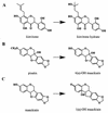
Isoflavonoid phytoalexins from legumes and some of their fungal degradation products. Kievitone, pisatin, and maackiain are isoflavonoid phytoalexins produced by bean, pea, and chickpea. Kievitone is detoxified by the bean pathogen Fusarium solani f. sp. phaseoli, while pisatin and maackiain are degraded by isolates of Nectria haematococca (Fusarium solani), which are pathogenic to pea or chickpea, respectively.

Similar articles

Cited by

References

    1. Adrian M, Jeandet P, Veneau J, Weston L A, Bessis R. Biological activity of resveratrol, a stilbenic compound from grapevines, against Botrytis cinerea, the causal agent for gray mould. J Chem Ecol. 1997;23:1689–1702.
    1. Adrian M, Rajaei H, Jeandet P, Veneau J, Bessis R. Resveratrol oxidation in Botrytis cinerea conidia. Phytopathology. 1998;88:472–476. - PubMed
    1. Ardi R, Kobiler I, Jacoby B, Keen N T, Prusky D. Involvement of epicatechin biosynthesis in the activation of the mechanism of resistance of avocado fruits to Colletotrichum gloeosporioides. Physiol Mol Plant Pathol. 1998;53:269–285.
    1. Argandoña V H, Corcuera L J. Distribution of hydroxamic acids in Zea mays tissues. Phytochemistry. 1985;24:177–178.
    1. Argandoña V H, Zúniga G E, Corcuera L J. Distribution of gramine and hydroxamic acids in barley and wheat leaves. Phytochemistry. 1987;26:1917–1918.

Publication types