Role of alpha(v) integrins in adenovirus cell entry and gene delivery
- PMID: 10477314
- PMCID: PMC103752
- DOI: 10.1128/MMBR.63.3.725-734.1999
Role of alpha(v) integrins in adenovirus cell entry and gene delivery
Abstract
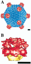
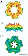
Adenoviruses (Ad) are a significant cause of acute infections in humans; however, replication-defective forms of this virus are currently under investigation for human gene therapy. Approximately 20 to 25% of all the gene therapy trials (phases I to III) conducted over the past 10 years involve the use of Ad gene delivery for treatment inherited or acquired diseases. At present, the most promising applications involve the use of Ad vectors to irradicate certain nonmetastatic tumors and to promote angiogenesis in order to alleviate cardiovascular disease. While specific problems of using Ad vectors remain to be overcome (as is true for almost all viral and nonviral delivery methods), a distinct advantage of Ad is the extensive knowledge of its macromolecular structure, genome organization, sequence, and mode of replication. Moreover, significant information has also been acquired on the interaction of Ad particles with distinct host cell receptors, events which strongly affect virus tropism. This review provides an overview of the structure and function of Ad attachment (coxsackievirus and Ad receptor [CAR]) and internalization (alpha(v) integrins) receptors and discusses their precise role in virus infection and gene delivery. Recent structure studies of integrin-Ad complexes by cryoelectron microscopy are also highlighted. Finally, unanswered questions arising from the current state of knowledge of Ad-receptor interactions are presented in the context of improving Ad vectors for future human gene therapy applications.
Figures


Similar articles
-
Reduction of natural adenovirus tropism to mouse liver by fiber-shaft exchange in combination with both CAR- and alphav integrin-binding ablation.J Virol. 2003 Dec;77(24):13062-72. doi: 10.1128/jvi.77.24.13062-13072.2003. J Virol. 2003. PMID: 14645563 Free PMC article.
-
Adenovirus interaction with distinct integrins mediates separate events in cell entry and gene delivery to hematopoietic cells.J Virol. 1996 Jul;70(7):4502-8. doi: 10.1128/JVI.70.7.4502-4508.1996. J Virol. 1996. PMID: 8676475 Free PMC article.
-
Modified adenoviral vectors ablated for coxsackievirus-adenovirus receptor, alphav integrin, and heparan sulfate binding reduce in vivo tissue transduction and toxicity.Hum Gene Ther. 2006 Mar;17(3):264-79. doi: 10.1089/hum.2006.17.264. Hum Gene Ther. 2006. PMID: 16544976
-
Integrin-Targeting Strategies for Adenovirus Gene Therapy.Viruses. 2024 May 13;16(5):770. doi: 10.3390/v16050770. Viruses. 2024. PMID: 38793651 Free PMC article. Review.
-
Tropism and transduction of oncolytic adenovirus 5 vectors in cancer therapy: Focus on fiber chimerism and mosaicism, hexon and pIX.Virus Res. 2018 Sep 15;257:40-51. doi: 10.1016/j.virusres.2018.08.012. Epub 2018 Aug 17. Virus Res. 2018. PMID: 30125593 Review.
Cited by
-
Identification of sites in adenovirus hexon for foreign peptide incorporation.J Virol. 2005 Mar;79(6):3382-90. doi: 10.1128/JVI.79.6.3382-3390.2005. J Virol. 2005. PMID: 15731232 Free PMC article.
-
Regulatable gene expression systems for gene therapy.Curr Gene Ther. 2006 Aug;6(4):421-38. doi: 10.2174/156652306777934829. Curr Gene Ther. 2006. PMID: 16918333 Review.
-
Adenovirus-activated PKA and p38/MAPK pathways boost microtubule-mediated nuclear targeting of virus.EMBO J. 2001 Mar 15;20(6):1310-9. doi: 10.1093/emboj/20.6.1310. EMBO J. 2001. PMID: 11250897 Free PMC article.
-
Emerging roles for ubiquitin in adenovirus cell entry.Biol Cell. 2012 Mar;104(3):188-98. doi: 10.1111/boc.201100096. Epub 2012 Feb 27. Biol Cell. 2012. PMID: 22251092 Free PMC article. Review.
-
The transduction of Coxsackie and Adenovirus Receptor-negative cells and protection against neutralizing antibodies by HPMA-co-oligolysine copolymer-coated adenovirus.Biomaterials. 2011 Dec;32(35):9536-45. doi: 10.1016/j.biomaterials.2011.08.069. Epub 2011 Sep 28. Biomaterials. 2011. PMID: 21959008 Free PMC article.
References
-
- Acharya R, Fry E, Stuart D, Fox G, Rowlands D, Brown F. The three-dimensional structure of foot-and-mouth disease virus at 2.9 Å resolution. Nature. 1989;337:709–716. - PubMed
-
- Akke M, Liu J, Cavanagh J, Erickson H P, Palmer A G., III Pervasive conformational fluctuations on microsecond time scales in a fibronectin type III domain. Nat Struct Biol. 1998;5:55–59. - PubMed
-
- Bergelson J M, Cunningham J A, Droguett G, Kurt-Jones E A, Krithivas A, Hong J S, Horwitz M S, Crowell R L, Finberg R W. Isolation of a common receptor for coxsackie B viruses and adenoviruses 2 and 5. Science. 1997;275:1320–1323. - PubMed
Publication types
MeSH terms
Substances
Grants and funding
LinkOut - more resources
Full Text Sources
Other Literature Sources
Miscellaneous

