. 1999 Sep 6;190(5):607-15.
doi: 10.1084/jem.190.5.607.

Alteration of interleukin 4 production results in the inhibition of T helper type 2 cell-dominated inflammatory bowel disease in T cell receptor alpha chain-deficient mice

Affiliations

Alteration of interleukin 4 production results in the inhibition of T helper type 2 cell-dominated inflammatory bowel disease in T cell receptor alpha chain-deficient mice

H Iijima et al. J Exp Med. .

Abstract

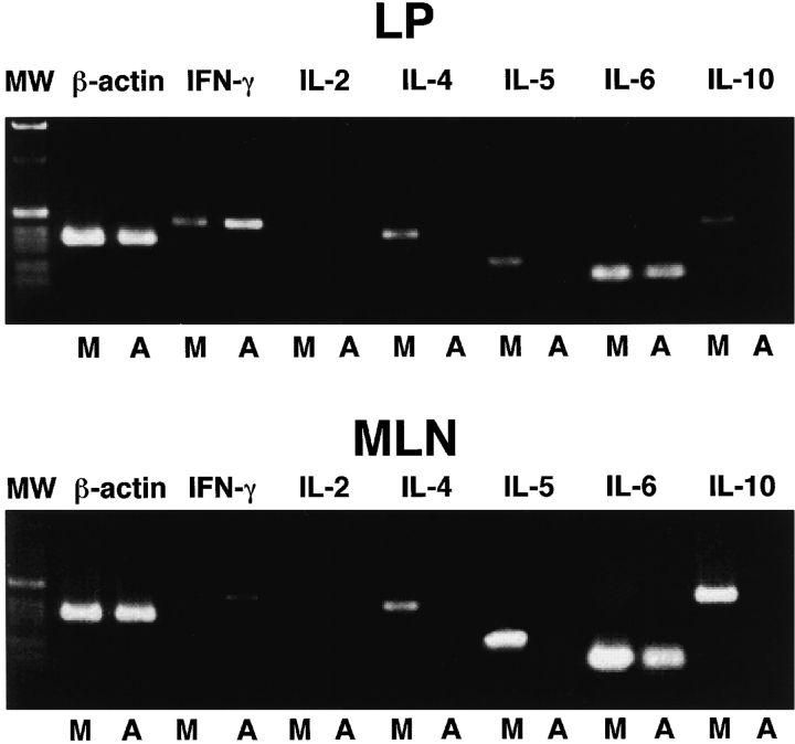
T cell receptor alpha chain-deficient (TCR-alpha(-/-)) mice are known to spontaneously develop inflammatory bowel disease (IBD). The colitis that develops in these mice is associated with increased numbers of T helper cell (Th)2-type CD4(+)TCR-betabeta (CD4(+)betabeta) T cells producing predominantly interleukin (IL)-4. To investigate the role of these Th2-type CD4(+)betabeta T cells, we treated TCR-alpha(-/-) mice with anti-IL-4 monoclonal antibody (mAb). Approximately 60% of TCR-alpha(-/-) mice, including those treated with mock Ab and those left untreated, spontaneously developed IBD. However, anti-IL-4 mAb-treated mice exhibited no clinical or histological signs of IBD, and their levels of mucosal and systemic Ab responses were lower than those of mock Ab-treated mice. Although TCR-alpha(-/-) mice treated with either specific or mock Ab developed CD4(+)betabeta T cells, only those treated with anti-IL-4 mAb showed a decrease in Th2-type cytokine production at the level of mRNA and protein and an increase in interferon gamma-specific expression. These findings suggest that IL-4-producing Th2-type CD4(+)betabeta T cells play a major immunopathological role in the induction of IBD in TCR-alpha(-/-) mice, a role that anti-IL-4 mAb inhibits by causing Th2-type CD4(+)betabeta T cells to shift to the Th1 type.


Figures

Figure 1
Figure 1
Comparison of Ig levels in serum and fecal extracts of TCR-α−/− mice treated with anti–IL-4 mAb (hatched bars) or rat IgG2b (mock Ab, black bars). (A) The levels of IgA, IgG, and IgM Abs in serum and fecal extracts were analyzed by ELISA. (B) The levels of IgG subclass Ab were also analyzed by ELISA. Data represent the mean ± SEM from eight mice per group. *Significantly different from each other (P < 0.01) by Student's t test.
Figure 1
Figure 1
Comparison of Ig levels in serum and fecal extracts of TCR-α−/− mice treated with anti–IL-4 mAb (hatched bars) or rat IgG2b (mock Ab, black bars). (A) The levels of IgA, IgG, and IgM Abs in serum and fecal extracts were analyzed by ELISA. (B) The levels of IgG subclass Ab were also analyzed by ELISA. Data represent the mean ± SEM from eight mice per group. *Significantly different from each other (P < 0.01) by Student's t test.
Figure 7
Figure 7
Histological analysis of TCR-α−/− mice treated with or without anti–IL-4. Rectums of TCR-α−/− mice treated with anti–IL-4 mAb or mock Ab were resected and routine histology, including fixing with formalin, embedding in paraffin, and staining with hematoxylin and eosin, was performed.
Figure 7
Figure 7
Histological analysis of TCR-α−/− mice treated with or without anti–IL-4. Rectums of TCR-α−/− mice treated with anti–IL-4 mAb or mock Ab were resected and routine histology, including fixing with formalin, embedding in paraffin, and staining with hematoxylin and eosin, was performed.
Figure 2
Figure 2
Enumeration of Ab-producing cells in systemic and mucosal lymphoid tissues from mice treated with anti–IL-4 mAb (hatched bars) or mock Ab (black bars). Mononuclear cells isolated from SP, MLNs, and colonic LP (LPL) of TCR-α−/− mice treated with anti–IL-4 mAb or rat IgG2b (mock Ab) were examined by isotype-specific ELISPOT. Data represent the mean ± SEM from five mice per group of three separate experiments. *Significantly different from each other (P < 0.01) by Student's t test.
Figure 3
Figure 3
Flow cytometric analysis of CD4+ββ T cells in the colonic LP of TCR-α−/− mice treated with/without anti–IL-4 mAb. Mononuclear cells from the colonic LP of TCR-α−/− mice treated with anti–IL-4 mAb or mock Ab were isolated and costained with anti-CD4 (L3T4) and anti–TCR-β (H57-597) mAbs. Flow cytometric analysis was performed by FACScan™.
Figure 4
Figure 4
Cytokine-specific mRNA expression by CD4+ββ T cells in the mucosal compartment of TCR-α−/− mice treated with or without anti–IL-4 mAb. CD4+ββ T cells in the MLNs and colonic LP of TCR-α−/− mice treated with anti–IL-4 mAb (A) or mock Ab (M) were purified by flow cytometry, and cytokine-specific mRNA expression was analyzed by Th1 and Th2 cytokine-specific RT-PCR. The far left column (MW) shows a 1-kb DNA ladder.
Figure 5
Figure 5
Cytokine production by colonic lymphocytes isolated from mice with anti–IL-4 mAb (hatched bars) or mock Ab (black bars) treatment. Colonic lymphocytes were stimulated in vitro with precoated anti-CD3∈ mAb (a-CD3) or anti–TCR-β mAb (a-TCRβ) for 72 h. Culture supernatants were then collected and cytokine production was analyzed by cytokine ELISA. Data represent the mean ± SEM from two mice per group of three different experiments.
Figure 6
Figure 6
Body weight of TCR-α−/− mice treated with/without anti–IL-4 mAb treatment. Body weight of TCR-α−/− mice treated with anti–IL-4 mAb (BVD4-1D11) or mock Ab (R35-38) was measured weekly for 25 wk. Data represent the mean ± SEM of eight mice per group.
Figure 8
Figure 8
Histological analysis of goblet cells in the intestinal epithelium of TCR-α−/− mice treated with or without anti–IL-4. Rectums of TCR-α−/− mice treated with anti–IL-4 mAb or mock Ab were resected and the periodic acid–Schiff–alcian blue procedure was performed, which includes fixing with formalin, embedding in paraffin, and staining with periodic acid–Schiff–alcian blue.
Figure 8
Figure 8
Histological analysis of goblet cells in the intestinal epithelium of TCR-α−/− mice treated with or without anti–IL-4. Rectums of TCR-α−/− mice treated with anti–IL-4 mAb or mock Ab were resected and the periodic acid–Schiff–alcian blue procedure was performed, which includes fixing with formalin, embedding in paraffin, and staining with periodic acid–Schiff–alcian blue.

References

    1. Powrie P. T cells in inflammatory bowel diseaseprotective and pathogenic roles. Immunity. 1995;3:171–174. - PubMed
    1. Mombaerts P., Mizoguchi E., Grusby M.J., Glimcher L.H., Bhan A.K., Tonegawa S. Spontaneous development of inflammatory bowel disease in T cell receptor mutant mice. Cell. 1993;75:274–282. - PubMed
    1. Strober W., Ehrhardt R.O. Chronic intestinal inflammationan expected outcome in cytokine or T cell receptor mutant mice. Cell. 1993;75:203–205. - PubMed
    1. Sadlack B., Merz H., Schimpl A., Feller A.C., Horak I. Ulcerative colitis-like disease in mice with a disrupted interleukin-2 gene. Cell. 1993;75:253–261. - PubMed
    1. Watanabe M., Ueno Y., Yajima T., Okamoto S., Hayashi T., Yamazaki M., Iwao Y., Ishii H., Habu S., Uehira M. Interleukin 7 transgenic mice develop chronic colitis with diseased interleukin 7 protein accumulation in the colonic mucosa. J. Exp. Med. 1998;187:389–402. - PMC - PubMed

Publication types

MeSH terms