. 1999 Sep 6;190(5):629-38.
doi: 10.1084/jem.190.5.629.

The requirement of membrane lymphotoxin for the presence of dendritic cells in lymphoid tissues

Affiliations

The requirement of membrane lymphotoxin for the presence of dendritic cells in lymphoid tissues

Q Wu et al. J Exp Med. .

Abstract

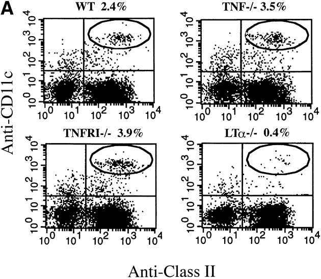
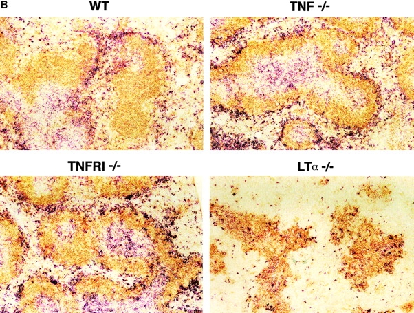
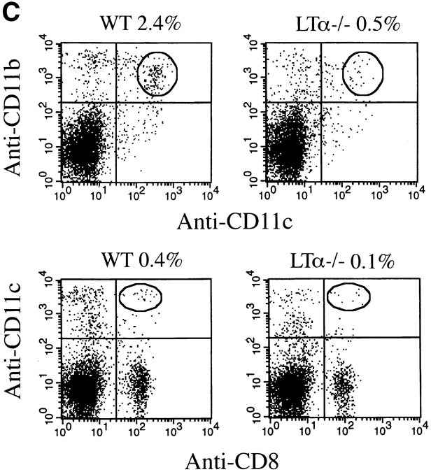
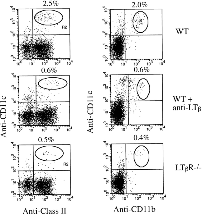
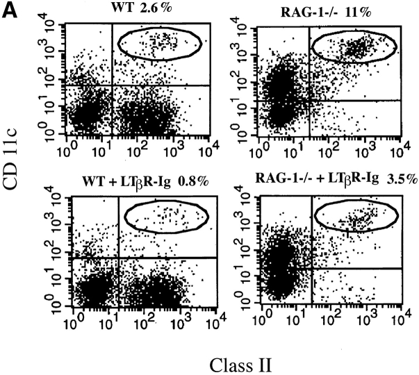
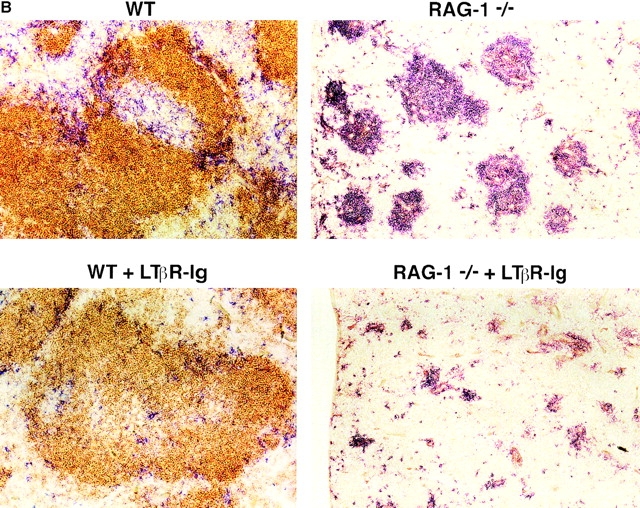
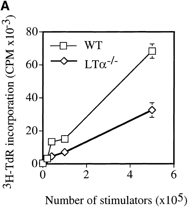
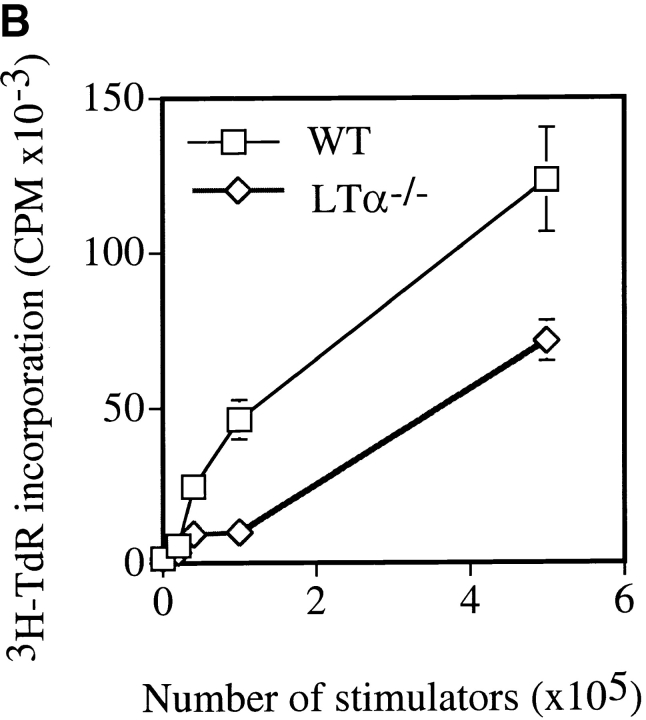
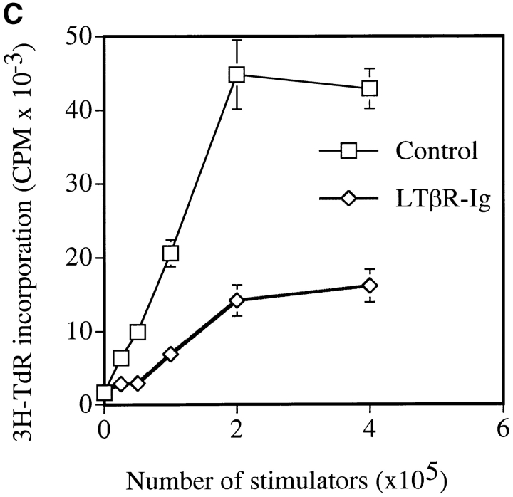
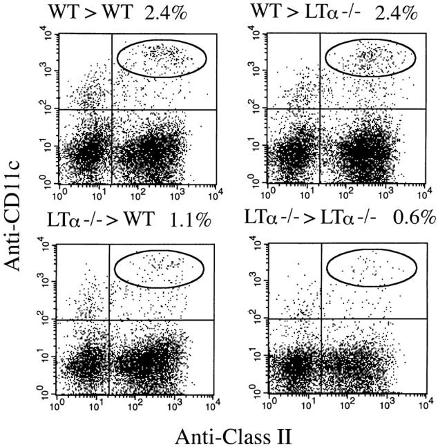
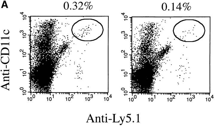
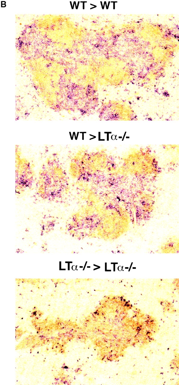
Although several cytokines, including tumor necrosis factor (TNF), can promote the growth of dendritic cells (DCs) in vitro, the cytokines that naturally regulate DC development and function in vivo have not been well defined. Here, we report that membrane lymphotoxin (LT), instead of TNF, regulates the migration of DCs in the spleen. LTalpha(-/-) mice, lacking membrane LTalpha/beta and LTalpha(3), show markedly reduced numbers of DCs in the spleen. Unlike wild-type mice and TNF(-/-) mice that have densely clustered DCs in the T cell zone and around the marginal zone, splenic DCs in LTalpha(-/-) mice are randomly distributed. The reduced number of DCs in lymphoid tissues of LTalpha(-/-) mice is associated with an increased number of DCs in nonlymphoid tissues. The number of splenic DCs in LTalpha(-/-) mice is restored when additional LT-expressing cells are provided. Blocking membrane LTalpha/beta in wild-type mice markedly diminishes the accumulation of DCs in lymphoid tissues. These data suggest that membrane LT is an essential ligand for the presence of DCs in the spleen. Mice deficient in TNF receptor, which is the receptor for both soluble LTalpha(3) and TNF-alpha(3) trimers, have normal numbers of DCs. However, LTbetaR(-/-) mice show reduced numbers of DCs, similar to the mice lacking membrane LT alpha/beta. Taken together, these results support the notion that the signaling via LTbetaR by membrane LTalpha/beta is required for the presence of DCs in lymphoid tissues.


Figures

Figure 1
Figure 1
Determination of DC number in mice deficient in LTα, TNF, or TNFR. Spleens from 6–10-wk-old wt (WT), TNF−/−, TNFR−/−, and LTα−/− mice were collected. (A) Splenocytes were stained for the DC marker (CD11c) and class II marker (I-Ab) as indicated. (B) The frozen spleen sections were stained for anti-CD11c antibody for DC (red) and anti-B220 antibody for B cells (brown). Data from one of five representative experiments is shown. (C) Both lymphoid and myeloid DC subsets are reduced in LTα−/− mice. The splenocytes were stained for DC subsets with CD11b/CD11c and CD11c/CD8α as indicated.
Figure 1
Figure 1
Determination of DC number in mice deficient in LTα, TNF, or TNFR. Spleens from 6–10-wk-old wt (WT), TNF−/−, TNFR−/−, and LTα−/− mice were collected. (A) Splenocytes were stained for the DC marker (CD11c) and class II marker (I-Ab) as indicated. (B) The frozen spleen sections were stained for anti-CD11c antibody for DC (red) and anti-B220 antibody for B cells (brown). Data from one of five representative experiments is shown. (C) Both lymphoid and myeloid DC subsets are reduced in LTα−/− mice. The splenocytes were stained for DC subsets with CD11b/CD11c and CD11c/CD8α as indicated.
Figure 3
Figure 3
Signaling via LTβR by LTα1β2 is required for the presence of DCs in the spleen. Splenocytes from mice treated with anti-LTβ antibody or LTβR−/− mice were enzymatically treated and stained for the DC marker (CD11c) and class II marker (I-Ab) (left panels) or CD11c and CD11b (right panels).
Figure 2
Figure 2
Reduced numbers of DCs in the mice treated with LTβR–Ig. Wild-type (WT) mice (left) and RAG-1−/− mice (right) were treated with 50 μg human LFA-3–Ig (top) or LTβR–Ig (bottom). The spleens were collected 10 d after treatment. (A) The number of splenic DCs determined by flow cytometry analysis. (B) Distribution of DC clusters determined by immunohistology. The frozen spleen sections from these mice were stained for anti-CD11c antibody (red) and anti-B220 antibody (brown). Data from one of six experiments is presented.
Figure 2
Figure 2
Reduced numbers of DCs in the mice treated with LTβR–Ig. Wild-type (WT) mice (left) and RAG-1−/− mice (right) were treated with 50 μg human LFA-3–Ig (top) or LTβR–Ig (bottom). The spleens were collected 10 d after treatment. (A) The number of splenic DCs determined by flow cytometry analysis. (B) Distribution of DC clusters determined by immunohistology. The frozen spleen sections from these mice were stained for anti-CD11c antibody (red) and anti-B220 antibody (brown). Data from one of six experiments is presented.
Figure 6
Figure 6
Impaired ability to stimulate allogenic T cells by splenocytes from LTα−/− mice or wt mice treated with LTβR–Ig in mixed lymphocyte reaction assay. Responder LN cells from BALB/c mice were cultured with a titration of irradiated splenocytes from C57BL/6 or LTα−/− mice prepared by mechanic pressure (A), collagenase digestion (B), or from mice pretreated with LTβR–Ig for 10 d (C). Data from one of four experiments is presented.
Figure 6
Figure 6
Impaired ability to stimulate allogenic T cells by splenocytes from LTα−/− mice or wt mice treated with LTβR–Ig in mixed lymphocyte reaction assay. Responder LN cells from BALB/c mice were cultured with a titration of irradiated splenocytes from C57BL/6 or LTα−/− mice prepared by mechanic pressure (A), collagenase digestion (B), or from mice pretreated with LTβR–Ig for 10 d (C). Data from one of four experiments is presented.
Figure 5
Figure 5
Determination of the distribution of splenic DCs by BM-derived cells in an LT-dependent fashion. BM reconstitution was performed according to previously described methods 12. 6 wk after BM reconstitution, spleens were collected and treated by using collagenase 24. The cells were stained for anti-CD11c and anti–class II antibody. Data from one of three experiments is presented.
Figure 4
Figure 4
Migration of wt DCs into the spleens of LTα−/− mice in the absence or presence of LT-expressing cells. (A) Impaired migration of DCs in LTα−/− mice. BMDCs (5 × 10⁶) from Ly5.1 mice expanded in vitro in the presence of GM-CSF and IL-4 were transferred to sublethally irradiated LTα−/− mice (right) and C57BL/6 mice (left). The spleens were collected 24 h later and stained for Ly5.1 and CD11c. One of four experiments is represented. CD11c cells from donor origin are comparable in both groups (∼0.25%). (B) Restoration of the splenic DCs in LTα−/− mice 10 d after cotransfer of LT-expressing splenocytes. 5 × 10⁷ splenocytes from wt mice prepared by gentle pressure through a cell strainer were transferred (>) into sublethally irradiated wt (WT) or LTα−/− mice as previously reported 12. The spleens were harvested 10 d after transfer, and frozen sections were stained with anti-B220 (brown) and anti-CD11c (red) to visualize the number and distribution of DCs.
Figure 4
Figure 4
Migration of wt DCs into the spleens of LTα−/− mice in the absence or presence of LT-expressing cells. (A) Impaired migration of DCs in LTα−/− mice. BMDCs (5 × 10⁶) from Ly5.1 mice expanded in vitro in the presence of GM-CSF and IL-4 were transferred to sublethally irradiated LTα−/− mice (right) and C57BL/6 mice (left). The spleens were collected 24 h later and stained for Ly5.1 and CD11c. One of four experiments is represented. CD11c cells from donor origin are comparable in both groups (∼0.25%). (B) Restoration of the splenic DCs in LTα−/− mice 10 d after cotransfer of LT-expressing splenocytes. 5 × 10⁷ splenocytes from wt mice prepared by gentle pressure through a cell strainer were transferred (>) into sublethally irradiated wt (WT) or LTα−/− mice as previously reported 12. The spleens were harvested 10 d after transfer, and frozen sections were stained with anti-B220 (brown) and anti-CD11c (red) to visualize the number and distribution of DCs.

References

    1. Ware C.F., VanArsdale T.L., Crowe P.D., Browning J.L. The ligands and receptors of the lymphotoxin system. Curr. Top. Microbiol. Immunol. 1995;198:175–218. - PubMed
    1. Fu Y.-X., Chaplin D.D. Development and maturation of secondary lymphoid tissues. Annu. Rev. Immunol. 1999;17:399–433. - PubMed
    1. Browning J.L., Ngam E.A., Lawton P., DeMarinis J., Tizard R., Chow E.P., Hession C., O'Brine G.B., Foley S.F., Ware C.F. Lymphotoxin beta, a novel member of the TNF family that forms a heteromeric complex with lymphotoxin on the cell surface. Cell. 1993;72:847–856. - PubMed
    1. Browning J.L., Sizing I.D., Lawton P., Bourdon P.R., Rennert P.D., Majeau G.R., Ambrose C.M., Hession C., Miatkowski K., Griffiths D.A. Characterization of lymphotoxin-alpha-beta complexes on the surface of mouse lymphocytes. J. Immunol. 1997;159:3288–3298. - PubMed
    1. Crowe P.D., VanArsdale T.L., Walter B.N., Ware C.F., Hession C., Ehrenfels B., Browning J.L., Din W.S., Goodwin R.G., Smith C.A. A lymphotoxin-beta-specific receptor. Science. 1994;264:707–710. - PubMed

Publication types

MeSH terms