. 1999 Sep 6;190(5):691-704.
doi: 10.1084/jem.190.5.691.

Somatic mutation and light chain rearrangement generate autoimmunity in anti-single-stranded DNA transgenic MRL/lpr mice

Affiliations

Somatic mutation and light chain rearrangement generate autoimmunity in anti-single-stranded DNA transgenic MRL/lpr mice

F Brard et al. J Exp Med. .

Abstract

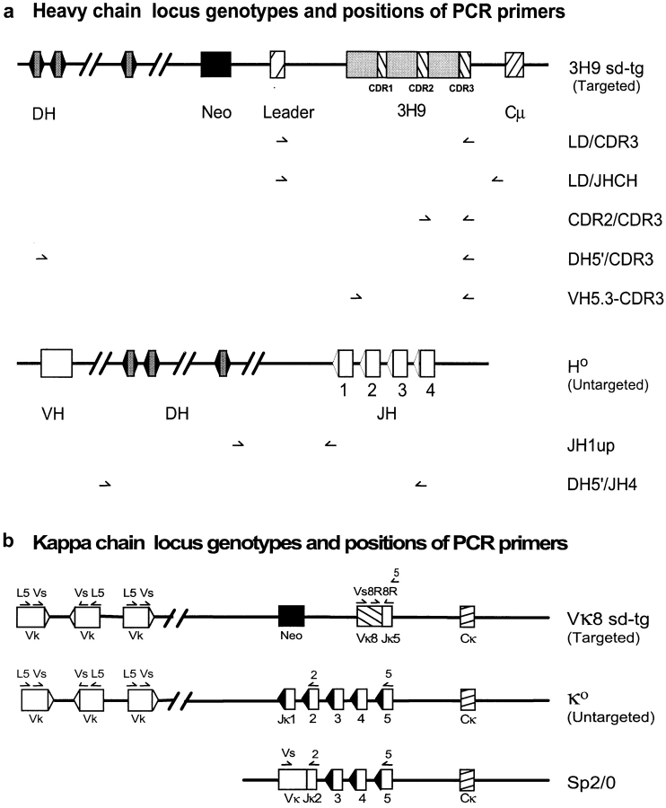
Antibodies to single-stranded (ss)DNA are expressed in patients with systemic lupus erythematosus and in lupus-prone mouse models such as the MRL/Mp-lpr/lpr (MRL/lpr) strain. In nonautoimmune mice, B cells bearing immunoglobulin site-directed transgenes (sd-tgs) that code for anti-ssDNA are functionally silenced. In MRL/lpr autoimmune mice, the same sd-tgs are expressed in peripheral B cells and these autoantibodies gain the ability to bind other autoantigens such as double-stranded DNA and cell nuclei. These new specificities arise by somatic mutation of the anti-ssDNA sd-tgs and by secondary light chain rearrangement. Thus, B cells that in normal mice are anergic can be activated in MRL/lpr mice, which can lead to the generation of pathologic autoantibodies. In this paper, we provide the first direct evidence for peripheral rearrangement in vivo.


Figures

Figure 1
Figure 1
Positions of PCR primers in IgH and Igκ loci in 3H9Vκ8 mice. Locations of primers used to assess the rearrangement status of both targeted and untargeted H chain (a) and L chain (b) alleles are shown. H chain primers: LD, CDR2, and CDR3 are primers specific for the targeted H chain 12 22. JHCH binds in the JH-CH intron of both H chain alleles (see Materials and Methods). DH5′ is a degenerate primer that binds 5′ of DH gene segments (except DSP.2 and DFL16.2) and can be used to detect D gene invasion 22 on the targeted allele and partial DJ rearrangements on the untargeted allele. VH5.3 is another degenerate primer specific for the FW3 of ∼65% of VH genes; VH5.3 is used to detect VH replacement 22. The JH1 up primer set is used to detect unrearranged untargeted alleles. Any DJ rearrangement on this allele results in deletion of the JH1up sequence. L chain primers: 8R primers are specific for the targeted L chain 12 23. Both Vs 28 and L5 48 are forward Vκ primers; the former detects 80% of Vκ genes, and the latter 50–60%. Vs and L5 in combination with Jκ2 or Jκ5 reverse primers are used to detect Vκ-Jκ rearrangements. Sp2/0 L chain locus: Sp2/0 has been used as the hybridoma fusion partner 25. It harbors a nonproductive Vκ-Jκ2 rearrangement that is bound by Vs but not L5. Figures are not drawn to scale.
Figure 4
Figure 4
Nucleotide and deduced amino-acid sequences of the Vκ8 sd-tg in IgG-secreting hybridomas. The Vκ8 sd-tg sequence is used as a reference. Identities are indicated with dashes. CDRs are defined according to Kabat et al. 29. Mutations shared by at least two sequences are shaded. Only mutated Vκ8 sequences are presented (10 out of 46). Six sequences display a stop codon at the beginning of the CDR1.
Figure 2
Figure 2
Immunofluorescence staining of HEp-2 cells by 3H9Vκ8/lpr serum and hybridoma supernatants. HEp-2 cells were incubated with supernatant from hybridomas (15, 28, 38, 50, 83, and 79) or 1:500 diluted serum from 3H9Vκ8/lpr and 3H9Vκ8/BALB/c mice. Serum Ig from 3H9Vκ8/lpr mice and all hybrids but 79 showed similar ANA patterns compatible with DNA or DNA–protein complex staining. Hybridoma 79 and sera from 3H9Vκ8/BALB/c mice gave the same negative pattern.
Figure 5
Figure 5
Nucleotide and deduced amino-acid sequences of Vκ23 from B cell clone members. Hybridomas that had a nonsense mutation in their Vκ8 gene were tested for the presence of another L chain by sequencing L chain mRNA. All six hybridomas express a Vκ23 L chain. These sequences are compared with two Vκ23 germline genes, Vκ23GL1 and Vκ23GL2, obtained from MRL/lpr tail DNA. Identities are indicated with dashes. Mutations shared by at least two members are shaded. T and C nucleotides located at the V-J junction are likely to result from the germline segments and therefore are not considered as mutations or N-additions. The CDRs are defined according to Kabat et al. 29. In all sequences, the Vκ23 gene is rearranged to a Jκ2′ gene segment. Jκ2′ is an MRL/lpr allelic variant of Jκ2 that occupies the same chromosomal position as Jκ2 relative to other Jκs. It has a serine at position 100 (boxed) instead of the glycine of Jκ2. The usage of a Jκ2′ segment demonstrates that the Vκ gene rearranged on the untargeted allele of the MRL/lpr mouse.
Figure 3
Figure 3
Nucleotide and deduced amino-acid sequences of hybridoma H chains. (A) Sequences of the 3H9 sd-tg in IgG-secreting hybridomas (50, 79, 15, 83, 28, 38, 46, and 16). The sequence of the 3H9 sd-tg is shown. Identities are indicated with dashes. CDRs are defined according to Kabat et al. 29. Unmutated sequences from IgG-secreting hybridomas are not presented in this figure. Mutations shared by at least two sequences are shaded except for the first two, which result from the degenerate nature of the VH5.3 primer used in amplification. (B) VH replacement sequences. In these hybridomas (57, 85, 13, 1, and 2), the 3H9 sd-tg has been replaced by an upstream VH gene, via the embedded heptamer at the end of CDR3 in 3H9 22. The sequence of the 3H9 sd-tg is used as a reference for the hybridomas 57, 85, and 13 because the replacing genes belong to the same J558 gene family. Boxed residues at positions 53, 57, and 65 are specific to the 3H9 gene (mutations). Identical nucleotides between hybridomas 57 and 85 are shaded when different from 3H9. These two hybrids represent potential members of the same clone (see text). The sequence of hybridoma 13 is also compared with the unmutated H chain sequence of mAb 19.1.2, which uses a more related J558 gene (sequence data available from EMBL/GenBank/DDBJ under accession number AJ223534). Identical nucleotides between hybridoma 13 and the reference J558 gene are also shaded. The D segment used in hybridoma 13 has homology with DQ52 (5′ boundary) and DSP2.6-c (3′ boundary) gene segments, probably resulting from a D–D fusion. Sequences from hybridomas 1 and 2 are compared with two different genes from the 7183 gene family, EMBL/GenBank/DDBJ accession numbers AF003722 and Z22078, respectively. In hybridoma 1, the D segment has homology with both the DSP2.9 (5′ boundary) and the DSP2.10-c (3′ boundary) gene segments, and in hybridoma 2, with both the DSP2.10 (5′ boundary) and the DFL16.1E (3′ boundary) gene segments. The regions of homology are underlined and identical nucleotides are in bold.
Figure 3
Figure 3
Nucleotide and deduced amino-acid sequences of hybridoma H chains. (A) Sequences of the 3H9 sd-tg in IgG-secreting hybridomas (50, 79, 15, 83, 28, 38, 46, and 16). The sequence of the 3H9 sd-tg is shown. Identities are indicated with dashes. CDRs are defined according to Kabat et al. 29. Unmutated sequences from IgG-secreting hybridomas are not presented in this figure. Mutations shared by at least two sequences are shaded except for the first two, which result from the degenerate nature of the VH5.3 primer used in amplification. (B) VH replacement sequences. In these hybridomas (57, 85, 13, 1, and 2), the 3H9 sd-tg has been replaced by an upstream VH gene, via the embedded heptamer at the end of CDR3 in 3H9 22. The sequence of the 3H9 sd-tg is used as a reference for the hybridomas 57, 85, and 13 because the replacing genes belong to the same J558 gene family. Boxed residues at positions 53, 57, and 65 are specific to the 3H9 gene (mutations). Identical nucleotides between hybridomas 57 and 85 are shaded when different from 3H9. These two hybrids represent potential members of the same clone (see text). The sequence of hybridoma 13 is also compared with the unmutated H chain sequence of mAb 19.1.2, which uses a more related J558 gene (sequence data available from EMBL/GenBank/DDBJ under accession number AJ223534). Identical nucleotides between hybridoma 13 and the reference J558 gene are also shaded. The D segment used in hybridoma 13 has homology with DQ52 (5′ boundary) and DSP2.6-c (3′ boundary) gene segments, probably resulting from a D–D fusion. Sequences from hybridomas 1 and 2 are compared with two different genes from the 7183 gene family, EMBL/GenBank/DDBJ accession numbers AF003722 and Z22078, respectively. In hybridoma 1, the D segment has homology with both the DSP2.9 (5′ boundary) and the DSP2.10-c (3′ boundary) gene segments, and in hybridoma 2, with both the DSP2.10 (5′ boundary) and the DFL16.1E (3′ boundary) gene segments. The regions of homology are underlined and identical nucleotides are in bold.
Figure 6
Figure 6
Genealogical relationships between antinuclear B cells from a 3H9Vκ8/lpr mouse. Genealogical trees based on the coding sequences of the 3H9 (A), Vκ8 (B), and Vκ23 (C) genes from the clonally related hybridomas are shown. The trees are constructed from patterns of shared and unique mutations by assuming that shared mutations represent single events and not independent parallel mutations and by assuming the minimum number of mutational events. To construct the 3H9 and Vk8 trees, it was sometimes necessary to assume that a specific nucleotide underwent two different mutations. For example, in the 3H9 tree we assumed all members of the clone originally had the silent C→T mutation in the serine of FW3 and that subsequently hybridoma 79 underwent a T→A mutation at the same position. This explains why there are eight shared mutations in the stem of the 3H9 tree, whereas Fig. 3 A shows only seven coding region mutations shared by all members of the clone. The length of the line between each B cell is proportional to the number of mutations (given in parentheses). Note the similar shapes and branch morphology of the three trees.

Similar articles

Cited by

References

    1. Tan E.M. Antinuclear antibodiesdiagnostic markers and clues to the basis of systemic autoimmunity Pediatr. Infect. Dis. J. 7Suppl1988. S3 S9 - PubMed
    1. Hardin J.A. The lupus autoantigens and the pathogenesis of systemic lupus erythematosus. Arthritis Rheum. 1986;29:457–460. - PubMed
    1. Shlomchik M.J., Marshak-Rothstein A., Wolfowicz C.B., Rothstein T.L., Weigert M.G. The role of clonal selection and somatic mutation in autoimmunity. Nature. 1987;328:805–811. - PubMed
    1. Shlomchik M., Mascelli M., Shan H., Radic M.Z., Pisetsky D., Marshak-Rothstein A., Weigert M. Anti-DNA antibodies from autoimmune mice arise by clonal expansion and somatic mutation. J. Exp. Med. 1990;171:265–292. - PMC - PubMed
    1. Craft J.E., Hardin J.A. Linked sets of antinuclear antibodieswhat do they mean? J. Rheumatol. 14Suppl1987. 106 109 - PubMed

Publication types

MeSH terms