. 1999 Sep 6;146(5):1033-44.
doi: 10.1083/jcb.146.5.1033.

Microtubule targeting of substrate contacts promotes their relaxation and dissociation

Affiliations

Microtubule targeting of substrate contacts promotes their relaxation and dissociation

I Kaverina et al. J Cell Biol. .

Abstract

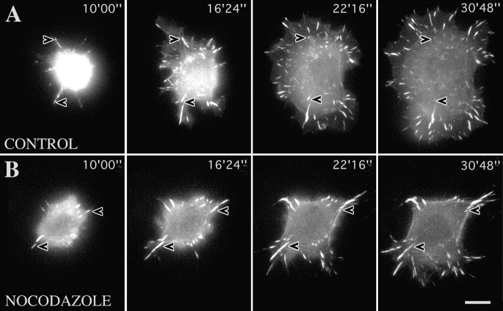
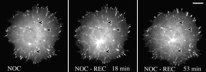
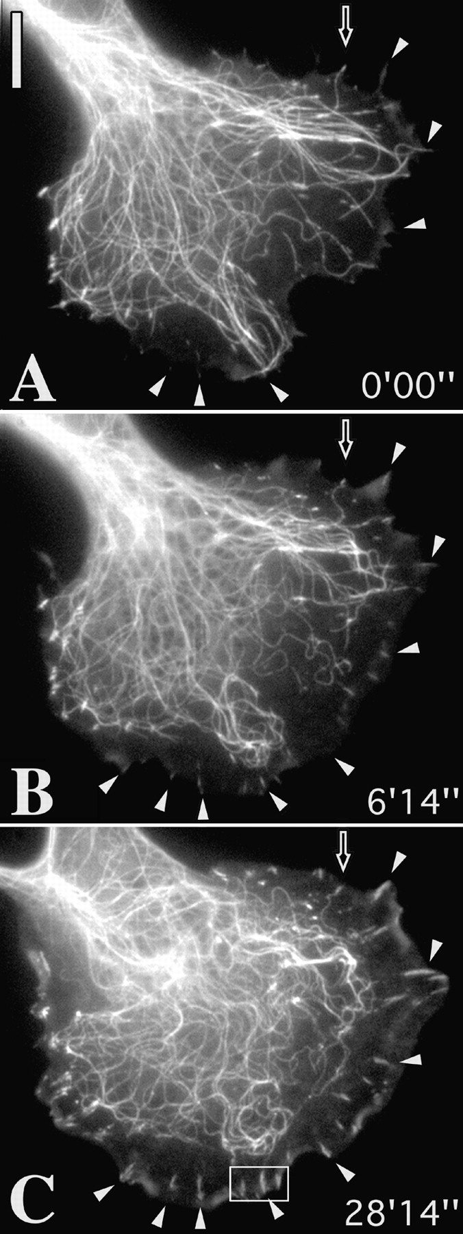
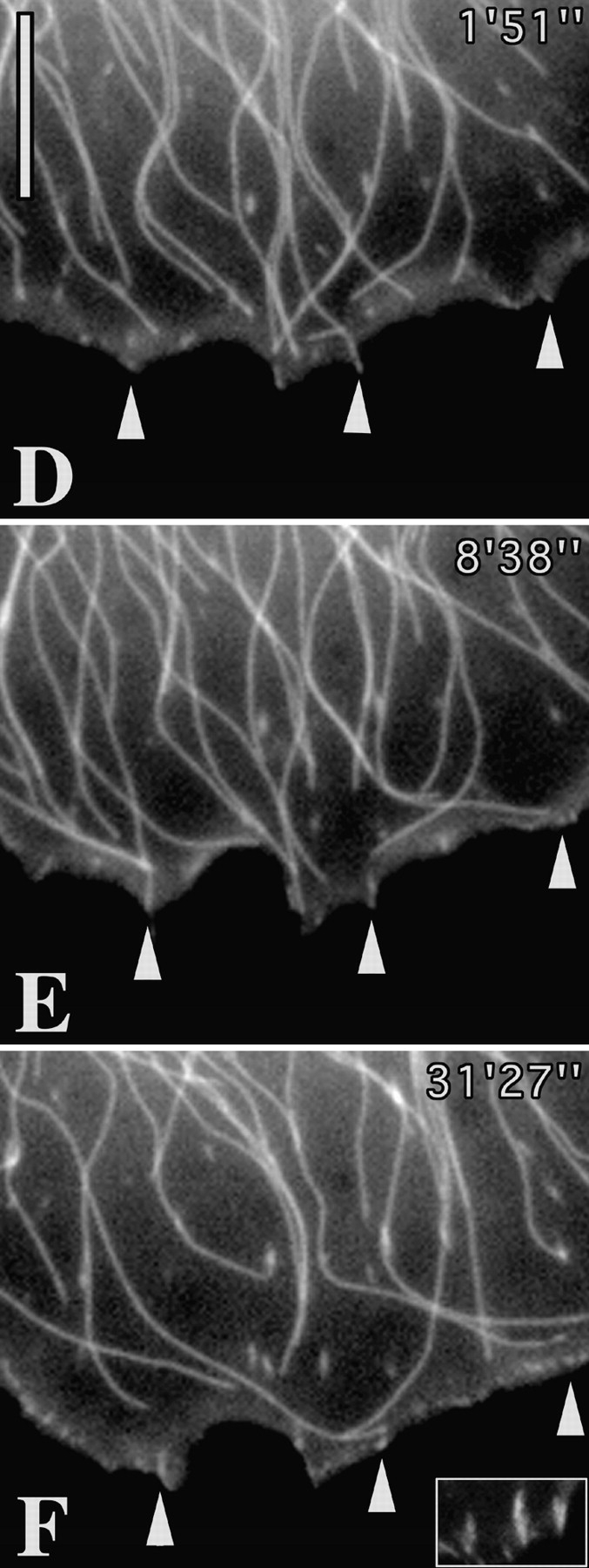
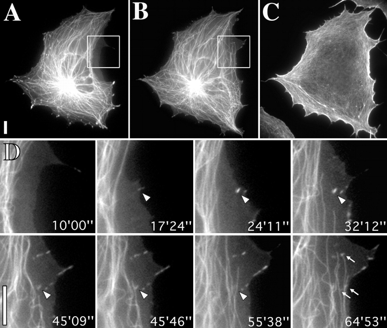
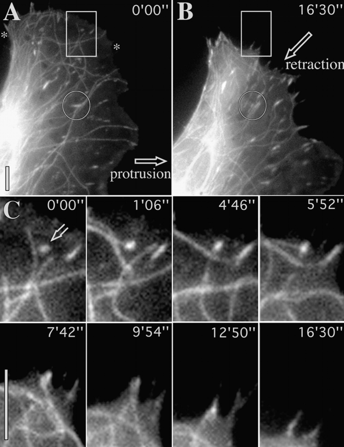
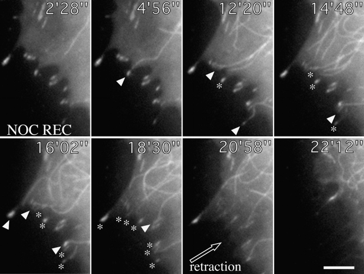
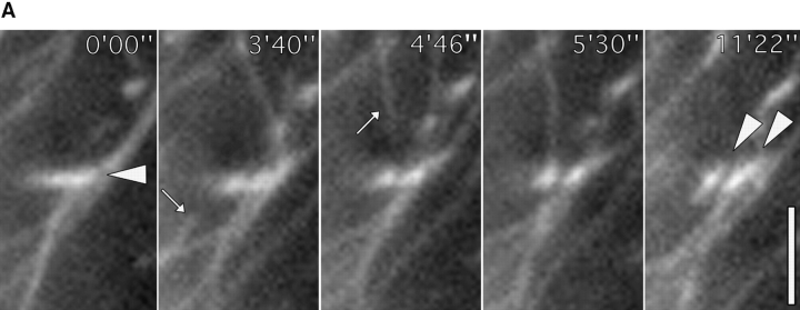
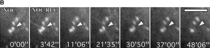
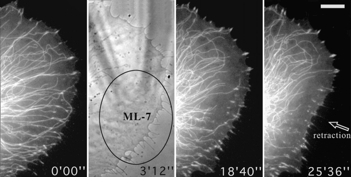
We recently showed that substrate contact sites in living fibroblasts are specifically targeted by microtubules (Kaverina, I., K. Rottner, and J.V. Small. 1998. J. Cell Biol. 142:181-190). Evidence is now provided that microtubule contact targeting plays a role in the modulation of substrate contact dynamics. The results are derived from spreading and polarized goldfish fibroblasts in which microtubules and contact sites were simultaneously visualized using proteins conjugated with Cy-3, rhodamine, or green fluorescent protein. For cells allowed to spread in the presence of nocodazole the turnover of contacts was retarded, as compared with controls and adhesions that were retained under the cell body were dissociated after microtubule reassembly. In polarized cells, small focal complexes were found at the protruding cell front and larger adhesions, corresponding to focal adhesions, at the retracting flanks and rear. At retracting edges, multiple microtubule contact targeting preceded contact release and cell edge retraction. The same effect could be observed in spread cells, in which microtubules were allowed to reassemble after local disassembly by the application of nocodazole to one cell edge. At the protruding front of polarized cells, focal complexes were also targeted and as a result remained either unchanged in size or, more rarely, were disassembled. Conversely, when contact targeting at the cell front was prevented by freezing microtubule growth with 20 nM taxol and protrusion stimulated by the injection of constitutively active Rac, peripheral focal complexes became abnormally enlarged. We further found that the local application of inhibitors of myosin contractility to cell edges bearing focal adhesions induced the same contact dissociation and edge retraction as observed after microtubule targeting. Our data are consistent with a mechanism whereby microtubules deliver localized doses of relaxing signals to contact sites to retard or reverse their development. We propose that it is via this route that microtubules exert their well-established control on cell polarity.


Figures

Figure 1
Figure 1
Substrate contact turnover during cell spreading is potentiated by microtubules. A: Video sequence of a spreading fish fibroblast transfected with EGFP-zyxin. (B) As in A but for a cell pretreated with and replated in the presence of 2.5 μg/ml nocodazole. Arrowheads indicate the position of representative contacts throughout the two sequences. Times are given in minutes and seconds. Bar, 10 μm. Video available at http://www.jcb.org/cgi/content/full/146/5/1033/F1/DC1
Figure 2
Figure 2
Focal adhesions are dissociated during recovery from nocodazole. Control image shows fish fibroblast cotransfected with EGFP-tubulin and EGFP-zyxin pretreated with and replated in 2.5 μg/ml nocodazole (NOC) for 1 h. Subsequent frames show recovery after nocodazole washout. Time is given in minutes and seconds. Arrowheads indicate some of the adhesion sites that were dissociated during microtubule reassembly. Bar, 10 μm.
Figure 3
Figure 3
Elimination of targeting leads to contact growth. (A–C) Sequences of a cell coinjected with Rh-tubulin and Rh-vinculin and pretreated with 20 μM taxol for 30 min (A) to stabilize microtubules, before injection of L61 Rac at time 0:00. Arrowheads indicate peripheral contact sites that grew in the protruding cell edge induced by Rac. Hollow arrowhead indicates a contact that remained in association with a microtubule end and that did not grow. (D–F) control cell injected with Rac but not treated with taxol. Contact sites are targeted and do not enlarge. Bars, 10 μm. Videos available at http://www.jcb. org/cgi/content/full/146/5/1033/ F3/DC1
Figure 3
Figure 3
Elimination of targeting leads to contact growth. (A–C) Sequences of a cell coinjected with Rh-tubulin and Rh-vinculin and pretreated with 20 μM taxol for 30 min (A) to stabilize microtubules, before injection of L61 Rac at time 0:00. Arrowheads indicate peripheral contact sites that grew in the protruding cell edge induced by Rac. Hollow arrowhead indicates a contact that remained in association with a microtubule end and that did not grow. (D–F) control cell injected with Rac but not treated with taxol. Contact sites are targeted and do not enlarge. Bars, 10 μm. Videos available at http://www.jcb. org/cgi/content/full/146/5/1033/ F3/DC1
Figure 4
Figure 4
Rho-kinase independent contact sites are also targeted by microtubules. (A–C) low magnification view of cell incubated in 100 μM Y-27632. A and B correspond to the beginning and end of the sequence and C to the phalloidin-labeled image obtained after fixation immediately after the sequence. Cell was transiently cotransfected with tubulin-EGFP and zyxin-EGFP. D, video sequence of inset region in A and B showing targeting of newly formed contact sites. Zyxin-containing site marked with arrowhead, which appeared at time 17′24″ was targeted by a microtubule between 45′ and 46′ and had disappeared by 55′. Arrows indicate other sites that were targeted by microtubule ends. Bars, 10 μM. Videos available at http://www.jcb. org/cgi/content/full/146/5/1033/ F4/DC1
Figure 5
Figure 5
Repetitive targeting of peripheral focal adhesions precedes cell edge retraction. (A and B) First and last frames of a video sequence of a locomoting fish fibroblast that was coinjected with Rh-vinculin and Rh-tubulin. During the sequence, the region between the asterisks was retracted towards the cell body. (C) Video sequence of inset in A and B showing the typical fate of a contact site. Sequential frames show six targeting events at contact site (indicated by open arrow at 0′00″) by microtubules approaching from different directions. Inward retraction began after the 4th event. Circle in A indicates inset region shown in Fig. 7 A. Bars, 10 μM. Videos available at http://www.jcb.org/cgi/content/full/146/5/1033/F5/DC1
Figure 6
Figure 6
Contact targeting and retraction after recovery from local application of nocodazole. Cell shown was cotransfected with zyxin-EGFP and tubulin-EGFP and treated on one edge with 50 μg/ml nocodazole applied through a microneedle to locally depolymerize microtubules. Video sequences show recovery after removal of needle at time 0′00″. Arrowheads indicate sequential targeting events at peripheral contact sites and asterisks the positions of contacts that had already been targeted. Time is in minutes and seconds. Bar, 5 μM. Video available at http://www.jcb.org/cgi/content/full/146/5/1033/F6/DC1
Figure 7
Figure 7
(A) Contact site remodeling. A shows video sequence of medial focal adhesion outlined by a circle in Fig. 5A and Fig. B. This contact site was targeted by microtubules from below and above (approaching microtubules are indicated by arrows at 3′40″ and 4′46″), and then split into two contacts oriented at around 45° to the parent contact (see arrowheads at 0′00″ and 11′22″). (B) Tailoring of contact group. B shows video sequence of a group of contacts in a cell cotransfected with zyxin-EGFP and tubulin-EGFP during recovery from nocodazole. The second contact from bottom was selectively targeted by microtubules at 3′42″ and 21′35″ and had disappeared at 48′. Bars: (A) 3 μM; (B) 5 μM. Videos available at http://www.jcb. org/cgi/content/full/146/5/1033/ F7/DC1
Figure 7
Figure 7
(A) Contact site remodeling. A shows video sequence of medial focal adhesion outlined by a circle in Fig. 5A and Fig. B. This contact site was targeted by microtubules from below and above (approaching microtubules are indicated by arrows at 3′40″ and 4′46″), and then split into two contacts oriented at around 45° to the parent contact (see arrowheads at 0′00″ and 11′22″). (B) Tailoring of contact group. B shows video sequence of a group of contacts in a cell cotransfected with zyxin-EGFP and tubulin-EGFP during recovery from nocodazole. The second contact from bottom was selectively targeted by microtubules at 3′42″ and 21′35″ and had disappeared at 48′. Bars: (A) 3 μM; (B) 5 μM. Videos available at http://www.jcb. org/cgi/content/full/146/5/1033/ F7/DC1
Figure 8
Figure 8
Locally applied ML-7 destabilizes microtubules and induces the dissociation of peripheral contact sites. Figure shows video sequence of a cell cotransfected with zyxin-EGFP and tubulin-EGFP that was treated topically, from time 0′00″ with a local application of 2 mM ML-7 through a microneedle (visible in the phase contrast image at 3′12″), over the region indicated by the ellipse. Bar, 10 μM. Video available at http://www.jcb.org/cgi/content/full/146/5/1033/F8/DC1
Figure 9
Figure 9
Schematic illustration of substrate contact dynamics in a moving fibroblast. (Top) Substrate contacts are initiated in the protruding and ruffling lamellipodium (Lam, ruf). Two classes of primary contacts are depicted: punctate focal complexes (fc) and linear contacts associated with some microspike bundles (ms/c). Their formation is associated with the activation of Rac and Cdc42, respectively. Each type of primary contact can develop into a precursor of a focal adhesion (pFA). Further abbreviations: iFA, intermediate focal adhesion in the body of the cell; tFA, focal adhesion at a trailing cell edge. (Bottom) Four types of contact sites with different strengths of anchorage to the substrate (anchors) are depicted and correspond to those in the upper diagram. All sites rely on contractility for their maintenance, indicated by different levels of myosin II–dependent tension (T and t). For precursor (pFA) and mature focal adhesions (iFA, tFA), myosin activation depends on Rho-kinase. The contractility of focal complexes is Rho-kinase independent. Microtubules (MT) interface with contact sites and modulate their turnover by locally inhibiting contractility. The relaxing dose is controlled by the total number and frequency of microtubule targeting events. Focal complexes may or may not be targeted by microtubules.

Similar articles

Cited by

References

    1. Bershadsky A.D., Vasiliev J.M. Cytoskeleton 1988. Plenum Press; New York: pp. 298 pp
    1. Bershadsky A., Chausovsky A., Becker E., Lyubimova A., Geiger B. Involvement of microtubules in the control of adhesion-dependent signal transduction. Curr. Biol. 1996;6:1279–1289. - PubMed
    1. Best A., Ahmed S., Kozma R., Lim L. The Ras-related GTPase Rac1 binds tubulin. J. Biol. Chem. 1996;271:3756–3762. - PubMed
    1. Chen W.-T. Surface changes during retraction-induced spreading of fibroblasts. J. Cell Sci. 1981;49:1–13. - PubMed
    1. Chrzanowska-Wodnicka M., Burridge K. Rho-stimulated contractility drives the formation of stress fibers and focal adhesions. J. Cell Biol. 1996;133:1403–1415. - PMC - PubMed

Publication types

MeSH terms