. 1999 Sep 6;146(5):1107-16.
doi: 10.1083/jcb.146.5.1107.

Regulation of cell contraction and membrane ruffling by distinct signals in migratory cells

Affiliations

Regulation of cell contraction and membrane ruffling by distinct signals in migratory cells

D A Cheresh et al. J Cell Biol. .

Abstract

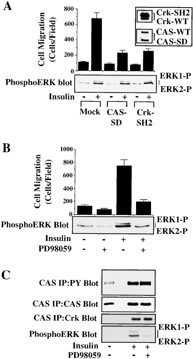
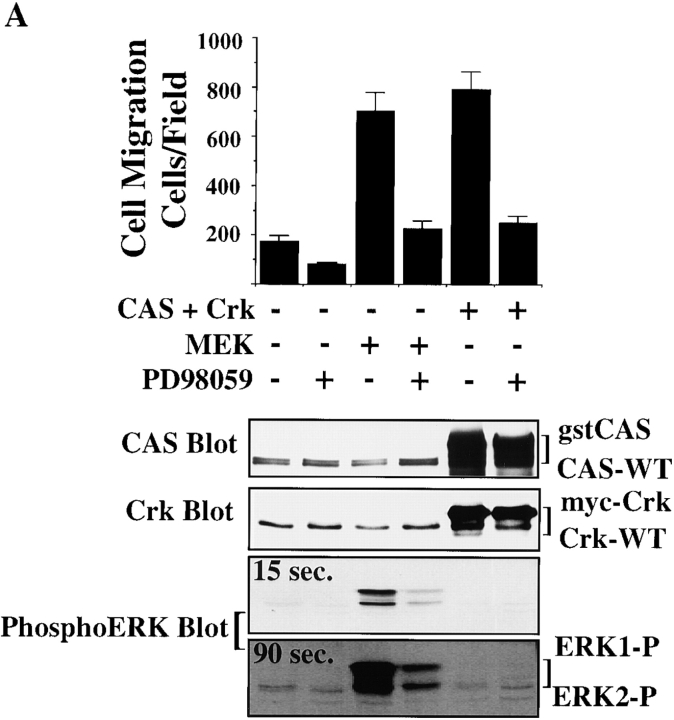
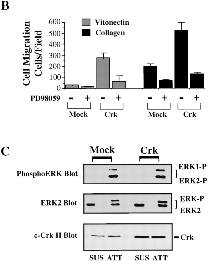
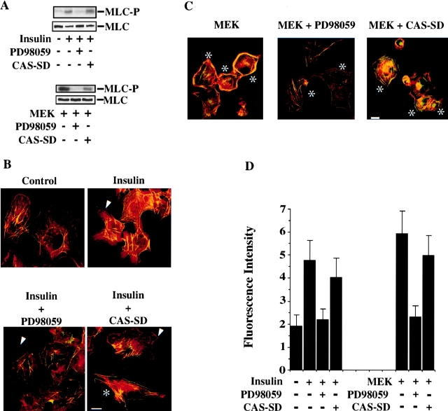
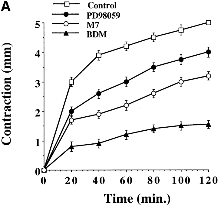
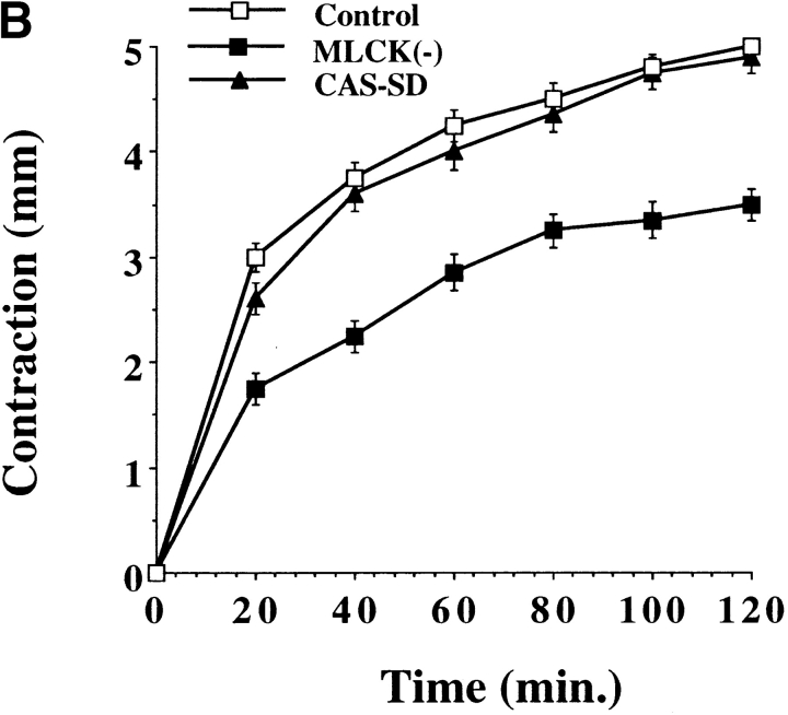
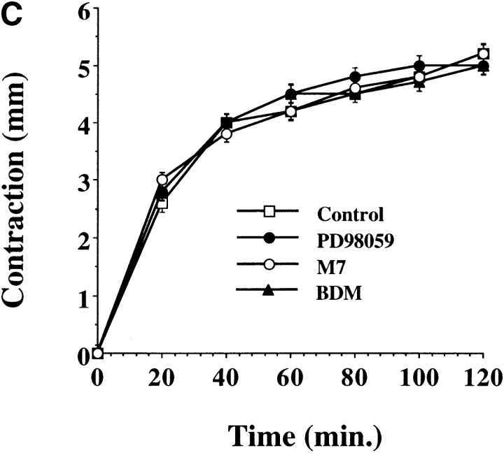
Cell migration and wound contraction requires assembly of actin into a functional myosin motor unit capable of generating force. However, cell migration also involves formation of actin-containing membrane ruffles. Evidence is provided that actin-myosin assembly and membrane ruffling are regulated by distinct signaling pathways in the migratory cell. Interaction of cells with extracellular matrix proteins or cytokines promote cell migration through activation of the MAP kinases ERK1 and ERK2 as well as the molecular coupling of the adaptor proteins p130CAS and c-CrkII. ERK signaling is independent of CAS/Crk coupling and regulates myosin light chain phosphorylation leading to actin-myosin assembly during cell migration and cell-mediated contraction of a collagen matrix. In contrast, membrane ruffling, but not cell contraction, requires Rac GTPase activity and the formation of a CAS/Crk complex that functions in the context of the Rac activating protein DOCK180. Thus, during cell migration ERK and CAS/Crk coupling operate as components of distinct signaling pathways that control actin assembly into myosin motors and membrane ruffles, respectively.


Figures

Figure 1
Figure 1
ERK activation and CAS/Crk coupling are separate signaling events necessary for cytokine-induced cell migration. (A) Serum-starved COS-7 cells were allowed to migrate for 3 h on vitronectin-coated membranes in the presence or absence of insulin (10 μg/ml) after transient transfection with a β-gal reporter construct, along with either the empty expression vector (Mock) or with expression vectors encoding gst-tagged CAS without its substrate domain (CAS-SD), or myc-tagged Crk with a mutated SH2 domain (Crk-SH2). CAS-SD and Crk-SH2 have been shown to prevent CAS/Crk coupling and downstream signals (Matsuda et al. 1993; Feller et al. 1994; Klemke et al. 1998). The number of transfected cells migrating were enumerated by counting cells on the underside of the membrane that coexpress the β-gal vector as described in Materials and Methods. An aliquot of cells treated as described for the migration experiment above was lysed in detergent and immunoblotted with antibodies to either the phosphorylated/activated form of ERK1/ERK2 (lower panel), Crk, or CAS (top right). Note that Crk-SH2 shows reduced mobility compared with wild-type endogenous Crk (Crk-WT) as the result of the molecular tag. (B) Serum-starved COS-7 cells were allowed to migrate in the presence or absence of the MEK inhibitor PD98059 (25 μM) with or without insulin (10 μg/ml). Cell migration and ERK1/ERK2 activity in these cells were determined as described above. Similar findings were obtained with fibronectin and collagen type I–coated membranes (data not shown). Each bar represents the mean ± SEM of at least three independent experiments. (C) Serum-starved COS-7 cells pretreated with or without the MEK inhibitor PD98059 (50 μM) for 2 h were exposed to insulin (10 μg/ml) for 5 min before being lysed in detergent. CAS was immunoprecipitated and then immunoblotted with antibodies to either phosphotyrosine, Crk, or CAS. The detergent lysates from these cells were also examined for changes in ERK1/ERK2 activity as described above. The result shown is representative from at least three independent experiments.
Figure 2
Figure 2
CAS/Crk-induced cell migration does not result from increased ERK activity. (A) Upper panel, serum-starved COS-7 cells were allowed to migrate for 3 h in the presence or absence of the MEK inhibitor PD98059 (25 μM) on vitronectin-coated membranes after transient transfection with a β-gal reporter construct, along with either the empty expression vector or with expression vectors encoding gst-tagged wild-type CAS and myc-tagged c-Crk or mutationally activated MEK. The number of transfected cells migrating was enumerated by counting cells on the underside of the membrane that coexpress the β-gal vector as described in Materials and Methods. Each bar represents the mean ± SEM of at least three independent experiments. Lower panels, total cell lysates prepared from cells treated as described for the migration experiments above were immunoblotted with antibodies that recognize the activated form of ERK1/ERK2 proteins, Crk, or CAS. Note that the gstCAS and myc-Crk proteins show reduced mobility compared with endogenous wild-type forms of these proteins as the result of the molecular tags. Immunoblots were exposed to film for 15 and 90 s after treatment with enhanced chemiluminescence reagents as described in Materials and Methods. (B) Serum-starved FG cells stably transfected with Crk or mock-transfected with the empty vector were allowed to migrate on vitronectin or collagen-coated membranes for 3 h in the presence or absence of PD98059 (25 μM) and quantified as described in Materials and Methods. Each bar represents the mean ± SEM of at least three independent experiments. (C) Cells treated as described for the migration experiment above were either held in suspension (SUS) or allowed to attach (ATT) to collagen-coated culture dishes for 30 min, then lysed in detergent and immunoblotted with either antibodies that specifically recognize the activated form of ERK1/ERK2 proteins (upper panel), ERK2 (middle panel), or Crk (lower panel). The upper band recognized by the ERK2 antibody represents the phosphorylated/activated form of this protein (ERK2-P) that has reduced mobility as a result of being phosphorylated. Similar results were obtained with ERK1 protein (data not shown).
Figure 2
Figure 2
CAS/Crk-induced cell migration does not result from increased ERK activity. (A) Upper panel, serum-starved COS-7 cells were allowed to migrate for 3 h in the presence or absence of the MEK inhibitor PD98059 (25 μM) on vitronectin-coated membranes after transient transfection with a β-gal reporter construct, along with either the empty expression vector or with expression vectors encoding gst-tagged wild-type CAS and myc-tagged c-Crk or mutationally activated MEK. The number of transfected cells migrating was enumerated by counting cells on the underside of the membrane that coexpress the β-gal vector as described in Materials and Methods. Each bar represents the mean ± SEM of at least three independent experiments. Lower panels, total cell lysates prepared from cells treated as described for the migration experiments above were immunoblotted with antibodies that recognize the activated form of ERK1/ERK2 proteins, Crk, or CAS. Note that the gstCAS and myc-Crk proteins show reduced mobility compared with endogenous wild-type forms of these proteins as the result of the molecular tags. Immunoblots were exposed to film for 15 and 90 s after treatment with enhanced chemiluminescence reagents as described in Materials and Methods. (B) Serum-starved FG cells stably transfected with Crk or mock-transfected with the empty vector were allowed to migrate on vitronectin or collagen-coated membranes for 3 h in the presence or absence of PD98059 (25 μM) and quantified as described in Materials and Methods. Each bar represents the mean ± SEM of at least three independent experiments. (C) Cells treated as described for the migration experiment above were either held in suspension (SUS) or allowed to attach (ATT) to collagen-coated culture dishes for 30 min, then lysed in detergent and immunoblotted with either antibodies that specifically recognize the activated form of ERK1/ERK2 proteins (upper panel), ERK2 (middle panel), or Crk (lower panel). The upper band recognized by the ERK2 antibody represents the phosphorylated/activated form of this protein (ERK2-P) that has reduced mobility as a result of being phosphorylated. Similar results were obtained with ERK1 protein (data not shown).
Figure 3
Figure 3
ERK-induced cell migration requires CAS/Crk and Rac activity. (A) Upper panel, serum-starved COS-7 cells were allowed to migrate for 3 h on vitronectin-coated membranes after transient transfection with a β-gal reporter construct, along with either the empty expression vector or with expression vectors encoding mutationally activated MEK, or MEK cotransfected with dominant negative CAS (CAS-SD). The number of transfected cells migrating were enumerated by counting cells on the underside of the membrane that coexpress the β-gal vector as described in Materials and Methods. Each bar represents the mean ± SEM of at least three independent experiments. Lower panels, cells treated as described for the migration experiment above were lysed in detergent and immunoblotted with antibodies to the activated form of ERK1/ERK2 (middle panel) or ERK2 (lower panel). The upper band recognized by the ERK2 antibody represents the phosphorylated/activated form of this protein (ERK2-P) that has reduced mobility as a result of being phosphorylated. Similar results were obtained with ERK1 protein (data not shown). (B) Serum-starved COS-7 cells were allowed to migrate for 3 h on vitronectin-coated membranes after transient transfection with a β-gal reporter construct, along with either the empty expression vector or with expression vectors encoding mutationally activated MEK, or MEK together with dominant negative myc-tagged Rac (RacN17) in the presence or absence of insulin (10 μg/ml) in the lower chamber. An aliquot of cells transfected with RacN17 and lysed and immunoblotted with an antibody to Rac is shown (top right). Note that RacN17 migrates slower as the result of the myc tag compared with endogenous wild-type Rac (Rac-Wt). Each bar represents the mean ± SEM of at least three independent experiments.
Figure 4
Figure 4
CAS/Crk association, but not ERK activation, is required for Rac-dependent membrane ruffling. (A) Serum-starved COS-7 cells in the presence or absence of insulin (10 μg/ml for 15 min) were stained with rhodamine-conjugated phalloidin, then analyzed by confocal imaging for F-actin (red) containing membrane ruffles after being transfected with either the empty vector (control) or the vector encoding dominant negative CAS (CAS-SD) along with a reporter vector encoding green fluorescent protein (GFP) to identify transfected cells. In some cases, control cells were pretreated for 2 h with 50 μM of PD98059 to inhibit ERK activity before being exposed to insulin as described above. Photomicrographs were taken with a Bio-Rad Labs 1024 laser and a Zeiss Axiovert microscope (400×). Arrowheads indicate cells with prominent F-actin membrane ruffles. (B) COS-7 cells treated as described above were scored for membrane ruffles as described in Materials and Methods. Results are expressed as the percentage of total transfected cells (i.e., green cells) that displayed prominent F-actin membrane ruffles and are the mean ± SEM of three separate experiments. (C) COS cells were transfected with expression vectors encoding CAS, Crk, and dominant negative RacN17, along with a reporter vector encoding GFP, then examined for actin membrane ruffles as describe above. Photomicrographs of CAS/Crk cells (400×) and RacN17 expressing cells (600×) were taken with a Bio-Rad Labs 1024 laser and a Zeiss Axiovert microscope. Arrowheads indicate cells with prominent F-actin membrane ruffles. (D) COS-7 cells transfected with either wild-type DOCK180, gst-tagged CAS and myc-tagged Crk, or CAS and Crk, together with DOCK180 and/or myc-tagged RacN17 were examined for cell migration as described above. An aliquot of cells transfected with CAS/Crk and DOCK180 (lane 2) or cells mock-transfected with the empty vectors (lane 1) as described for the migration experiment above were lysed in detergent and immunoblotted with antibodies to the phosphorylated/activated form of ERK1/ERK2 as described above (top left). Note that in these experiments cells were transfected with CAS/Crk vectors at DNA levels that give half-maximal migration. Each bar represents the mean ± SEM of at least three independent experiments. (E) An aliquot of cells treated as for the migration experiment above were lysed in detergent then immunoblotted with antibodies to CAS, Crk, DOCK180, or myc to detect myc-tagged RacN17. Lane 1, control cells transfected with the empty vectors. Lane 2, cells transfected with the vector containing the cDNA as indicated. Note that the gstCAS and mycCrk proteins show reduced mobility compared with endogenous wild-type forms of these proteins as the result of the molecular tag. Bars, 10 μm.
Figure 5
Figure 5
ERK, but not CAS/Crk signaling, promotes myosin light chain phosphorylation and actin-myosin colocalization. (A) Myosin light chains (MLC) immunoprecipitated with an anti-myosin IIB antibody from COS-7 cells metabolically labeled with [32P]orthophosphate and treated with or without insulin (10 μg/ml) for 5 min as described in Materials and Methods. In some cases, 32P-labeled cells were pretreated for 2 h with or without the MEK inhibitor PD98059 or transfected with mutationally activated MEK, and/or CAS-SD before immunoprecipitation of myosin IIB and SDS-PAGE as described above. MLC-P denotes phosphorylated light chain. MLC shows light chains stained with Coomassie before autoradiography to confirm that equal amounts of protein were precipitated in these experiments. (B) Serum-starved COS-7 cells either pretreated with the MEK inhibitor PD98059 (25 μM) or transfected with CAS-SD along with a β-gal reporter construct to identify transfected cells. Cells were stimulated with insulin (10 μg/ml) for 10 min, then exposed briefly to detergent to remove insoluble actin-myosin, fixed, and costained for actin and myosin IIB under conditions that preserve the association of these protein in cells as described in Materials and Methods (Cramer and Mitchison 1995). Immunofluorescent laser confocal imaging was performed as described in Materials and Methods. Rhodamine-phalloidin (red) was used to visualize F-actin, whereas rabbit anti-myosin IIB and secondary FITC-conjugated goat anti–rabbit specific antibodies were used to visualize myosin (green). Yellow staining is the colocalization of red and green staining in the merged image. An asterisk indicates transfected cells as detected by immunostaining with a mouse anti-β-galactosidase antibody and a secondary antibody conjugated with Cy5 (blue, not shown). Arrowhead shows actin-containing membrane ruffles. (C) COS-7 cells transfected with mutationally activated MEK and/or CAS-SD along with a β-gal reporter construct to identify transfected cells and examined for actin-myosin association as described above. In some cases, MEK+ transfected cells were exposed to 25 μM PD98059 for 2 h to block MEK activation of ERK before staining for actin and myosin. Photomicrographs were taken with a Bio-Rad Labs 1024 laser and Zeiss Axiovert microscope (600×). An asterisk indicates transfected cells and arrow shows membrane ruffles. (D) Insoluble myosin content of COS-7 cells treated as described in B and C. Cells were stained with anti-myosin IIB and FITC-conjugated secondary antibodies and the total amount of green fluorescence intensity (×106) determined per cell area (μm2) as described in Materials and Methods. Each bar represents the mean ± SEM of 30–40 cells of 6–8 different fields of three independent experiments. Bars: (B and C) 10 μm.
Figure 6
Figure 6
ERK activity, but not CAS/Crk association, is necessary for cell contraction of collagen gels. Three-dimensional collagen gels containing COS-7 cells were released from the culture dish and allowed to contract for various times as described in Materials and Methods. (A) COS-7 cells in collagen gels pretreated for 60 min with the MEK inhibitor PD98059 (50 μM), the MLCK inhibitor M7 (1 μM), or an inhibitor of myosin ATPase activity (BDM, 10 mM). (B) COS-7 cells transfected with either the empty expression vector (control) or the vector containing CAS without a substrate domain (CAS-SD) or a dominant negative form of MLCK (MLCK−) (Klemke et al. 1997). Contraction is presented as change in diameter (starting-final) measured in millimeters. (C) Cells treated as described in A were washed to remove inhibitors then cultured an additional 24 h before being allowed to contract the collagen matrix for various times as described in Materials and Methods. Each point represents the mean ± SEM of three independent experiments.
Figure 6
Figure 6
ERK activity, but not CAS/Crk association, is necessary for cell contraction of collagen gels. Three-dimensional collagen gels containing COS-7 cells were released from the culture dish and allowed to contract for various times as described in Materials and Methods. (A) COS-7 cells in collagen gels pretreated for 60 min with the MEK inhibitor PD98059 (50 μM), the MLCK inhibitor M7 (1 μM), or an inhibitor of myosin ATPase activity (BDM, 10 mM). (B) COS-7 cells transfected with either the empty expression vector (control) or the vector containing CAS without a substrate domain (CAS-SD) or a dominant negative form of MLCK (MLCK−) (Klemke et al. 1997). Contraction is presented as change in diameter (starting-final) measured in millimeters. (C) Cells treated as described in A were washed to remove inhibitors then cultured an additional 24 h before being allowed to contract the collagen matrix for various times as described in Materials and Methods. Each point represents the mean ± SEM of three independent experiments.

References

    1. Adelstein R.S. Regulation of contractile proteins by phosphorylation. J. Clin. Invest. 1983;72:1863–1866. - PMC - PubMed
    1. Altun-Glutekin Z.F., Chandrian S., Bougert C., Ishizaki I., Narumiya S., de Graaf P., Van Bergen en Henegouwen P., Hanafusa H., Wagner J.A., Birge R.B. Activation of Rho-dependent cell spreading and focal adhesion biogenesis by the v-Crk adaptor protein. Mol. Cell Biol. 1998;18:3044–3058. - PMC - PubMed
    1. Anand-Apte B., Zetter B.R., Viswanathan A., Qui R., Chen J., Ruggeri R., Symons M. Platelet-derived growth factor and fibronectin-stimulated migration are differentially regulated by the Rac and extracellular signal-regulated kinase pathways. J. Biol. Chem. 1997;272:30688–30692. - PubMed
    1. Aplin A.E., Howe A., Alahari S.K., Juliano R.L. Signal transduction and signal modulation by cell adhesion receptorsthe role of integrins, cadherins immunoglobulin-cell adhesion molecules, and selectins. Pharm. Rev. 1998;50:197–263. - PubMed
    1. Blaukat A., Ivankovic-Dikic I., Gronroos E., Dolfi F., Tokiwa G., Vuori K., Dikic I. Adaptor proteins Grb2 and Crk couple Pyk2 with activation of specific mitogen-activated kinase cascades. J. Biol. Chem. 1999;274:14893–14901. - PubMed

Publication types

MeSH terms

Substances