. 1999 Sep 15;19(18):7757-69.
doi: 10.1523/JNEUROSCI.19-18-07757.1999.

Differential expression of brain-derived neurotrophic factor, neurotrophin-3, and neurotrophin-4/5 in the adult rat spinal cord: regulation by the glutamate receptor agonist kainic acid

Affiliations

Differential expression of brain-derived neurotrophic factor, neurotrophin-3, and neurotrophin-4/5 in the adult rat spinal cord: regulation by the glutamate receptor agonist kainic acid

I A Scarisbrick et al. J Neurosci. .

Abstract

Previous in vitro studies indicate that select members of the neurotrophin gene family, namely brain-derived neurotrophic factor (BDNF), neurotrophin-3 (NT-3), and neurotrophin-4/5 (NT-4/5), contribute to survival and differentiation of spinal cord motoneurons. To investigate the potential roles of these factors in the adult spinal cord, we examined their cellular localization and regulation after systemic exposure to an excitotoxic stimulus, kainic acid (KA). Of the neurotrophins examined, NT-4/5 mRNA was most robustly expressed in the lumbosacral spinal cord of the normal adult rat, including expression by neurons throughout the gray matter, and in a subpopulation of white and gray matter glia. Both BDNF and NT-3 mRNAs were also densely expressed by alpha motoneurons of lamina IX, but were detected at lower levels elsewhere in the gray matter. NT-3 mRNA was additionally expressed by spinal cord glia, but was less widespread compared to NT-4/5. In response to systemic administration of KA, NT-4/5 and BDNF mRNAs were dramatically upregulated in a spatially and temporally restricted fashion, whereas levels of NT-3 mRNA were unchanged. These results provide strong in vivo evidence to support the idea that BDNF, NT-3, and in particular, NT-4/5, play a role in the normal function of the adult spinal cord. Furthermore, our results indicate that the actions of BDNF and NT-4/5 participate in the response of the cord to excitotoxic stimuli, and that those of NT-4/5 and NT-3 include both neurons and glia.


Figures
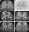
Fig. 1.
Fig. 1.
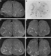
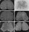
NT-4/5 mRNA expression was robust throughout all regions of the adult rat lumbosacral spinal cord and was differentially upregulated by systemic KA. Dark-field photomicrographs show the autoradiographic localization of hybridization to NT-4/5 mRNA in representative transverse sections at the L3/L4 level of the spinal cord of a control rat (A), and in parallel sections of paired experimental rats at 4 (C), 24 (D), 72 (E), or 168 hr (F) after intraperitoneal administration of KA. Bright-field photomicrograph (B), shows the appearance of Nissl-stained cellular elements in the dorsal (DF), lateral (LF), and ventral funiculi (VF) of the white matter, and in laminae I–X of the gray matter, in a parallel section from a control animal. There was a significant increase in density of autoradiographic grains (seen here as white under dark-field illumination), overlying the dorsal horn (DH), and in the white matter funiculi, of spinal cord sections from animals at 4 hr (C) relative to control (A) (Fig. 8B). NT-4/5 mRNA labeling was also elevated in the dorsal horn relative to controls when examined at 72 (E) and 168 hr (F) after KA administration (Fig. 5). Large arrows indicate position of lamina IX motoneurons. Small arrowsindicate NT-4/5 mRNA hybridization by white matter glia.VH, Ventral horn. Scale bar, 500 μm.
Fig. 2.
Fig. 2.
BDNF mRNA expression was dense in association with the α motoneurons of the adult rat lumbosacral spinal cord and upregulated therein by systemic KA. Dark-field photomicrographs show the autoradiographic localization of hybridization to BDNF mRNA in representative transverse sections through the lumbar region of the spinal cord of a control rat (A), and in parallel sections of paired experimental rats at 4 (C), 24 (D), 72 (E), or 168 hr (F), after intraperitoneal administration of KA. Bright-field photomicrograph (B) shows the appearance of Nissl-stained cellular elements in the dorsal (DF), lateral (LF), and ventral funiculi (VF) of the white matter, and in laminae I–X of the gray matter, in a parallel section from a control animal. There was an increase in density of autoradiographic grains overlying the motoneurons of lamina IX of the ventral horn of the spinal cord at 72 hr (E), after kainic acid administration relative to control (A) (Fig. 5).Arrows indicate position of lamina IX motoneurons.DH, Dorsal horn; VH, ventral horn. Scale bar, 500 μm.
Fig. 3.
Fig. 3.
NT-3 mRNA expression was dense in association with the α motoneurons of the adult rat lumbosacral spinal cord, and unchanged by systemic KA. Dark-field photomicrographs show the autoradiographic localization of hybridization to NT-3 mRNA in representative transverse sections at the L3/L4 level of the spinal cord of a control rat (A), and in parallel sections of paired experimental rats at 4 (C), 24 (D), 72 (E), or 168 hr (F), after intraperitoneal administration of KA. Bright-field photomicrograph (B) shows the appearance of Nissl-stained cellular elements in the dorsal (DF), lateral (LF), and ventral funiculi (VF) of the white matter, and in laminae I–X of the gray matter, in a parallel section from a control animal. Arrows indicate position of lamina IX motoneurons. DH, Dorsal horn; VH, ventral horn. Scale bar, 500 μm.
Fig. 4.
Fig. 4.
Quantification of the relative abundance of NT-4/5, BDNF, and NT-3 mRNA labeling in the dorsal and ventral regions of the spinal cord gray matter, compared with the white matter, in the normal adult rat lumbosacral spinal cord. The values plotted represent the ratio of the mean ± SE of the relative optical density, measured from film autoradiographs, of the [35S]-labeled riboprobes in the each region of the control spinal cord examined, that is the white matter (WM), dorsal gray matter (DG), or ventral gray matter (VG), compared with the white matter (n = 6). NT-4/5 mRNA labeling was 3.5-fold higher in the dorsal and ventral horns of the spinal cord gray matter relative to the white matter of the normal adult spinal cord. Compared with NT-4/5, BDNF and NT-3 mRNA labeling was more evenly distributed across the white and gray layers of the spinal cord, but each was elevated by ∼1.2-fold in the gray relative to the white matter of the normal adult rat.
Fig. 5.
Fig. 5.
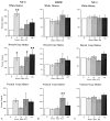
Quantification of kainic acid-induced changes in the expression of NT-4/5, BDNF, and NT-3 mRNAs in the white and gray matter of the adult rat lumbosacral spinal cord. Bar graphs show densitometric measurements of film autoradiograms of (A) α-[35S]-NT-4/5 cRNA labeling, (B) α-[35S]-BDNF cRNA labeling, or (C) α-[35S]-NT-3 cRNA labeling in the white matter, and in the dorsal or ventral gray regions, of the spinal cord of rats who were killed at 4, 24, 72, or 168 hr after intraperitoneal injection of KA. Measurements in each region from KA-treated animals were expressed as a percentage of values from paired control animals. The values plotted represent group mean ± SE. NT-4/5(n = 4, 4 hr; n = 6, 24 hr;n = 3, 72 and 168 hr), BDNF(n = 4, 4 hr; n = 5, 24 hr;n = 3, 72 and 168 hr), and NT-3(n = 5, 4 hr; n = 7, 24 hr;n = 3, 72 and 168 hr). Significant differences in mRNA labeling between treated and control groups were shown by ANOVA (p < 0.01).Stars indicate significant differences from control values (★p < 0.05; ★★p< 0.01; Student–Newman–Keuls post hoc test).
Fig. 6.
Fig. 6.
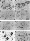
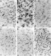
NT-4/5, BDNF, and NT-3 mRNA expression was dense in the lumbosacral α motoneurons of lamina IX, but only BDNF mRNA levels were significantly altered by systemic KA. Bright-field photomicrographs show the autoradiographic localization of hybridization to NT-4/5 (A, B), BDNF (C, D), and NT-3 (E,F) mRNAs within lamina IX of the ventral horn of control animals (A, C, E) and paired experimental animals that were killed at 72 hr after KA administration (B, D,F). Although the level of NT-4/5 and NT-3 mRNA hybridization in the ventral horn was unchanged after KA administration, the level of BDNF mRNA hybridization was elevated by 2.6-fold at the 72 hr time point (p < 0.01; Fig. 5). In addition to dense autoradiographic signal in association with the α motoneurons of lamina IX (arrows), the NT-4/5, and to a lesser extent the NT-3, riboprobe produced significant levels of signal in association with gray matter glia (A, B, E,arrowheads). G shows the appearance of α motoneurons in a Nissl-stained section from a control animal, andH shows the typical background level of autoradiographic signal produced by the 35S-labeled-NT-4/5 sense stand cRNA (arrows indicate α motoneurons). Scale bar, 25 μm.
Fig. 7.
Fig. 7.
NT-4/5 mRNA expression was dense in the dorsal horn of the lumbosacral spinal cord and upregulated by systemic KA. Bright-field photomicrographs show the autoradiographic localization of hybridization of NT-4/5 (A), BDNF (D) and NT-3 (E) mRNAs within laminae I–IV of the dorsal horn of control animals (arrows). The amount of NT-4/5 mRNA hybridization was significantly elevated at 4 (B), 72 (Fig.1E), and 168 hr (C) after KA administration. The level of BDNF and NT-3 mRNA hybridization in the dorsal horn was unchanged at each time point examined after KA administration (arrowheads show examples of labeled cells) (see Fig. 5 for quantification). Bright-field photomicrograph (F), shows the appearance of Nissl-stained cellular elements in lamina I–IV of the dorsal horn in a parallel section from a control animal. Scale bar, 25 μm.
Fig. 8.
Fig. 8.
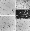
High levels of NT-4/5 mRNA expression were associated with glia of the dorsal funiculus of the lumbosacral spinal cord white matter, and the level of hybridization was dramatically upregulated by systemic KA. Bright-field photomicrographs show the autoradiographic localization of hybridization to NT-4/5 mRNA within the dorsal funiculus of the adult rat spinal cord of a control rat (A), and of a paired experimental rat that was killed at 4 hr (B) after kainic acid administration (arrowheads show examples of labeled cells). A significant elevation in the level of NT-4/5 cRNA hybridization was observed in the spinal cord white matter at 4 hr (B) after systemic KA administration (Fig. 5). In contrast, significantly lower levels of hybridization were produced by the BDNF (C) and NT-3 (D) riboprobes in association with glia of the dorsal funiculus of control animals, and this level was not altered after exposure to KA (Figs. 2,3). Scale bar, 25 μm.
Fig. 9.
Fig. 9.
Significant levels of NT-4/5 and NT-3 mRNA expression were associated with glia of the lateral funiculi of the normal adult rat lumbosacral spinal cord white matter. Bright-field photomicrographs show the autoradiographic localization of hybridization produced by the NT-4/5 (A), NT-3 (B), and BDNF (E)35S-labeled riboprobes in the lateral funiculus of control rats (arrowheads show examples of labeled cells). Virtually all cells in the lateral funiculus of the normal spinal cord white matter were double-labeled (examples atarrowheads), for DIG-labeled NT-4/5 cRNA (C) and immunofluorescence for the oligodendrocyte marker Rip (D). Scale bars:A, B, E, 25 μm;C, D, 50 μm.

References

    1. Agrawal SK, Fehlings MG. Role of NMDA and non-NMDA ionotropic glutamate receptors in traumatic spinal cord axonal injury. J Neurosci. 1997;17:1055–1063. - PMC - PubMed
    1. Aloe L. Intracerebral pretreatment with nerve growth factor prevents irreversible brain lesions in neonatal rats injected with ibotenic acid. Biotechnology. 1987;5:1085–1086.
    1. Barres BA, Koroshetz WJ, Swartz KJ, Chun LL, Corey DP. Ion channel expression by white matter glia: the O-2A glial progenitor cell. Neuron. 1990;4:507–524. - PubMed
    1. Barres BA, Raff MC, Gaese F, Bartke I, Dechant G, Barde Y-A. A crucial role for neurotrophin-3 in oligodendrocyte development. Nature. 1994;367:371–375. - PubMed
    1. BDNF Study Group (Phase III) A controlled trial of recombinant methionyl human BDNF in ALS. Neurology. 1999;52:1427–1433. - PubMed

Publication types

MeSH terms