. 1999 Sep 15;19(18):8009-26.
doi: 10.1523/JNEUROSCI.19-18-08009.1999.

Ultrastructural localization of full-length trkB immunoreactivity in rat hippocampus suggests multiple roles in modulating activity-dependent synaptic plasticity

Affiliations

Ultrastructural localization of full-length trkB immunoreactivity in rat hippocampus suggests multiple roles in modulating activity-dependent synaptic plasticity

C T Drake et al. J Neurosci. .

Abstract

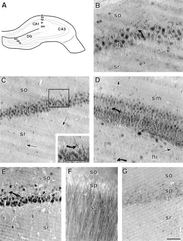
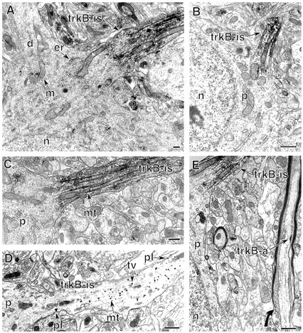
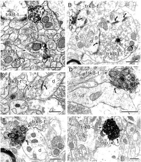
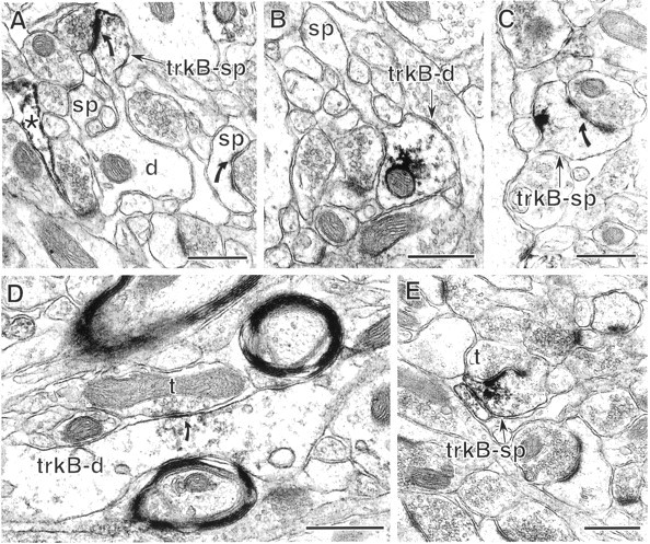
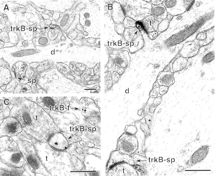
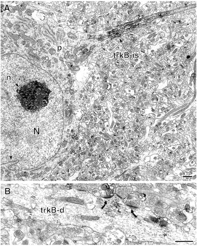
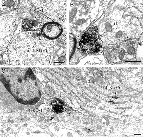
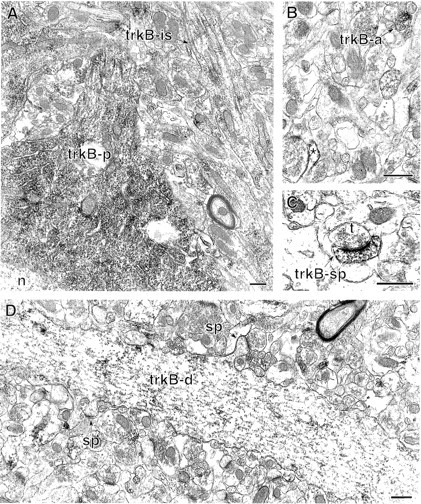
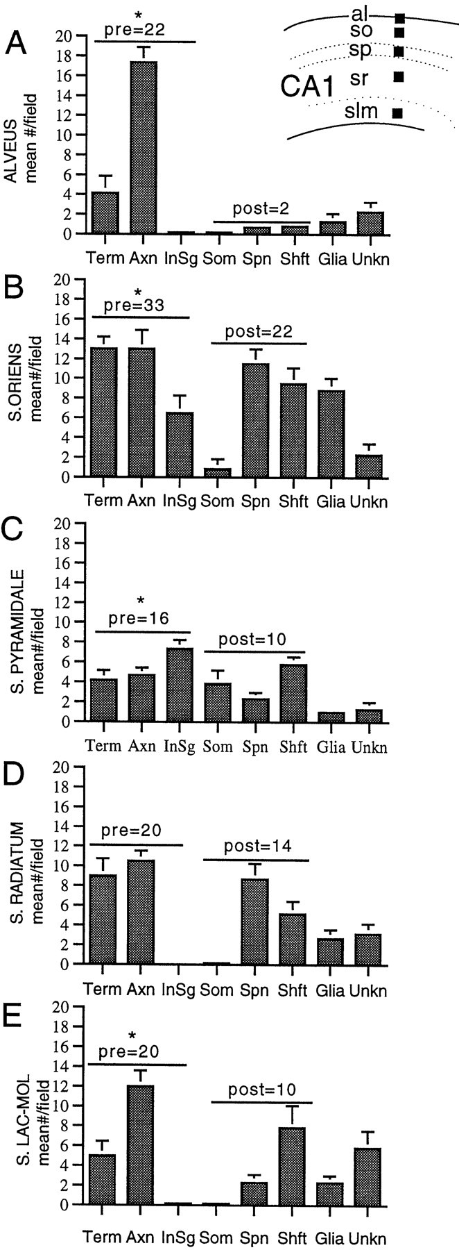
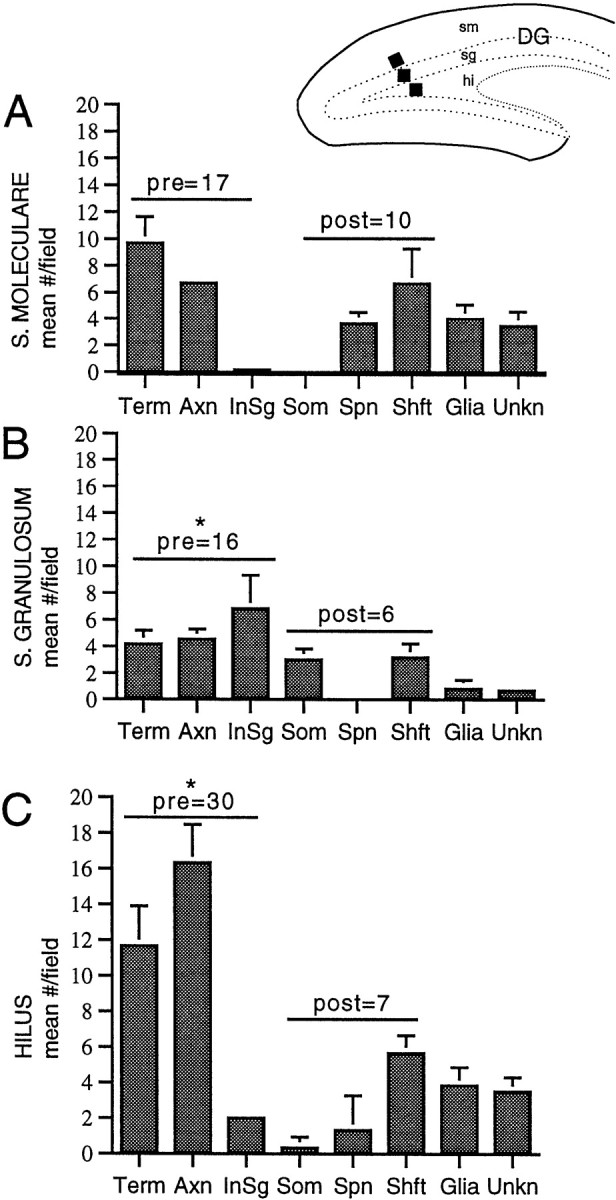
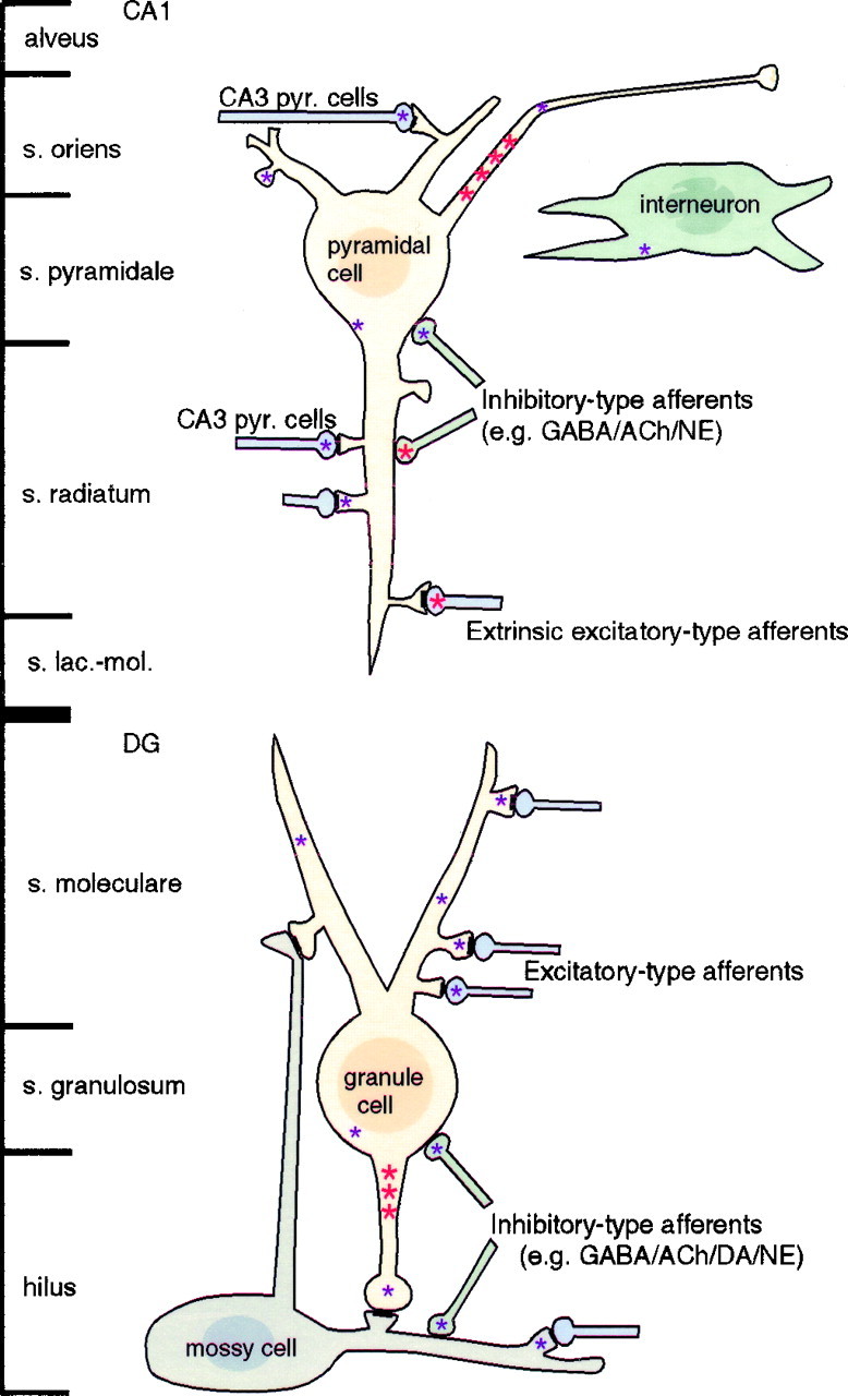
Neurotrophins acting at the trkB receptor have been shown to be important modulators of activity-dependent plasticity in the hippocampus, but the mechanisms underlying these effects are not yet well understood. To identify the cellular and subcellular targets of trkB ligands in the adult rat hippocampal formation, full-length trkB receptor immunoreactivity (trkB-IR) was localized using electron microscopy. trkB-IR was present in the glutamatergic pyramidal and granule cells. Labeling in these neurons appeared as discrete clusters and was primarily in axons, excitatory-type axon terminals, and dendritic spines and to a lesser extent in somata and dendritic shafts. trkB-IR was commonly found on the plasma membrane of dendritic spines, whereas in other subcellular regions trkB-IR was often intracellular. Labeling was strikingly dense within axon initial segments, suggesting extensive receptor trafficking. trkB-IR was not confined to pyramidal and granule cells. Dense trkB-IR was found in occasional interneuron axon initial segments, some axon terminals forming inhibitory-type synapses onto somata and dendritic shafts, and excitatory-type terminals likely to originate extrahippocampally. This suggests that trkB is contained in some GABAergic interneurons, neuromodulatory (e.g., cholinergic, dopaminergic, and noradrenergic) afferents, and/or glutamatergic afferents. These data indicate that full-length trkB receptor activation may modulate glutamatergic pathways of the trisynaptic circuit both presynaptically at axon terminals and initial segments and postsynaptically at dendritic spines and shafts. Signaling via catalytic trkB may also presynaptically affect inhibitory and modulatory neurons. A pan-trkB antibody labeled the same neuronal populations as the full-length-specific trkB antiserum, but the labels differed in density at various subcellular sites. These findings provide an ultrastructural foundation for further examining the mechanisms through which neurotrophins acting at trkB receptors contribute to synaptic plasticity.


Figures

Fig. 1.
Fig. 1.
Light microscopy and specificity of trkB-IR.A, Schematic of a coronal section through the hippocampal formation (adapted from Swanson, 1995). al, Alveus; hi, hilus; sg, st. granulosum;slm, st. lacunosum-moleculare; so, st. oriens; sp, st. pyramidale; sm, st. moleculare; sr, st. radiatum. B, In the CA3 region of hippocampus, trkB-IR is present in neuronal somata and smooth processes (example indicated by the curved arrow) extending from neurons in st. pyramidale, and in small punctate structures in st. radiatum and st. oriens. C, In the CA1 region of hippocampus, trkB-IR is present in neuronal somata and in processes extending into st. oriens (inset, curved arrow), as well as in scattered fine beaded processes (straight arrows) in st. radiatum. D, In dentate gyrus, st. granulosum and the hilus contain darkly labeled somata and smooth, tapering processes (curved arrows) extending from somata. Fine punctate processes (straight arrows) are present in the hilus and st. moleculare.E, In the CA1 region, different processing conditions (4% paraformaldehyde fixation and trkB antiserum diluted 1:200) result in trkB labeling that is darker with increased diffuse background but is otherwise like that seen with acrolein and paraformaldehyde fixation and trkB antiserum diluted 1:2000 (compare with C). F, Pyramidal cell apical dendrites are prominently labeled in the CA1 region of acrolein- and paraformaldehyde-fixed tissue incubated in an antibody (trkB23–36, diluted 1:500) raised against a peptide common to both the full-length and truncated trkB receptors. G, The CA1 region of an adsorption control section contains virtually no trkB labeling (compare with C). Scale bars, 50 μm.
Fig. 2.
Fig. 2.
Dense trkB-IR is present in the axon initial segments of principal cells. A, A labeled pyramidal cell axon initial segment (trkB-is) contains intense trkB-IR. The somatic perikaryon (p) contains clusters of labeling affiliated with endoplasmic reticulum (er) and mitochondria (m). The nucleus (n) and a primary dendrite (d) lack labeling. B, In the dentate gyrus, intense trkB-IR is in an axon initial segment (trkB-is) of a granule cell. The somatic perikaryon (p) and nucleus (n) are unlabeled. C, Higher magnification of a labeled pyramidal cell axon initial segment (trkB-is) shows peroxidase labeling associated with the plasmalemma and microtubule fascicles (mt). D, TrkB–immunogold labeling is affiliated with bundled microtubules (mt), membranes of tubulovesicular organelles (tv), and occasionally the plasma membrane (pl) of a pyramidal cell axon initial segment (trkB-is).E, A pyramidal cell axon initial segment (trkB-is) contains dense, abundant trkB labeling. In contrast, a nearby axon (trkB-a), which is at the initiation of myelination (curved arrow) and likely originates from a pyramidal cell, contains a small patch of labeling along the plasma membrane. Scale bars, 0.5 μm.
Fig. 3.
Fig. 3.
Excitatory-type axon terminals contain trkB-IR.A, Two terminals (trkB-t) in st. radiatum contain sparse trkB–immunogold labeling. The lower, sparsely labeled terminal forms an asymmetric (excitatory-type) synapse (curved arrow) with a dendritic spine (sp). B, In the hilus of dentate gyrus, a cluster of trkB-IR is present in mossy fiber terminals (trkB-t) identified by their large size and numerous asymmetric synapses (curved arrows) with dendrites (d). C, A trkB–immunogold-labeled terminal (trkB-t) in st. oriens contains immunogold–silver particles affiliated with small synaptic vesicles and less commonly with the plasma membrane (open arrow). This terminal forms an asymmetric synapse (curved arrow) with an unlabeled dendrite (d). D, A terminal (trkB-t) in the hilus contains dense trkB-IR and forms an asymmetric synapse (curved arrow) with the shaft of an unlabeled dendrite (d). E, F, In st. lacunosum-moleculare, terminals with dense trkB-IR form asymmetric synapses (curved arrows) with an unlabeled dendritic shaft (E) and an unlabeled dendritic spine (F). Scale bars, 0.5 μm.
Fig. 4.
Fig. 4.
In the CA1 region, dendritic trkB-IR is primarily in spines and to a lesser extent in dendritic shafts. A, In st. radiatum, a dendritic spine contains trkB-IR (trkB-sp) along the postsynaptic density (curved arrow) and plasmalemma. In contrast, no labeling is detected in the parent dendrite of the labeled spine (d) or in another spine (sp) receiving an asymmetric synapse. An astrocytic process (*) in this field is also labeled.B, In CA1 st. radiatum, a dendritic shaft (trkB-d) contains labeling in the cytoplasm and along a small patch of plasma membrane and extends an unlabeled spine (sp). C, A spine (trkB-sp) in st. radiatum contains a cluster of trkB-IR along the plasmalemma opposite the postsynaptic density (curved arrow).D, A dendritic shaft (trkB-d) in st. lacunosum-moleculare contains trkB-IR at and near a symmetric synapse (curved arrow) from an unlabeled terminal (t). E, A labeled dendritic spine (trkB-sp) in st. lacunosum-moleculare receives a asymmetric synapse from an unlabeled terminal (t). The peroxidase reaction product is present along the postsynaptic density, nearby plasmalemma, and cytoplasmic organelles. Scale bars, 0.5 μm.
Fig. 5.
Fig. 5.
In dentate gyrus, trkB-IR is occasionally detected in spiny dendrites. A, In st. moleculare, a dendrite (d) extends one trkB-labeled spine (trkB-sp) and one unlabeled spine (s). B, In the hilus, two spines with trkB–immunoperoxidase labeling (trkB-sp) receive asymmetric synapses from unlabeled terminals (t). Immunoperoxidase reaction product is present along postsynaptic densities, the plasmalemma, and intracellular organelles. The dendritic shaft (d) giving rise to the upper labeled spine lacks trkB-IR. C, TrkB-immunogold labeling in a dendritic spine (trkB-sp) in the hilus. This spine receives synapses from two unlabeled terminals (t). Immunogold particles are located along the plasmalemma near one postsynaptic density and over an unidentifiable intracellular organelle. A nearby terminal (trkB-t) also contains trkB-IR affiliated with synaptic vesicles. Scale bars, 0.5 μm.
Fig. 6.
Fig. 6.
Some nonprincipal neurons contain intense trkB-IR.A, An interneuron in st. oriens of the CA1 region contains intense trkB-IR in the axon initial segment (trkB-is). This nonprincipal cell exhibits the nuclear infolding (arrow) characteristic of interneurons. The somatic perikaryon (p) lacks detectable trkB-labeling. N, Nucleus, n, nucleolus.B, An interneuron dendrite (trkB-d) in st. oriens contains diffuse trkB-IR. Several terminals, one of which is also trkB-labeled, form asymmetric synapses (curved arrows) with the trkB-labeled dendrite. Scale bars, 1 μm.
Fig. 7.
Fig. 7.
Some inhibitory-type axon terminals in the hippocampus and dentate gyrus contain dense trkB-IR. A, A labeled axon terminal (trkB-t) in st. pyramidale of the CA1 region forms an inhibitory-type synapse (parallel arrows) with a large labeled dendrite (trkB-d) containing a small patch of trkB immunoprecipitate (arrow). B, In the hilus of the dentate gyrus, an intensely labeled terminal (trkB-t) and an unlabeled terminal (t) form symmetric synapses (parallel arrows) with an unlabeled dendrite (d). Other surfaces of the labeled terminal appose glial processes (*). C, At the hilus–st. granulosum interface in the dentate gyrus, a densely labeled terminal (trkB-t) forms a symmetric synapse (parallel arrows) with a granule cell perikaryon (p) containing trkB-immunolabeled rough endoplasmic reticulum (trkB-er). Scale bars 0.5 μm.
Fig. 8.
Fig. 8.
The distribution of trkB23–36labeling overlaps that of the full-length-specific trkB antibody but is concentrated in different subcellular compartments. A. In st. pyramidale of the CA1 region, dense trkB23–36–immunogold labeling is present in a pyramidal cell perikaryon (trkB-p). The nucleus (n) is unlabeled. An adjacent axon initial segment (trkB-is) contains light immunoreactivity along microtubule bundles. B, In st. radiatum, trkB23–36 is present in an axon (trkB-a) and a glial process (*). C, A trkB23–36-labeled dendritic spine (trkB-sp) in st. radiatum receives an asymmetric synapse from an unlabeled terminal (t). D, TrkB23–36 labeling in a spiny dendrite in st. radiatum. The dendritic shaft (trkB-d) contains dense immunoreactivity and extends two unlabeled spines (sp). Scale bars, 0.05 μm.
Fig. 9.
Fig. 9.
The number and type of profiles with trkB-IR vary between laminae in the CA1 region. Inset, From each animal (n = 3), 55 × 55 μm fields (black squares show location) were selected in two thin sections (each from a different 40 μm section) in the alveus (al), st. oriens (so), st. pyramidale (sp), st. radiatum (sr), and st. lacunosum-moleculare (slm). A–E, Mean distribution ± SEM of trkB-IR in terminals, axons, axon intial segments, somata, spines, shafts, glia, and unknown profiles in each lamina. Values within graphs indicate the sum of the means of presynaptic profiles (terminals, axons, and axon initial segments) and postsynaptic profiles (dendritic spines, shafts, and somata). As indicated by the asterisks, the number of trkB-labeled presynaptic profiles was significantly greater than labeled postsynaptic profiles in the alveus (p = 0.002), st. oriens (p = 0.002), st. pyramidale (p = 0.041),and st. lacunosum-moleculare (p = 0.026). Labeled presynaptic and postsynaptic profiles were of similar abundance in st. radiatum (p = 0.093).
Fig. 10.
Fig. 10.
The number and type of profiles with trkB-IR varies between laminae in the dentate gyrus. Inset, For each animal (n = 3), 55 × 55 μm fields (black squares show location) were selected in two thin sections (each from a different 40 μm section) from the subgranular zone of the hilus (hi), suprapyramidal blade of st. granulosum (sg), and inner one-third of st. moleculare (sm). A–C, Mean number ± SEM of trkB-IR in terminals, axons, axon intial segments, somata, spines, shafts, glia, and unknown profiles in each lamina. Values within graphs indicate the sum of the means of presynaptic profiles (terminals, axons, and axon initial segments) and postsynaptic profiles (dendritic spines, shafts, and somata). As indicated by theasterisks, trkB-labeled presynaptic profiles were significantly more numerous than labeled postsynaptic profiles in st. granulosum and the hilus (p = 0.026 and 0.004, respectively) and were of similar abundance in st. moleculare (p = 0.240).
Fig. 11.
Fig. 11.
Summary of sites containing specific trkB labeling in the CA1 region of hippocampus and the dentate gyrus (DG). Intense labeling (large red asterisks) and sparse labeling (small purple asterisks) were sometimes observed in different compartments of the same cells. Not shown: intensely labeled terminals of unidentified origin in the hilus.

References

    1. Abraham WC, Bear MF. Metaplasticity: the plasticity of synaptic plasticity. Trends Neurosci. 1996;19:126–130. - PubMed
    1. Alderson RF, Alterman AL, Barde YA, Lindsay RM. Brain-derived neurotrophic factor increases survival and differentiated functions of rat septal cholinergic neurons in culture. Neuron. 1990;5:297–306. - PubMed
    1. Alkondon M, Pereira EF, Barbosa CT, Albuquerque EX. Neuronal nicotinic acetylcholine receptor activation modulates gamma-aminobutyric acid release from CA1 neurons of rat hippocampal slices. J Pharmacol Exp Ther. 1997;283:1396–1411. - PubMed
    1. Allendoerfer KL, Cabelli RJ, Escandon E, Kaplan DR, Nikolics K, Shatz CJ. Regulation of neurotrophin receptors during the maturation of the mammalian visual system. J Neurosci. 1994;14:1795–1811. - PMC - PubMed
    1. Altar CA, DiStefano PS. Neurotrophin trafficking by anterograde transport. Trends Neurosci. 1998;21:433–437. - PubMed

Publication types

MeSH terms

Substances

LinkOut - more resources