

Review
doi: 10.1128/JB.181.18.5543-5550.1999.
Holliday junction processing in bacteria: insights from the evolutionary conservation of RuvABC, RecG, and RusA
Affiliations
- PMID: 10482492
- PMCID: PMC94071
- DOI: 10.1128/JB.181.18.5543-5550.1999
Item in Clipboard
Review
Holliday junction processing in bacteria: insights from the evolutionary conservation of RuvABC, RecG, and RusA
J Bacteriol.
1999 Sep.
No abstract available
Figures
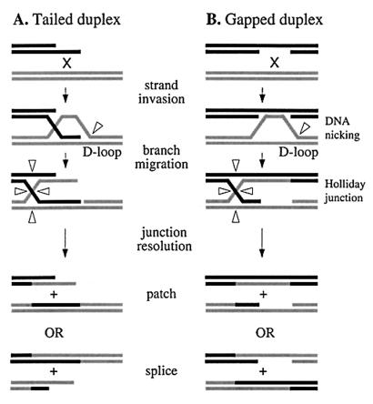
Schematic diagram showing molecular pathways for homologous recombination by single-strand invasion at ends (A) and gaps (B). Arrowheads indicate sites of DNA strand cutting.
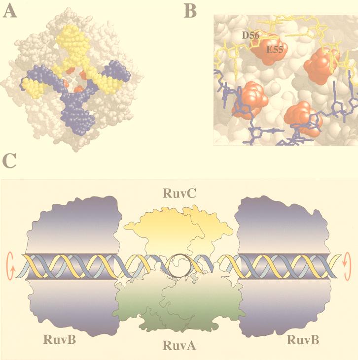
Holliday junction processing by RuvABC. (A) Structure of the RuvA tetramer complexed with a Holliday junction (produced with RasMol) (18). The acidic pins are shown in red. (B) Locations of the acidic pin residues (Glu55 and Asp56; colored red) of RuvA, showing their proximity to the phosphate backbone at the junction core. (C) Model of branch migration and resolution by the RuvABC resolvasome. A tetramer of RuvA holds the Holliday junction in a square planar conformation (side view). RuvB hexamer rings encircle duplexes on each side of the RuvA-junction complex. Branch migration is achieved by drawing the duplexes through these rings, generating heteroduplex DNA. A RuvC dimer bound to the upper face of the junction scans for specific target sequences as the DNA passes across RuvA. Symmetrically related incisions are introduced to yield nicked duplexes which can be sealed by DNA ligase. Outlines of RuvA and RuvC were taken from the crystal structures of these proteins (3, 38). The RuvB structure is based on electron micrographs of RuvAB-junction complexes (36). An animated model of branch migration by RuvAB can be viewed at the Krebs Institute web site (23a).
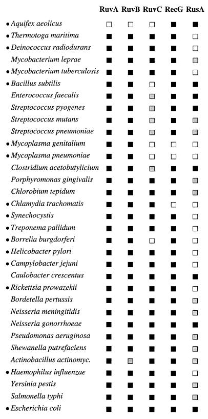
Occurrence of Holliday junction-processing enzymes in eubacteria. Searches were performed with gapped BLAST and PSI-BLAST programs (2) and databases provided by the National Center for Biotechnology Information (32a), The Institute for Genomic Research (46a), and the Sanger Centre (40a). The definition of a homolog (or ortholog) of each gene was based on the following four criteria: (i) sequence similarity searches using the E. coli proteins and subsequent searches with the identified homologs, (ii) analysis based on the known crystal structures of RuvA and RuvC and information derived from mutant versions of the five enzymes site-directed, (iii) experimental evidence confirming the appropriate activities of predicted homologs, and (iv) conservation of distinctive motifs belonging to the family of proteins. The random expectation values (2) for the E. coli protein against the most dissimilar homolog were as follows: Mycoplasma genitalium RuvA (6 × 10⁻⁴), Mycoplasma pneumoniae RuvB (2 × 10−49), Synechocystis sp. strain RuvC (5 × 10⁻⁶), and Campylobacter jejuni RecG (2 × 10−48). Specific features used to identify each homolog included the following: the pair of helix-hairpin-helix motifs and the conserved acidic pin residues of RuvA (37), high conservation of the unusual helicase domain of RuvB, four essential acidic residues required for catalysis and conserved residues around helices 3 and 4 of RuvC (17, 40), and similarities in the helicase and N-terminal domains of RecG and the unique motif it shares with TRCF. TRCF and RecG have different N- and C-terminal domains, sharing considerable homology (38% identity between the E. coli proteins) only in the central helicase motifs (42). RusA homologs are highly divergent and were identified by searching with each member of the family. With the exception of Clostridium acetobutylicium, all contained three aspartic acid residues required for Holliday junction resolution in a conserved C-terminal domain of RusA (7). Several eukaryotic proteins that have been designated RuvB-like (TIP49, RUVBL1, and RUVBL2) were excluded due to the absence of several motifs characteristic of the bacterial RuvB family and the lack of evidence supporting their involvement in recombination. Details of alignments and the accession numbers of identified homologs are available on request. Species are placed in order relative to the phylogenetic tree shown in Fig. 4. Small black circles identify the organisms for which the genome sequence has been completed. The presence (black squares) and absence (white squares) of RuvA, RuvB, RuvC, RecG, and RusA homologs are indicated; a grey square indicates that these homologs were not found in that species, but the genome sequence is incomplete. Abbreviations: Synechocystis, Synechocystis sp. strain PCC6803; Actinobacillus actinomyc., Actinobacillus actinomycetemcomitans.

Phylogenetic tree of eubacteria based on 16s rRNA sequences. The tree contains all the organisms listed in Fig. 3 and was obtained from the Ribosomal Database Project II (30) at its website (38a). The Aquifex sequence comes from Aquifex pyrophilus and not Aquifex aeolicus, because the latter was unavailable. Horizontal branch lengths are drawn to scale; the bar is 0.1 nucleotide replacement per site in length. Species for which homologs of RuvC are absent or yet to be detected are shown in bold type. Abbreviations: Porphyromonas, Porphyromonas gingivalis; Actinobacillus actinomyc., Actinobacillus actinomycetemcomitans.
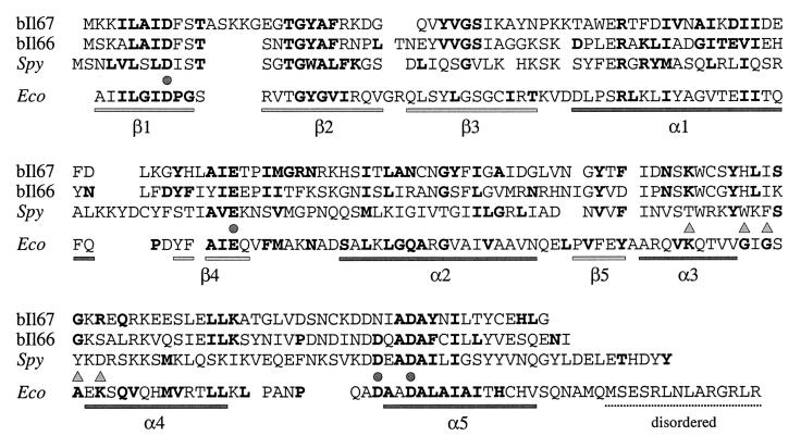
Alignment of L. lactis and S. pyogenes phage RuvC homologs. Protein sequences from L. lactis phages bIL66 and bIL67 were chosen as representatives of the larger family of phage proteins. The bIL66 RuvC has percent identity values of 98, 96, and 93 to homologs from sk1, 712, and bIL170, respectively. bIL67 RuvC protein is 93% identical to those from c2 and vML3. Residues in boldtype in the E. coli RuvC sequence (Eco) represent those conserved among the eubacterial RuvC family (the structure of E. coli RuvC is represented under the sequence). Residues in boldtype in the bIL66, bIL67, and Spy (S. pyogenes) RuvC homologs represent matches with the E. coli RuvC sequence. Circles indicate the four acidic residues required for catalysis by E. coli RuvC; triangles mark residues thought to be involved in sequence specificity of Holliday junction resolution (3, 17). Residues considered similar are as follows: A and G; D and E; F, I, L, M, V, W, and Y; K and R; N and Q; and S and T.
References
-
- Ariyoshi M, Vassylyev D G, Iwasaki H, Nakamura H, Shinagawa H, Morikawa K. Atomic structure of the RuvC resolvase: a Holliday junction-specific endonuclease from E. coli. Cell. 1994;78:1063–1072. - PubMed
-
- Bennett R J, Dunderdale H J, West S C. Resolution of Holliday junctions by RuvC resolvase: cleavage specificity and DNA distortion. Cell. 1993;74:1021–1031. - PubMed
-
- Bennett R J, West S C. Structural analysis of the RuvC-Holliday junction complex reveals an unfolded junction. J Mol Biol. 1995;252:213–226. - PubMed
Publication types
MeSH terms
Substances
LinkOut - more resources
Full Text Sources
Other Literature Sources
Molecular Biology Databases
