. 1999 Sep;181(18):5652-61.
doi: 10.1128/JB.181.18.5652-5661.1999.

Genomic subtraction identifies Salmonella typhimurium prophages, F-related plasmid sequences, and a novel fimbrial operon, stf, which are absent in Salmonella typhi

Affiliations

Genomic subtraction identifies Salmonella typhimurium prophages, F-related plasmid sequences, and a novel fimbrial operon, stf, which are absent in Salmonella typhi

M Emmerth et al. J Bacteriol. 1999 Sep.

Abstract

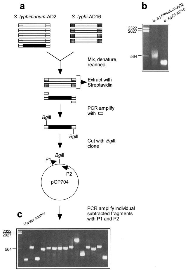
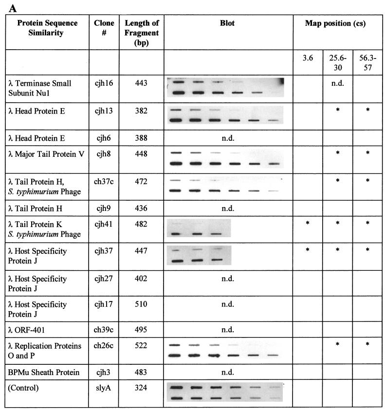
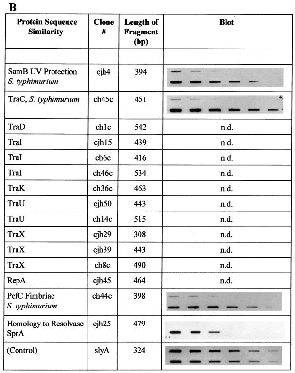
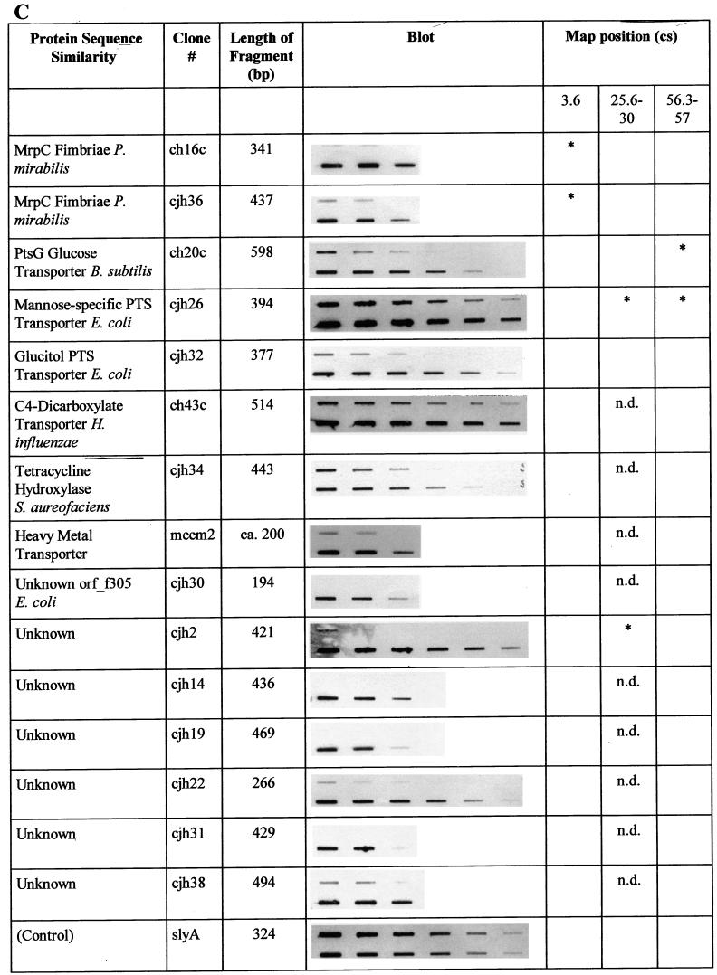
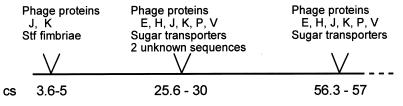
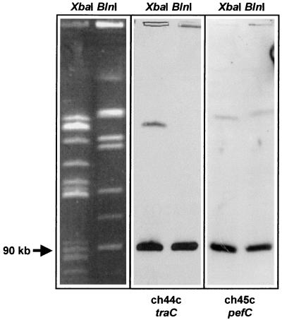
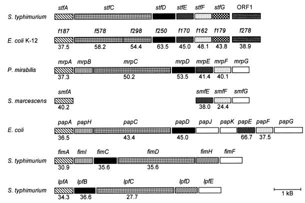
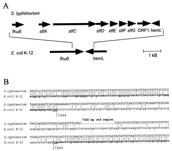
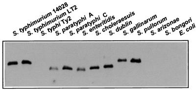
Salmonella typhimurium causes systemic and fatal infection in inbred mice, while the related serotype Salmonella typhi is avirulent for mammals other than humans. In order to identify genes from the virulent strain S. typhimurium ATCC 14028 that are absent in S. typhi Ty2, and therefore might be involved in S. typhimurium mouse virulence, a PCR-supported genomic subtractive hybridization procedure was employed. We have identified a novel putative fimbrial operon, stfACDEFG, located at centisome 5 of the S. typhimurium chromosome, which is absent in S. typhi, Salmonella arizonae, and Salmonella bongori but was detected in several other Salmonella serotypes. The fimbrial genes represent a genomic insertion in S. typhimurium compared to the respective region between fhuB and hemL in Escherichia coli K-12. In addition, the subtraction procedure yielded F plasmid-related sequences from the S. typhimurium virulence plasmid, a number of DNA fragments representing parts of lambdoid prophages and putative sugar transporters, and several fragments with unknown sequences. The majority of subtracted chromosomal sequences map to three distinct locations, around centisomes 5, 27, and 57.


Figures

FIG. 1
FIG. 1
Genomic subtraction procedure used to isolate S. typhimurium-specific DNA fragments. (a) Schematic representation of the subtraction procedure. A fragment of S. typhimurium DNA not present in S. typhi is shown in black. Biotinylated adapter sequences are indicated by hatching. (b) Agarose gel (sizes [in kilobases] of marker fragments are indicated) showing PCR-amplified S. typhimurium-AD2 fragments and S. typhi-AD16 fragments before subtraction. (c) Agarose gel showing individually amplified S. typhimurium DNA fragments after subtraction and cloning. The PCR product obtained by using the cloning vector without an insertion as the template is in the lane adjacent to the size marker lane. (See text for further details.)
FIG. 2
FIG. 2
Summary of the characterizations of S. typhimurium-specific subtracted DNA fragments. Protein sequence similarities were determined by BLAST analysis. Similarities were considered significant if BLAST scores were below 10−5. The slot blots shown demonstrate the absence of individual sequences in S. typhi. The upper row of each blot shows total genomic DNA from S. typhi Ty2 blotted in decreasing amounts, while the lower row contains blotted total genomic DNA from S. typhimurium ATCC 14028. The blots were individually hybridized with full-length DNA fragments isolated by subtraction (see text). The control blot was hybridized with a probe present in both organisms (slyA) and demonstrates equal amounts of target DNA in each row. Each row in each blot contains serial dilutions in the range of 2.5 to 0.04 μg of total genomic DNA, with equal amounts of DNA loaded in the corresponding upper and lower slots. The locations on the chromosomal map of S. typhimurium (17) (Fig. 3), as determined by hybridization of specific fragments to an ordered array of mapping phages (7), are indicated. An asterisk indicates a hybridization signal obtained with mapping phages representing one of three different chromosomal locations near cs 3.6, cs 25.6 to 30, and cs 56.3 to 57. n.d., not determined. Characterizations of sequences with similarities to phage proteins (A), sequences of the S. typhimurium virulence plasmid pSLT (B), and other sequences (C) are summarized. B. subtilis, Bacillus subtilis; H. influenzae, Haemophilus influenzae; S. aureofaciens, Streptomyces aureofaciens.
FIG. 2
FIG. 2
Summary of the characterizations of S. typhimurium-specific subtracted DNA fragments. Protein sequence similarities were determined by BLAST analysis. Similarities were considered significant if BLAST scores were below 10−5. The slot blots shown demonstrate the absence of individual sequences in S. typhi. The upper row of each blot shows total genomic DNA from S. typhi Ty2 blotted in decreasing amounts, while the lower row contains blotted total genomic DNA from S. typhimurium ATCC 14028. The blots were individually hybridized with full-length DNA fragments isolated by subtraction (see text). The control blot was hybridized with a probe present in both organisms (slyA) and demonstrates equal amounts of target DNA in each row. Each row in each blot contains serial dilutions in the range of 2.5 to 0.04 μg of total genomic DNA, with equal amounts of DNA loaded in the corresponding upper and lower slots. The locations on the chromosomal map of S. typhimurium (17) (Fig. 3), as determined by hybridization of specific fragments to an ordered array of mapping phages (7), are indicated. An asterisk indicates a hybridization signal obtained with mapping phages representing one of three different chromosomal locations near cs 3.6, cs 25.6 to 30, and cs 56.3 to 57. n.d., not determined. Characterizations of sequences with similarities to phage proteins (A), sequences of the S. typhimurium virulence plasmid pSLT (B), and other sequences (C) are summarized. B. subtilis, Bacillus subtilis; H. influenzae, Haemophilus influenzae; S. aureofaciens, Streptomyces aureofaciens.
FIG. 3
FIG. 3
Chromosomal mapping of the stf region. An internal fragment of stfC was hybridized to an ordered array of Mud-P22 mapping phages which individually package defined genomic segments (7). The signals obtained are correlated with the chromosomal start point (o) of DNA packaging, and the direction of DNA packaging of individual phages is indicated.
FIG. 4
FIG. 4
Schematic representation of the locations of subtracted DNA fragments on the chromosomal map of S. typhimurium. Map positions are indicated according to reference (17).
FIG. 5
FIG. 5
Genomic mapping of subtracted S. typhimurium DNA fragments representing parts of the S. typhimurium virulence plasmid genes traC and pefC. Probes were hybridized to a blot of total genomic DNA of S. typhimurium ATCC 14028 restricted with either XbaI or BlnI and separated by PFGE. Assignment of DNA fragment sizes was done by using a size standard (not shown) as well as by comparing fragment patterns to published S. typhimurium PFGE analyses (13, 14).
FIG. 6
FIG. 6
Comparison of genetic organization and similarities of deduced proteins of the stf genes and other fimbrial operons from various organisms. Direction of transcription is from left to right. Similarities of deduced proteins are indicated by different patterns. The identities of amino acid sequences with the stf-encoded proteins are given as percentages. S. marcescens, Serratia marcescens.
FIG. 7
FIG. 7
(A) Genomic organization of the stf region, located between fhuB and hemL in S. typhimurium. The genes encoding the stf fimbriae and their positions relative to fhuB and hemL in E. coli are indicated by arrows. (B) Alignment of DNA sequences flanking the stf region in S. typhimurium with fhuB and hemL sequences of E. coli. The 3′ ends of the E. coli fhuB and hemL genes are shown in bold. The exact positions of the underlined stop codons of the complete E. coli K-12 genome sequence are given in base pairs. Identical bases are indicated by vertical bars.
FIG. 8
FIG. 8
Distribution of stf sequences among different Salmonella serotypes and E. coli. PstI-restricted genomic DNA of the organisms indicated was hybridized with an internal probe of the stfC gene (see Materials and Methods).

References

    1. Bahrani F K, Mobley H L T. Proteus mirabilis MR/P fimbrial operon: genetic organization, nucleotide sequence, and conditions for expression. J Bacteriol. 1994;176:3412–3419. - PMC - PubMed
    1. Bäumler A J, Heffron F. Identification and sequence analysis of lpfABCDE, a putative fimbrial operon of Salmonella typhimurium. J Bacteriol. 1995;177:2087–2097. - PMC - PubMed
    1. Bäumler A J, Tsolis R M, Bowe F A, Kusters J G, Hoffmann S, Heffron F. The pef fimbrial operon of Salmonella typhimurium mediates adhesion to murine small intestine and is necessary for fluid accumulation in the infant mouse. Infect Immun. 1996;64:61–68. - PMC - PubMed
    1. Bäumler A J, Tsolis R M, Heffron F. Contribution of fimbrial operons to attachment to and invasion of epithelial cell lines by Salmonella typhimurium. Infect Immun. 1996;64:1862–1865. - PMC - PubMed
    1. Bäumler A J, Tsolis R M, Heffron F. The lpf fimbrial operon mediates adhesion of Salmonella typhimurium to murine Peyer’s patches. Proc Natl Acad Sci USA. 1996;93:279–283. - PMC - PubMed

Publication types

MeSH terms

Associated data

LinkOut - more resources