

doi: 10.1128/JB.181.18.5852-5854.1999.
A sequence downstream of the initiation codon is essential for cold shock induction of cspB of Escherichia coli
Affiliations
- PMID: 10482531
- PMCID: PMC94110
- DOI: 10.1128/JB.181.18.5852-5854.1999
Item in Clipboard
A sequence downstream of the initiation codon is essential for cold shock induction of cspB of Escherichia coli
J Bacteriol.
1999 Sep.
Abstract
Cold shock induction of cspB has been shown to be primarily regulated at the mRNA level. Here, we demonstrate that the induction of cspB at low temperature also requires the translational cis-acting element called the downstream box (DB). Full induction of cspB at low temperature is achieved in the presence of both the Shine-Dalgarno sequence and DB. We propose that the DB sequence functions as a translational enhancer for the biosynthesis of CspB to bypass the inhibitory effect in translation caused by cold shock.
Figures
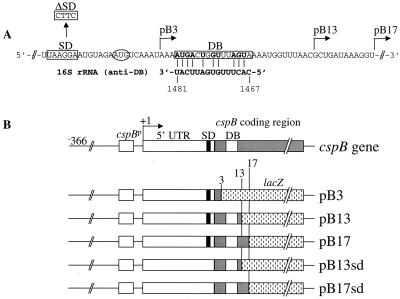
Construction of cspB-lacZ fusions. (A) cspB-DB–anti-DB complementarity. The cspB-DB sequence is boxed and encompasses the region from codons 5 to 9 (13). The AUG codon is circled, the SD sequence is boxed, and the L-shaped arrows show the positions where the cspB gene was fused to lacZ. (B) Translational cspB-lacZ fusion constructs. On the top, the E. coli cspB gene is depicted from its 5′-end. In pB3, pB13, and pB17, cspB is fused to lacZ at residue +177 (3rd codon), +200 (13th codon), and +212 (17th codon), respectively. The cspB DNA fragments were amplified by PCR using synthetic oligonucleotide primers containing a BamHI site at the 5′ end. A plasmid, pSJ7 (10) carrying the wild-type cspB gene was used as a template DNA to create the PCR fragments B3, B13, and B17. The 5′-end oligonucleotide primer used in each of the above PCR reactions is 5′-CCGGATCCAGCTTTAATATAGCT-3′. The 3′-end oligonucleotide primers for the PCR products B3, B13, and B17 are 5′-CCGGATCCAGATTTGACATTCTACA-3′, 5′-CCGGATCCAGGTTAAACCATTTT-3′, and 5′-CCGGATCCAGACCTTTATCAGCGTT-3′, respectively. A deletion of the SD sequence in the cspB gene was created by site-directed mutagenesis using the QuickChange site-directed mutagenesis kit (Stratagene). The PCR reaction was carried out using the pSJ7 plasmid as a template and the oligonucleotides Bsd1 (5′-GAAAGGCTCAAGTTACTTCATGTAGAATG-3′) and Bsd2 (5′-CATTCTAC ATGAAGTAACTTGAGCCTTTC-3′) to create pSJsd. Then, pSJsd was used as a template to make the PCR fragments B13sd and B17sd. The 5′ and 3′-end oligonucleotide primers used in these PCR reactions are the same as the one used for the PCR fragments B13 and B17. All the above PCR products were cloned at the BamHI site of the pRS414 vector (10, 15) to create the pB3, pB13, pB13sd, pB17, and pB17sd constructs.
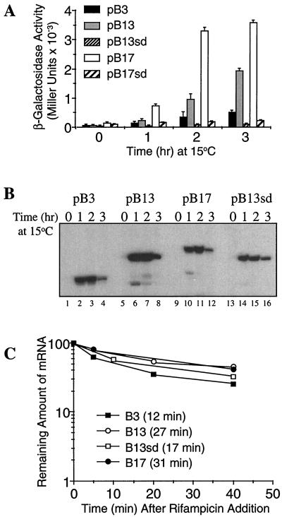
Enhancement of cspB translation by the DB. (A) β-Galactosidase activity of the cspB-lacZ constructs obtained before (time zero) and 1, 2, and 3 h after temperature shift from 37 to 15°C. E. coli AR137 cells transformed with pB3, pB13, pB13sd, pB17, and pB17sd were grown in Luria-Bertani medium, and at mid-log phase (optical density at 600 nm = 0.4) cultures were shifted from 37 to 15°C. β-Galactosidase activity was measured as described by Miller (12). (B) mRNA levels of pB3, pB13, pB13sd, and pB17 after temperature shift from 37 to 15°C. The cspB-lacZ mRNAs were detected by primer extension as described previously (13) at the same time points described above (panel A). (C) Stabilities of pB3, pB13, pB13sd, and pB17 mRNA. E. coli AR137 cells transformed with pB3, pB13, pB13sd, and pB17 were grown under the same conditions as those described above. At mid-log phase the culture was shifted to 15°C, and after 30 min rifampin was added to a final concentration of 0.2 mg/ml (time zero). Total RNA was extracted at 5, 10, 20, and 40 min after rifampin addition. The cspB-lacZ mRNA was detected by primer extension as described previously (13).
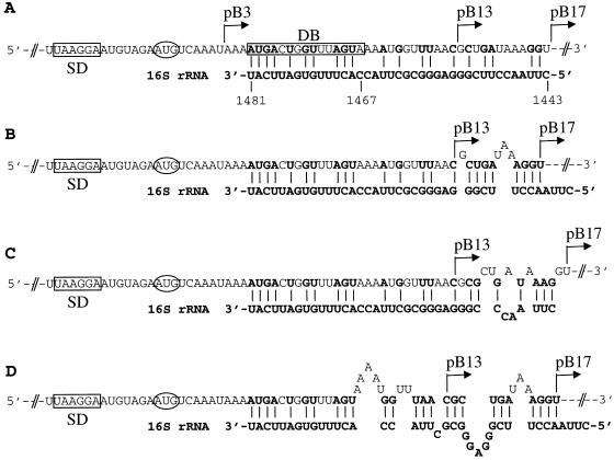
Additional base pairing between the cspB mRNA and the 16S rRNA. (A) Additional base pairs between the pB17 mRNA (within the sequence from codon 13 to 17) and the 16S rRNA with no mismatches. (B) pB17 mRNA (within the sequence from codon 13 to 17)-16S rRNA base pairs with two mismatches in the cspB mRNA. (C) Same as in panel B with three mismatches in the pB17 mRNA and one mismatch in the 16S rRNA. (D) Additional base pairs between pB17 mRNA and 16S rRNA with multiple mismatches in both of them allowed.
References
-
- Brandi A, Pietroni P, Gualerzi C O, Pon C L. Post-transcriptional regulation of CspA expression in Escherichia coli. Mol Microbiol. 1996;19:231–240. - PubMed
-
- Etchegaray J-P, Jones P G, Inouye M. Differential thermoregulation of two highly homologous cold-shock genes, cspA and cspB, of Escherichia coli. Genes Cell. 1996;1:171–178. - PubMed
-
- Etchegaray J-P, Inouye M. Translational enhancement by an element downstream of the initiation codon in Escherichia coli. J Biol Chem. 1999;274:10079–10085. - PubMed
-
- Fang L, Jiang W, Bae W, Inouye M. Promoter-independent cold-shock induction of cspA and its derepression at 37°C by mRNA stabilization. Mol Microbiol. 1997;23:355–364. - PubMed
Publication types
MeSH terms
Substances
Grants and funding
LinkOut - more resources
Full Text Sources
Molecular Biology Databases
Miscellaneous
