. 1999 Oct;73(10):8083-94.
doi: 10.1128/JVI.73.10.8083-8094.1999.

Mutagenesis of the NS2B-NS3-mediated cleavage site in the flavivirus capsid protein demonstrates a requirement for coordinated processing

Affiliations

Mutagenesis of the NS2B-NS3-mediated cleavage site in the flavivirus capsid protein demonstrates a requirement for coordinated processing

S M Amberg et al. J Virol. 1999 Oct.

Abstract

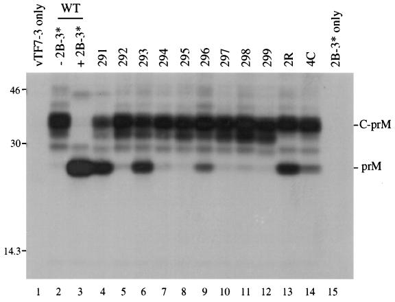
Analysis of flavivirus polyprotein processing has revealed the presence of a substrate for the virus-encoded NS2B-NS3 protease at the carboxy-terminal end of the C (capsid or core) protein. Cleavage at this site has been implicated in the efficient generation of the amino terminus of prM via signal peptidase cleavage. Yellow fever virus has four basic residues (Arg-Lys-Arg-Arg) in the P1 through P4 positions of this cleavage site. Multiple alanine substitutions were made for these residues in order to investigate the substrate specificity and biological significance of this cleavage. Mutants were analyzed by several methods: (i) a cell-free trans processing assay for direct analysis of NS2B-NS3-mediated cleavage; (ii) a trans processing assay in BHK-21 cells, using a C-prM polyprotein, for analysis of prM production; (iii) an infectivity assay of full-length transcripts to determine plaque-forming ability; and (iv) analysis of proteins expressed from full-length transcripts to assess processing in the context of the complete genome. Mutants that exhibited severe defects in processing in vitro and in vivo were incapable of forming plaques. Mutants that contained two adjacent basic residues within the P1 through P4 region were processed more efficiently in vitro and in vivo, and transcripts bearing these mutations were fully infectious. Furthermore, two naturally occurring plaque-forming revertants were analyzed and shown to have restored protein processing phenotypes in vivo. Finally, the efficient production of prM was shown to be dependent on the proteolytic activity of NS3. These data support a model of two coordinated cleavages, one that generates the carboxy terminus of C and another that generates the amino terminus of prM. A block in the viral protease-mediated cleavage inhibits the production of prM by the signal peptidase, inhibits particle release, and eliminates plaque formation.


Figures

FIG. 1
FIG. 1
Schematic diagram of the substrate used in the cell-free trans-processing assay. The YF polyprotein is shown at the top; the locations of the structural proteins (C, prM, and E) and the nonstructural proteins (NS1 through NS5) are indicated. Below the YF polyprotein is an expanded view of the C-prM cassette; the glycosylation sites (∗), the signal peptidase cleavage site (⧫), and the capsid dibasic cleavage site (↓) are noted, and the hydrophobic regions of the C-prM polyprotein are shaded. The substrate used for the protease assay, denoted anchC.3, is 51 amino acids in length. Its size and position relative to the C-prM polyprotein are shown. The sequence around the cleavage site is indicated, using the one-letter amino acid code.
FIG. 2
FIG. 2
Cell-free protease assay. Transcripts generated in vitro from mutant derivatives of pBS-anchC.3 (Tables 1 and 2) were translated in a cell-free system and radiolabeled by the inclusion of [35S]methionine. The resultant protein mixture was incubated with a Triton X-100 lysate from YF-infected SW-13 cells (lanes 4 to 15) or from mock-infected SW-13 cells (lane 3) and then separated by Tricine-SDS-PAGE. Visualization of the image was performed with a Bio-Rad Molecular Imager System. RNA was omitted from the translation mixture in lane 2. Lanes 3 and 4 show the wild-type (WT) substrate, while the substrates used in the other lanes are indicated. A set of 14C-labeled molecular mass standards was run in lane 1 (indicated on the left). The positions of the substrate and products are shown on the right. The wild-type substrate generates products of 31 and 20 amino acids (a.a.). However, the products seen for mutants 293 (lane 7) and 296 (lane 10) likely result from cleavage on the carboxy-terminal side of the basic pair of residues that generate products of 30 and 21 amino acids (293) or 29 and 22 amino acids (296).
FIG. 3
FIG. 3
Coexpression of C-prM mutants and NS2B-3181. Expression of the transfected plasmids was driven by a vaccinia virus recombinant that expresses T7 RNA polymerase (vTF7-3). The C-prM cassettes contained the mutations at the dibasic capsid site that are listed in Table 1. SDS lysates were immunoprecipitated with antiserum to either prM (A) or C (B). Mock infection, vTF7-3 alone, and pTM3-NS2B-3181 without pTM3-C-prM are shown in the first three lanes. Wild-type (WT) and mutant C-prM are indicated, either without (−) or with (+) cotransfection of pTM3-NS2B-3181. Positions of molecular mass standards ([14C]MW markers) are shown, with sizes in kilodaltons noted on the left (the 12.5- and 14.3-kDa markers comigrate on SDS–13% PAGE). The positions of C, prM, and the C-prM polyprotein are indicated on the right.
FIG. 4
FIG. 4
Coexpression of wild-type C-prM and NS2B-3181 components. Following infection with vTF7-3, BHK-21 cells were transfected with pTM3-C-prM alone (lane 3) or in combination with pTM3-NS2B (lane 4), pTM3-NS3181 (lane 5), pTM3-NS3181(S→A) (lane 6), pTM3-NS2B-3181 (lane 7), pTM3-NS2B plus pTM3-NS3181 (lane 8), or pTM3-NS2B plus pTM3-NS3181(S→A) (lane 9). Following immunoprecipitation with antiserum to prM (A) or a combination of antisera to NS2B and NS3 (B), proteins were separated by SDS–13% PAGE. Mock-infected cell lysate was immunoprecipitated in lane 1, and untransfected vTF7-3-infected cell lysate was immunoprecipitated in lane 2. The identities of the proteins are noted on the right side of each panel, and the positions of standards (in kilodaltons) are indicated on the left. A small amount of unprocessed NS2B-3181 can be seen in lane 7 of panel B. NS3181 has only a single internal methionine residue, while NS2B has four, contributing to the difference in band intensity.
FIG. 5
FIG. 5
Coexpression of C-prM mutants and NS2B-3181 following transfection of plasmids at a 100:1 ratio. One microgram of pTM3-C-prM derivative and 10 ng of pTM3-NS2B-3181 were transfected for each 35-mm-diameter dish (lanes 3 to 14). SDS lysates were immunoprecipitated with prM antiserum and separated by SDS–13% PAGE. Lanes: 1, vTF7-3 alone; 2 and 3, wild-type (WT) pTM3-C-prM with (lane 3) or without (lane 2) pTM3-NS2B-3181; 15, pTM3-NS2B-3181 in the absence of pTM3-C-prM plasmid. The positions of protein standards (in kilodaltons) are shown on the left, and the positions of prM and the C-prM polyprotein are indicated on the right.
FIG. 6
FIG. 6
Plaque assays of YF containing mutations at the capsid dibasic cleavage site. BHK-21 and SW-13 cells were transfected with the mutant transcripts shown, and serial 10-fold dilutions were seeded on 35-mm-diameter dishes in the presence of untransfected cells. Monolayers were seeded with 0.2 ml of the following dilutions: for BHK-21, 10−5 (wild type [WT]), 10−4 (291), 10−3 (left) and 10−4 (right) (293 and 296), and 10−1 (294); and for SW-13, 10−4 except for 294, which was a 10−1 dilution.
FIG. 7
FIG. 7
Analysis of C-prM processing in cells transfected with full-length mutant transcripts of YF cDNA. Transfected BHK-21 cells were labeled with [35S]methionine for 5 h (19 to 24 h postelectroporation), and SDS lysates were immunoprecipitated with prM (A) or C (B) antiserum. As a control, cells were infected with YF at a multiplicity of infection of 10 and labeled under the same conditions (16 to 21 h postinfection). Immunoprecipitation of YF-infected cell lysates was performed with only one-half the volume used for transfected-cell lysates. Lane 1, YF control; lane 2, cells electroporated with no RNA (TE only); lane 3, electroporation of wild-type (WT) transcript; lanes 4 to 14, mutant transcripts (as denoted). The positions of C, prM, and the C-prM polyprotein are indicated on the left, and those of the molecular mass standards (in kilodaltons) are shown on the right.
FIG. 8
FIG. 8
Analysis of envelope protein distribution in cells transfected with full-length mutant transcripts of YF cDNA. (A) Transfected BHK-21 cells were labeled with [35S]methionine for 6 h (18 to 24 h postelectroporation); media was harvested after the labeling period, solubilized with SDS, and immunoprecipitated with antiserum to the E protein. (B) SDS lysates of the cell monolayers from the above experiment were prepared and immunoprecipitated with E antiserum. (C) SDS lysates of the cell monolayers from the above experiment were prepared and immunoprecipitated with NS3 antiserum. Immunoprecipitation of medium or of lysate of cells transfected with no RNA is shown in the first lane of each panel. The positions of E and NS3 are indicated on the right side of their respective panels, and the positions of protein standards (in kilodaltons) are shown on the left. WT, wild type.

Similar articles

Cited by

References

    1. Allison S L, Stadler K, Mandl C W, Kunz C, Heinz F X. Synthesis and secretion of recombinant tick-borne encephalitis virus protein E in soluble and particulate form. J Virol. 1995;69:5816–5820. - PMC - PubMed
    1. Amberg S M, Nestorowicz A, McCourt D W, Rice C M. NS2B-3 proteinase-mediated processing in the yellow fever virus structural region: in vitro and in vivo studies. J Virol. 1994;68:3794–3802. - PMC - PubMed
    1. Ausubel F M, Brent R, Kingston R E, Moore D D, Seidman J G, Smith J A, Struhl K, editors. Current protocols in molecular biology. New York, N.Y: John Wiley & Sons; 1998.
    1. Barrett A J. Classification of peptidases. Methods Enzymol. 1994;244:1–15. - PubMed
    1. Bredenbeek, P. J., E. Kooi, B. D. Lindenbach, M. Lucassen, N. Huijkman, W. J. M. Spaan, and C. M. Rice. Unpublished data.

Publication types

MeSH terms

LinkOut - more resources