. 1999 Oct;73(10):8290-302.
doi: 10.1128/JVI.73.10.8290-8302.1999.

trans-dominant interference with human immunodeficiency virus type 1 replication and transmission in CD4(+) cells by an envelope double mutant

Affiliations

trans-dominant interference with human immunodeficiency virus type 1 replication and transmission in CD4(+) cells by an envelope double mutant

S S Chen et al. J Virol. 1999 Oct.

Abstract

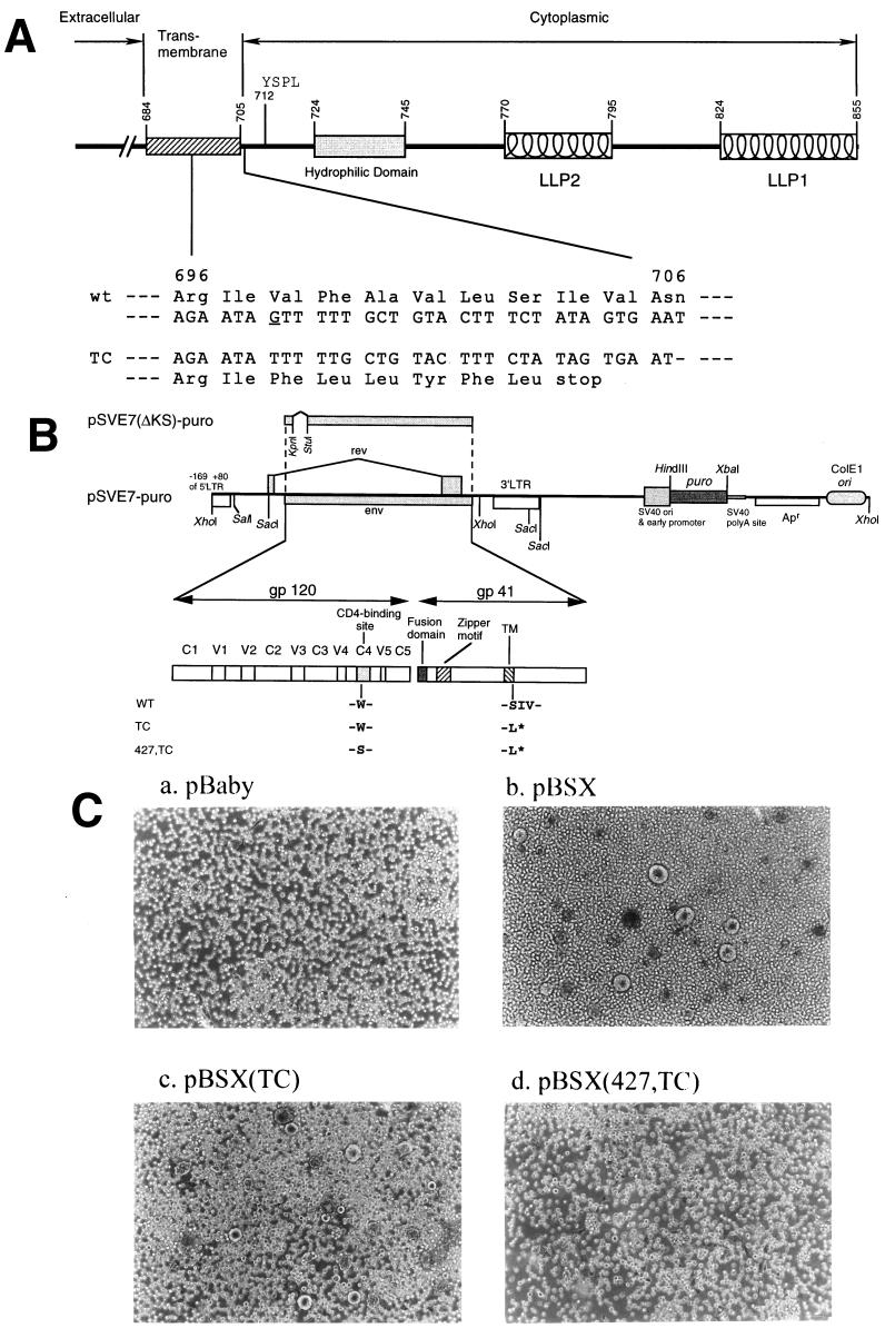
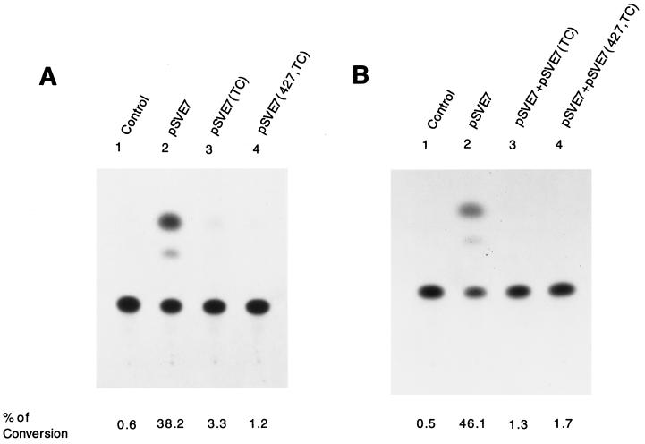
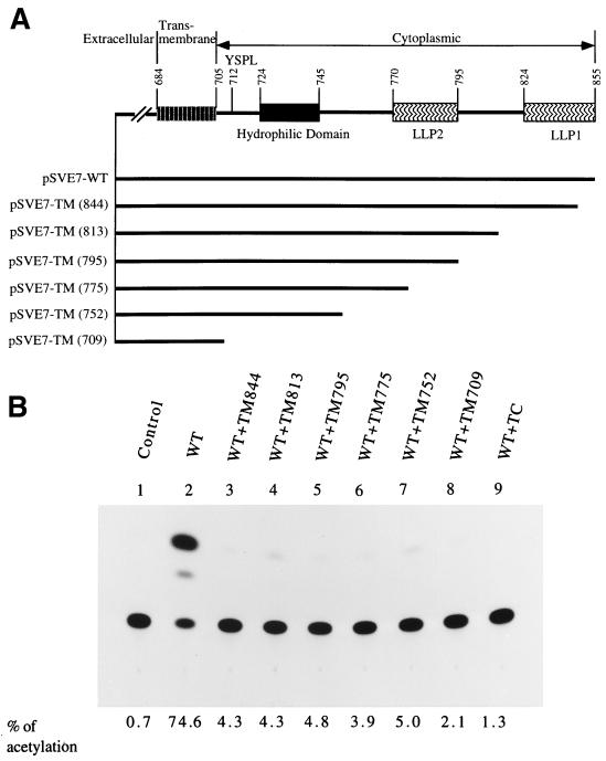
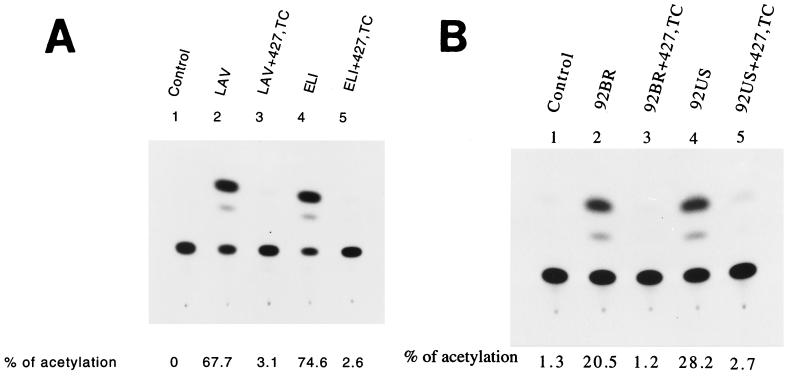
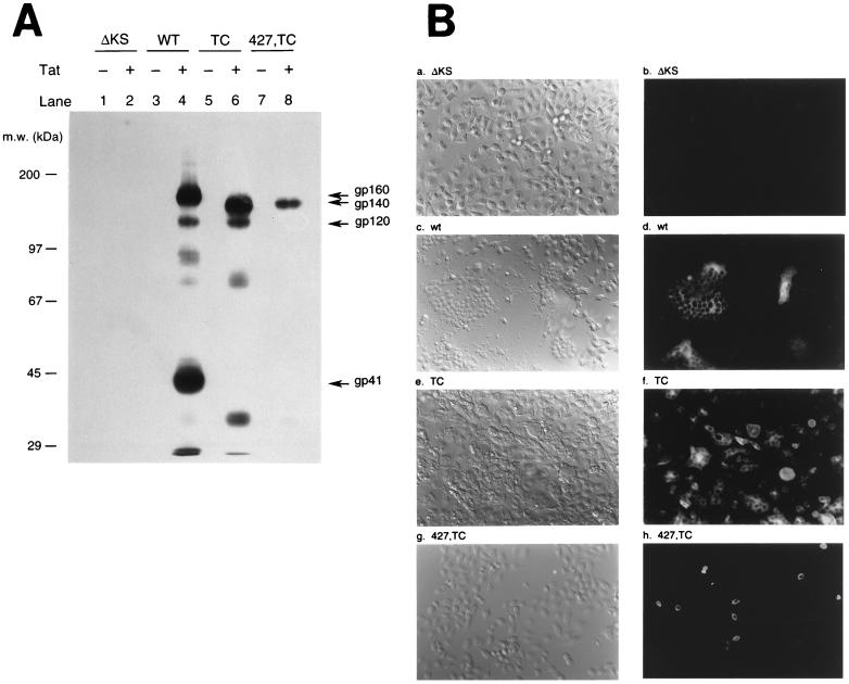
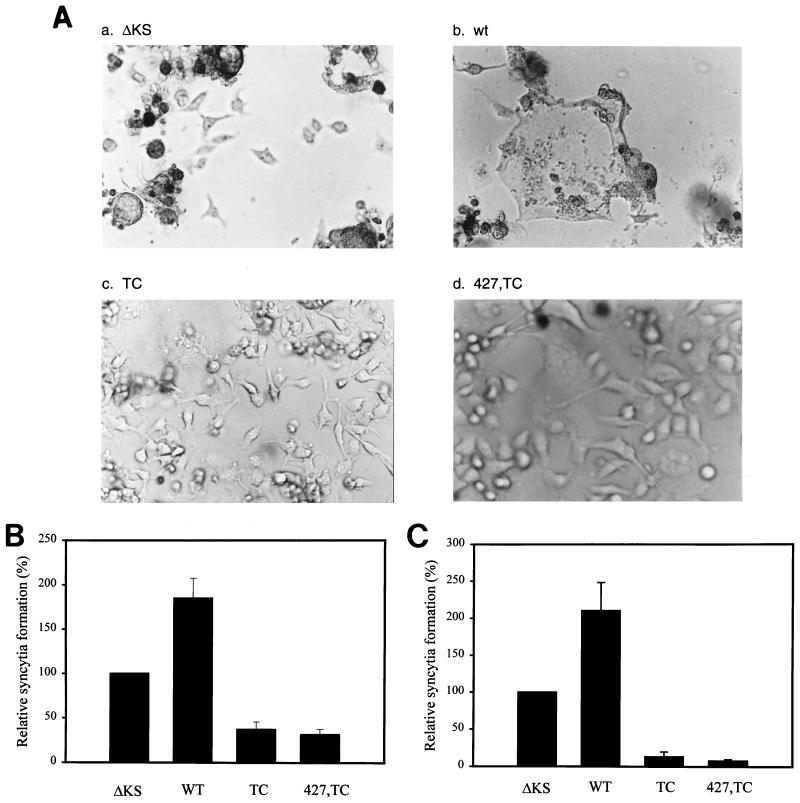
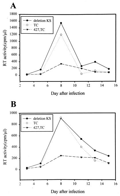
We previously reported that a human immunodeficiency virus type 1 (HIV-1) envelope (Env) mutant with the whole cytoplasmic domain deleted, denoted mutant TC, is able to dominantly interfere with wild-type (wt) virus infectivity. In the present study, the feasibility of developing a dominant negative mutant-based genetic anti-HIV strategy targeting the gp41 cytoplasmic domain was investigated. Mutants TC and 427,TC, a TC derivative with a Trp-to-Ser substitution introduced into residue 427 in the CD4-binding site, and a series of mutants with deletions in the cytoplasmic domain, effectively trans-dominantly interfered with wt Env-mediated viral infectivity, as demonstrated by an env trans-complementation assay. The syncytium formation-defective 427, TC double mutant not only inhibited heterologous LAV and ELI Env-mediated viral infectivity but also interfered with syncytium formation and infectivity mediated by the Env proteins of the two primary isolates 92BR and 92US. Stable HeLa-CD4-LTR-beta-gal clones that harbored Tat-controlled expression cassettes encoding the control DeltaKS, which had a deletion in the env gene, wt, or mutant env gene were generated. Viral transmission mediated by laboratory-adapted T-cell-tropic HXB2 and NL4-3 viruses was greatly reduced in the TC and 427,TC transfectants compared to that observed in the control DeltaKS and wt transfectants. Viral replication caused by HXB2 and NL4-3 viruses and by macrophage-tropic ConB and ADA-GG viruses was delayed or reduced in human CD4(+) T cells transfected with the 427,TC env construct compared to that observed in cells transfected with the control DeltaKS or TC env construct. The lack of significant interference by TC mutant was due neither to the lack of TC env gene integration into host DNA nor to the lack of TC Env expression upon Tat induction. These results indicate that this 427,TC Env double mutant has a role in the development of trans-dominant mutant-based genetic anti-HIV strategies.


Figures

FIG. 1
FIG. 1
Construction and characterization of Env mutants. (A) A schematic representation of the cytoplasmic domain truncation Env mutant. Structural motifs in gp41 cytoplasmic domains are shown, such as internalization signal YSPL and the tyrosine-based basolateral-targeting signal located at residue 712, a highly hydrophilic region, and two positively charged amphipathic α-helices marked LLP-1 and LLP-2. The amino acid residues are numbered according to their positions in the Env of the HXB2 strain. The region encompassing residues from 696 to 706 and its corresponding DNA sequence are indicated. The deletion of the G base as underlined located at nucleotide 8315 of the HXB2 sequence results in a frame shift of the open reading frame and a premature termination of translation after leucine at position 703 in the TM domain. (B) Construction of wt and mutant pSVE7-puro plasmids. A puromycin resistance expression cassette was inserted into the wt pSVE7. The KpnI-BamHI fragments isolated from various mutant pSVE7 plasmids were substituted for the corresponding sequence in wt pSVE7-puro to generate various mutant pSVE7-puro plasmids. The asterisk represents the stop codon in the TM domain as described in panel A. Single-letter amino acid codes are used. (C) Analysis of the syncytium-forming ability of Env mutants. COS-1 cells were transfected with 2 μg each of pBaby, wt, or mutant pBSX as indicated. Two days after transfection, 106 SupT1 cells were added into each transfected culture, and photographs were taken 18 h after coculture under a light microscope. Magnification, ×100.
FIG. 2
FIG. 2
Assessment of mutant Env proteins by an env trans-complementation assay. (A) Inability of mutant proteins to mediate virus-to-cell transmission. 293 cells were cotransfected with 10 μg of pHXBCATΔBgl and 10 μg each of the control ΔKS, wt, or mutant pSVE7. Cell-free viruses containing 2 × 10⁵ cpm of RT activity from each viral stock were used to challenge HeLa-CD4-LTR-β-gal cells, and CAT activity was measured. (B) Interference with wt Env-mediated viral infectivity by Env mutants. 293 cells were cotransfected with 10 μg each of pHXBCATΔBgl and wt pSVE7 in the presence or absence of 10 μg of mutant pSVE7 as described in Materials and Methods. Viruses containing 2 × 10⁵ cpm of RT activity from each transfection were used to determine Env-mediated virus-to-cell transmission. In panels A and B, the viruses used in the lanes marked control were produced from cotransfection of pHXBCATΔBgl and pSVE7(ΔKS). In panel B, the pSVE7(ΔKS) plasmid was added into transfection mixtures to maintain the total DNA amounts in all transfection reactions at the same levels.
FIG. 3
FIG. 3
Effect of coexpression with Env deletion mutants on wt Env-mediated viral infectivity. (A) A schematic representation of a series of Env mutants with deletions from the C terminus of the cytoplasmic domain. The numbers in parentheses indicate that a stop codon was introduced after the codon of the indicated residue. (B) Interference with wt Env-mediated infectivity by deletion mutants. 293 cells were cotransfected with 7.5 μg of pHXBCATΔBgl and 7.5 μg of wt pSVE7-puro in the presence or absence of 7.5 μg of mutant pSVE7-puro plasmids that encoded deletion mutants as indicated. Cell-free viruses containing 6.5 × 10⁴ cpm of RT activity from each viral supernatant were used to determine Env-mediated virus-to-cell transmission to HeLa-CD4-LTR-β-gal cells.
FIG. 4
FIG. 4
Interference by the 427,TC double mutant with viral infectivity. (A) Interference with viral infectivity mediated by heterologous T-tropic Env proteins. 293 cells were cotransfected with 7.5 μg of pHXBCATΔBgl and 5 μg each of pSVIII-LAV or pSVIII-ELI with or without 10 μg of pSVE7(427,TC). pSVIII-ELI encodes the Env of the HIV-1 ELI strain. Cell-free viruses containing 105 cpm of RT activity from each transfection were assayed for Env-mediated viral infectivity. (B) Interference with viral infectivity mediated by the Env proteins derived from primary isolates. 293 cells were cotransfected with 7.5 μg of pHXBCATΔBgl and 5 μg each of pSVIII-env expression plasmids that encoded Env proteins derived from primary isolates as indicated with or without 10 μg of pSVE7(427,TC). Viruses containing 105 cpm of RT activity from each viral supernatant were used to challenge P4-R5 MAGI indicator cells, and CAT activity was assayed.
FIG. 5
FIG. 5
Characterization of env stable transfectants. (A) Tat-dependent HIV-1 env gene expression in stable env transfectants. The four env transfectants were transfected with or without 5 μg of pIIIextat. Equal amount of cell lysates from each transfection were subjected to an SDS–7.5% PAGE, followed by Western blotting by using MAbs Chessie 8 and Chessie 13. m.w., molecular size. (B) Analysis of Tat-induced Env expression in env transfectants by indirect immunofluorescence microscopy. The four env transfectants as indicated were transfected with pIIIextat. Three days after transfection, cells were processed for immunofluorescence analysis as described in Materials and Methods. Cells were analyzed under a differential interference contrast microscope (panels a, c, e, and g) or fluorescence microscope (panels b, d, f, and h), both at a magnification of 200×.
FIG. 6
FIG. 6
Interference with wt Env-mediated viral transmission in mutant env transfectants. (A) Inhibition with exogenous Env-mediated cytopathicity. The four env transfectants as indicated were cotransfected with 10 μg of wt pSVE7-puro and 5 μg of pIIIextat. Three days posttransfection cultures were taken for photographs under a differential interference contrast microscope with a magnification of ×200. This experiment was performed three times, with similar results. Ten fields of each transfected culture were observed, all with similar results. A representative micrograph from each transfectant is shown. (B) Interference with homologous HXB2-mediated cell-to-cell-mediated transmission. The four env transfectants grown in 60-mm-diameter petri dishes with grids were cocultured with HXB2-transfected SupT1 cells. Three days after transfection, cultures were scored for syncytium formation. The degree of syncytium formation in the control ΔKS was arbitrarily set at 100%. The relative syncytium formation observed in the wt and mutant transfectants was expressed as the percentage of the numbers of syncytia observed relative to that found in the control ΔKS transfectant. The results from four individual experiments were averaged, and means and standard deviations were calculated. (C) Interference with heterologous NL4-3-mediated virus-to-cell transmission. The env transfectants as indicated were infected with the NL4-3 virus, and 3 days after infection, syncytia were scored. The diagram represents the degree of syncytium formation obtained in four independent experiments (means ± standard deviations).
FIG. 7
FIG. 7
Effect of Env truncation mutants on T-tropic viral replication in human CD4+ T cells. (A) Interference with viral replication and spread caused by the homologous HXB2 virus. The puromycin-selected CEM-SS cells transfected with various pSVE7-puro env constructs as indicated were infected with the HXB2 virus, and RT activity was monitored after infection. (B) Interference with NL4-3 virus-mediated viral replication and spread. CEM-SS cells transfected with env constructs as indicated in panel A were infected with the NL4-3 virus, and RT activity was monitored after infection. In panels A and B, similar results were obtained from at least three infection analyses. A representative result from each is shown. (C) Characterization of env-transfected CEM-SS cells. Left panel shows the presence of the env genes in transfected CEM-SS cells. Lysates obtained from equal volumes of env-transfected CEM-SS cells were analyzed by PCR by using env and β-globin primers as described in Materials and Methods. PCR products of each transfectant were mixed and resolved by 3% Nusieve agarose electrophoresis. The right panel shows Tat-induced Env expression in env-transfected CEM-SS cells. We transfected 107 CEM-SS cells harboring the env genes as indicated with 10 μg of pIIIextat. Three days after transfection, cell lysates were prepared and analyzed by Western blotting by using Chessie 13 and 902 MAbs. To indicate the migration position of the wt Env precursor, HIV-1-infected CEM-SS cell lysate was also analyzed in parallel (lane 4).
FIG. 8
FIG. 8
Interference with M-tropic viral replication in mutant env-transfected PM1 cells. PM1 cells were transfected with the pSVE7-puro env constructs as indicated by electroporation. The puromycin-selected transfected cells were infected with the ConB (A) or ADA-GG (B) virus, and RT activity was monitored after infection. Each experiment was performed three times, with similar results. A representative result for each is shown.

References

    1. Abacioglu Y H, Fouts T R, Laman J D, Claasen E, Pincus S H, Moore J P, Roby C A, Kamin-Lewis R, Lewis G K. Epitope mapping and topology of baculovirus-expressed HIV-1 gp160 determined with a panel of murine monoclonal antibodies. AIDS Res Hum Retroviruses. 1994;10:371–381. - PubMed
    1. Adachi A, Gendelman H E, Koenig S, Folks T, Willey R, Rabson A, Martin M A. Production of acquired immunodeficiency syndrome-associated retrovirus in human and nonhuman cells transfected with an infectious molecular clone. J Virol. 1986;59:284–291. - PMC - PubMed
    1. Ausubel F M, Brent R, Kingston R E, Moore D D, Seidman J G, Smith J A, Struhl K. Current protocols in molecular biology. Vol. 2. New York, N.Y: John Wiley & Sons; 1995.
    1. Buchschacher G L, Jr, Freed E O, Panganiban A T. Cells induced to express a human immunodeficiency virus type 1 envelope gene mutant inhibit the spread of wild-type virus. Hum Gene Ther. 1992;3:391–397. - PubMed
    1. Buchschacher G L, Jr, Freed E O, Panganiban A T. Effects of second-site mutation on dominant interference by a human immunodeficiency virus type 1 envelope glycoprotein mutant. J Virol. 1995;69:1344–1348. - PMC - PubMed

Publication types