

Prevalence of varicella-zoster virus DNA in dissociated human trigeminal ganglion neurons and nonneuronal cells
- PMID: 10482610
- PMCID: PMC112877
- DOI: 10.1128/JVI.73.10.8571-8577.1999
Prevalence of varicella-zoster virus DNA in dissociated human trigeminal ganglion neurons and nonneuronal cells
Abstract
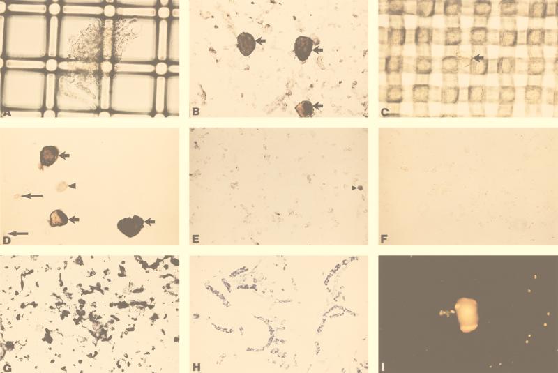
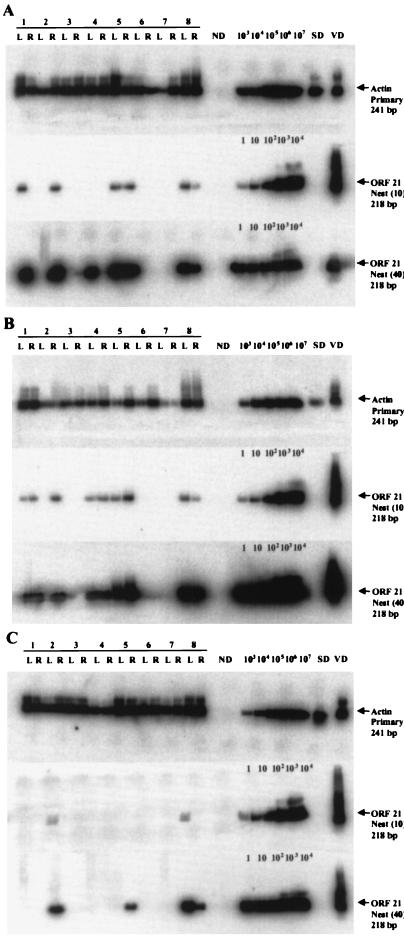
Previous analyses using in situ hybridization alone or together with PCR have yielded conflicting results regarding the cell type in which latent varicella-zoster virus (VZV) resides. We separated human trigeminal ganglia (TG) into neuronal and nonneuronal fractions, followed by primary and nested PCR to quantitate VZV DNA at the single cell level. Both TG from each of eight cadavers were dissociated and separated into neuronal and nonneuronal cell suspensions by differential filtration. Analysis of the neuron fraction (5,000 neurons per sample) revealed VZV DNA in 9 of 16 samples, with copy numbers ranging from 1 to 12, whereas only 2 of 16 nonneuronal cell samples were positive for VZV DNA, with 1 copy each. Further analysis of 10 samples of 100 neurons and the corresponding nonneuronal cell fractions from each TG of a single subject revealed VZV DNA in 3 of 10 samples of the left TG (range, 2 to 5 copies) and in 1 of 10 samples of the right TG (2 copies) but in none of the 20 nonneuronal cell fractions. These data indicate that latent VZV DNA is present primarily, if not exclusively, in neurons, at a frequency of two to five copies per latently infected neuron.
Figures


Similar articles
-
Varicella-zoster virus DNA in cells isolated from human trigeminal ganglia.J Virol. 2003 Jun;77(12):6979-87. doi: 10.1128/jvi.77.12.6979-6987.2003. J Virol. 2003. PMID: 12768016 Free PMC article.
-
Laser-capture microdissection: refining estimates of the quantity and distribution of latent herpes simplex virus 1 and varicella-zoster virus DNA in human trigeminal Ganglia at the single-cell level.J Virol. 2005 Nov;79(22):14079-87. doi: 10.1128/JVI.79.22.14079-14087.2005. J Virol. 2005. PMID: 16254342 Free PMC article.
-
Latent varicella-zoster virus is located predominantly in neurons in human trigeminal ganglia.Proc Natl Acad Sci U S A. 1998 Apr 14;95(8):4658-62. doi: 10.1073/pnas.95.8.4658. Proc Natl Acad Sci U S A. 1998. PMID: 9539794 Free PMC article.
-
Latency and reactivation of varicella zoster virus infections.Scand J Infect Dis Suppl. 1996;100:46-50. Scand J Infect Dis Suppl. 1996. PMID: 9163025 Review.
-
The problems of latent varicella zoster virus in human ganglia: precise cell location and viral content.J Neurovirol. 1999 Oct;5(5):445-8. doi: 10.3109/13550289909045372. J Neurovirol. 1999. PMID: 10568880 Review. No abstract available.
Cited by
-
Latent simian varicella virus reactivates in monkeys treated with tacrolimus with or without exposure to irradiation.J Neurovirol. 2010 Oct;16(5):342-54. doi: 10.3109/13550284.2010.513031. J Neurovirol. 2010. PMID: 20822371 Free PMC article.
-
Varicella-zoster virus DNA in cells isolated from human trigeminal ganglia.J Virol. 2003 Jun;77(12):6979-87. doi: 10.1128/jvi.77.12.6979-6987.2003. J Virol. 2003. PMID: 12768016 Free PMC article.
-
Varicella Zoster Virus in the Nervous System.F1000Res. 2015 Nov 26;4:F1000 Faculty Rev-1356. doi: 10.12688/f1000research.7153.1. eCollection 2015. F1000Res. 2015. PMID: 26918131 Free PMC article. Review.
-
Virus vasculopathy and stroke: an under-recognized cause and treatment target.Infect Disord Drug Targets. 2010 Apr;10(2):105-11. doi: 10.2174/187152610790963537. Infect Disord Drug Targets. 2010. PMID: 20166970 Free PMC article. Review.
-
Varicella zoster virus latency.Future Virol. 2011 Mar;6(3):341-355. doi: 10.2217/fvl.10.90. Future Virol. 2011. PMID: 21695042 Free PMC article.
References
-
- Cohrs R, Mahalingam R, Dueland A N, Wolf W, Wellish M, Gilden D H. Restricted transcription of varicella-zoster virus in latently infected human trigeminal and thoracic ganglia. J Infect Dis. 1992;166(Suppl. 1):24–29. - PubMed
-
- Davison A J, Scott J E. The complete DNA sequence of the varicella-zoster virus. J Gen Virol. 1986;67:1759–1786. - PubMed
Publication types
MeSH terms
Substances
Grants and funding
LinkOut - more resources
Full Text Sources
Other Literature Sources
Medical
Miscellaneous
