

African swine fever virus replication in the midgut epithelium is required for infection of Ornithodoros ticks
- PMID: 10482612
- PMCID: PMC112879
- DOI: 10.1128/JVI.73.10.8587-8598.1999
African swine fever virus replication in the midgut epithelium is required for infection of Ornithodoros ticks
Abstract
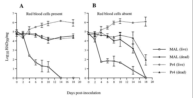
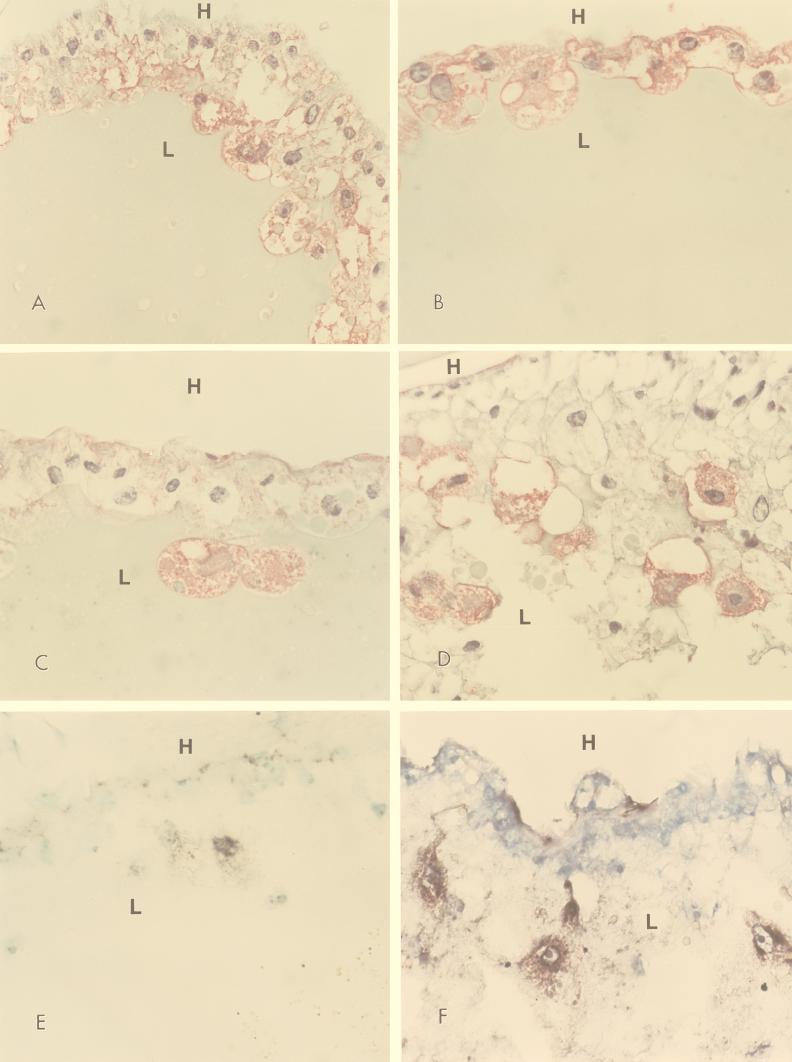
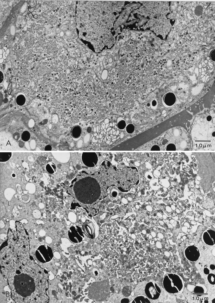
Although the Malawi Lil20/1 (MAL) strain of African swine fever virus (ASFV) was isolated from Ornithodoros sp. ticks, our attempts to experimentally infect ticks by feeding them this strain failed. Ten different collections of Ornithodorus porcinus porcinus ticks and one collection of O. porcinus domesticus ticks were orally exposed to a high titer of MAL. At 3 weeks postinoculation (p.i.), <25% of the ticks contained detectable virus, with viral titers of <4 log(10) 50% hemadsorbing doses/ml. Viral titers declined to undetectability in >90% of the ticks by 5 weeks p.i. To further study the growth defect, O. porcinus porcinus ticks were orally exposed to MAL and assayed at regular intervals p.i. Whole-tick viral titers dramatically declined (>1,000-fold) between 2 and 6 days p.i., and by 18 days p.i., viral titers were below the detection limit. In contrast, viral titers of ticks orally exposed to a tick-competent ASFV isolate, Pretoriuskop/96/4/1 (Pr4), increased 10-fold by 10 days p.i. and 50-fold by 14 days p.i. Early viral gene expression, but not extensive late gene expression or viral DNA synthesis, was detected in the midguts of ticks orally exposed to MAL. Ultrastructural analysis demonstrated that progeny virus was rarely present in ticks orally exposed to MAL and, when present, was associated with extensive cytopathology of phagocytic midgut epithelial cells. To determine if viral replication was restricted only in the midgut epithelium, parenteral inoculations into the hemocoel were performed. With inoculation by this route, a persistent infection was established although a delay in generalization of MAL was detected and viral titers in most tissues were typically 10- to 1,000-fold lower than those of ticks injected with Pr4. MAL was detected in both the salivary secretion and coxal fluid following feeding but less frequently and at a lower titer compared to Pr4. Transovarial transmission of MAL was not detected after two gonotrophic cycles. Ultrastructural analysis demonstrated that, when injected, MAL replicated in a number of cell types but failed to replicate in midgut epithelial cells. In contrast, ticks injected with Pr4 had replicating virus in midgut epithelial cells. Together, these results indicate that MAL replication is restricted in midgut epithelial cells. This finding demonstrates the importance of viral replication in the midgut for successful ASFV infection of the arthropod host.
Figures
Similar articles
-
African swine fever virus multigene family 360 genes affect virus replication and generalization of infection in Ornithodoros porcinus ticks.J Virol. 2004 Mar;78(5):2445-53. doi: 10.1128/jvi.78.5.2445-2453.2004. J Virol. 2004. PMID: 14963141 Free PMC article.
-
African swine fever virus infection in the argasid host, Ornithodoros porcinus porcinus.J Virol. 1998 Mar;72(3):1711-24. doi: 10.1128/JVI.72.3.1711-1724.1998. J Virol. 1998. PMID: 9499019 Free PMC article.
-
Pathogenesis of African swine fever virus in Ornithodoros ticks.Anim Health Res Rev. 2001 Dec;2(2):121-8. Anim Health Res Rev. 2001. PMID: 11831434 Review.
-
Differential vector competence of Ornithodoros soft ticks for African swine fever virus: What if it involves more than just crossing organic barriers in ticks?Parasit Vectors. 2020 Dec 9;13(1):618. doi: 10.1186/s13071-020-04497-1. Parasit Vectors. 2020. PMID: 33298119 Free PMC article.
-
Reviewing the Potential Vectors and Hosts of African Swine Fever Virus Transmission in the United States.Vector Borne Zoonotic Dis. 2019 Jul;19(7):512-524. doi: 10.1089/vbz.2018.2387. Epub 2019 Feb 19. Vector Borne Zoonotic Dis. 2019. PMID: 30785371 Free PMC article. Review.
Cited by
-
African Swine Fever Virus: An Emerging DNA Arbovirus.Front Vet Sci. 2020 May 13;7:215. doi: 10.3389/fvets.2020.00215. eCollection 2020. Front Vet Sci. 2020. PMID: 32478103 Free PMC article. Review.
-
A CRISPR/Cas12a Based Universal Lateral Flow Biosensor for the Sensitive and Specific Detection of African Swine-Fever Viruses in Whole Blood.Biosensors (Basel). 2020 Dec 10;10(12):203. doi: 10.3390/bios10120203. Biosensors (Basel). 2020. PMID: 33321741 Free PMC article.
-
Cloning and Expression of a Truncated Form of the p72 Protein of the African Swine Fever Virus (ASFV) for Application in an Efficient Indirect ELISA System.Pathogens. 2025 May 29;14(6):542. doi: 10.3390/pathogens14060542. Pathogens. 2025. PMID: 40559550 Free PMC article.
-
Epidemiological Characterization of African Swine Fever Dynamics in Ukraine, 2012-2023.Vaccines (Basel). 2023 Jun 25;11(7):1145. doi: 10.3390/vaccines11071145. Vaccines (Basel). 2023. PMID: 37514961 Free PMC article.
-
Expounding the role of tick in Africa swine fever virus transmission and seeking effective prevention measures: A review.Front Immunol. 2022 Dec 16;13:1093599. doi: 10.3389/fimmu.2022.1093599. eCollection 2022. Front Immunol. 2022. PMID: 36591310 Free PMC article. Review.
References
-
- Afonso C, Alcaraz C, Brun A, Sussman M D, Rock D L. Characterization of p30, a highly antigenic membrane and secreted protein of African swine fever virus. Virology. 1992;189:368–373. - PubMed
-
- Booth T F, Davies C R, Jones L D, Staunton D, Nuttall P A. Anatomical basis of Thogoto virus infection in BHK cell culture and in the Ixodid tick vector, Rhipicephalus appendiculatus. J Gen Virol. 1989;70:1093–1104. - PubMed
-
- Booth T F, Steele G M, Marriott A C, Nuttall P A. Dissemination, replication, and trans-stadial persistence of Dugbe virus (Nairovirus, Bunyaviridae) in the tick vector Amblyomma variegatum. Am J Trop Med Hyg. 1991;45:146–157. - PubMed
-
- Borca M V, Irusta P, Carrillo C, Afonso C L, Burrage T, Rock D L. African swine fever virus structural protein p72 contains a conformational neutralizing epitope. Virology. 1994;201:413–418. - PubMed
-
- Brown F. The classification and nomenclature of viruses: summary of results of meetings of the International Committee on Taxonomy of Viruses in Sendai, September 1984. Intervirology. 1986;25:141–143. - PubMed
MeSH terms
LinkOut - more resources
Full Text Sources
Research Materials
