Postinoculation PMPA treatment, but not preinoculation immunomodulatory therapy, protects against development of acute disease induced by the unique simian immunodeficiency virus SIVsmmPBj
- PMID: 10482616
- PMCID: PMC112883
- DOI: 10.1128/JVI.73.10.8630-8639.1999
Postinoculation PMPA treatment, but not preinoculation immunomodulatory therapy, protects against development of acute disease induced by the unique simian immunodeficiency virus SIVsmmPBj
Abstract
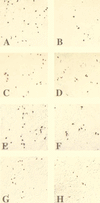
The fatal disease induced by SIVsmmPBj4 clinically resembles endotoxic shock, with the development of severe gastrointestinal disease. While the exact mechanism of disease induction has not been fully elucidated, aspects of virus biology suggest that immune activation contributes to pathogenesis. These biological characteristics include induction of peripheral blood mononuclear cell (PBMC) proliferation, upregulation of activation markers and Fas ligand expression, and increased levels of apoptosis. To investigate the role of immune activation and viral replication on disease induction, animals infected with SIVsmmPBj14 were treated with one of two drugs: FK-506, a potent immunosuppressive agent, or PMPA, a potent antiretroviral agent. While PBMC proliferation was blocked in vitro with FK-506, pig-tailed macaques treated preinoculation with FK-506 were not protected from acutely lethal disease. However, these animals did show some evidence of modulation of immune activation, including reduced levels of CD25 antigen and FasL expression, as well as lower tissue viral loads. In contrast, macaques treated postinoculation with PMPA were completely protected from the development of acutely lethal disease. Treatment with PMPA beginning as late as 5 days postinfection was able to prevent the PBj syndrome. Plasma and cellular viral loads in PMPA-treated animals were significantly lower than those in untreated controls. Although PMPA-treated animals showed acute lymphopenia due to SIVsmmPBj14 infection, cell subset levels subsequently recovered and returned to normal. Based upon subsequent CD4(+) cell counts, the results suggest that very early treatment following retroviral infection can have a significant effect on modifying the subsequent course of disease. These results also suggest that viral replication is an important factor involved in PBJ-induced disease. These studies reinforce the idea that the SIVsmmPBj model system is useful for therapy and vaccine testing.
Figures


Similar articles
-
IL-4 increases Simian immunodeficiency virus replication despite enhanced SIV immune responses in infected rhesus macaques.Int J Parasitol. 2002 May;32(5):543-50. doi: 10.1016/s0020-7519(01)00355-1. Int J Parasitol. 2002. PMID: 11943227
-
Replication of an acutely lethal simian immunodeficiency virus activates and induces proliferation of lymphocytes.J Virol. 1991 Sep;65(9):4902-9. doi: 10.1128/JVI.65.9.4902-4909.1991. J Virol. 1991. PMID: 1870205 Free PMC article.
-
Mutation of a diacidic motif in SIV-PBj Nef impairs T-cell activation and enteropathic disease.Retrovirology. 2011 Mar 2;8:14. doi: 10.1186/1742-4690-8-14. Retrovirology. 2011. PMID: 21366921 Free PMC article.
-
Development of virus-specific immune responses in SHIV(KU)-infected macaques treated with PMPA.Virology. 2001 Jan 5;279(1):97-108. doi: 10.1006/viro.2000.0710. Virology. 2001. PMID: 11145893
-
The very first year of PBJ.Porto Biomed J. 2017 Sep-Oct;2(5):135. doi: 10.1016/j.pbj.2017.04.006. Epub 2017 Sep 1. Porto Biomed J. 2017. PMID: 32258605 Free PMC article. Review. No abstract available.
Cited by
-
Structured treatment interruptions with tenofovir monotherapy for simian immunodeficiency virus-infected newborn macaques.J Virol. 2006 Jul;80(13):6399-410. doi: 10.1128/JVI.02308-05. J Virol. 2006. PMID: 16775328 Free PMC article.
-
Tenofovir treatment augments anti-viral immunity against drug-resistant SIV challenge in chronically infected rhesus macaques.Retrovirology. 2006 Dec 21;3:97. doi: 10.1186/1742-4690-3-97. Retrovirology. 2006. PMID: 17184540 Free PMC article.
-
Biologic studies of chimeras of highly and moderately virulent molecular clones of simian immunodeficiency virus SIVsmPBj suggest a critical role for envelope in acute AIDS virus pathogenesis.J Virol. 2001 Jul;75(14):6645-59. doi: 10.1128/JVI.75.14.6645-6659.2001. J Virol. 2001. PMID: 11413332 Free PMC article.
-
ALVAC-SIV-gag-pol-env-based vaccination and macaque major histocompatibility complex class I (A*01) delay simian immunodeficiency virus SIVmac-induced immunodeficiency.J Virol. 2002 Jan;76(1):292-302. doi: 10.1128/jvi.76.1.292-302.2002. J Virol. 2002. PMID: 11739694 Free PMC article.
-
Antiretroviral treatment start-time during primary SIV(mac) infection in macaques exerts a different impact on early viral replication and dissemination.PLoS One. 2010 May 11;5(5):e10570. doi: 10.1371/journal.pone.0010570. PLoS One. 2010. PMID: 20485497 Free PMC article.
References
-
- Birx D L, Lewis M G, Vahey M, Tencer K, Zack P M, Brown C R, Jahrling P B, Tosato G, Burke D, Redfield R. Association of interleukin-6 in the pathogenesis of acutely fatal SIVsmm/PBj14 in pigtailed macaques. AIDS Res Hum Retroviruses. 1993;9:1123–1129. - PubMed
-
- Dailey, P. Personal communication.
-
- Dailey P J, Zamroud M, Kelso R, Kolberg J, Urdea M. Quantitation of simian immunodeficiency virus (SIV) RNA in plasma of acute and chronically infected macaques using a branched DNA (bDNA) signal amplification assay. J Med Primatol. 1995;24:209.
Publication types
MeSH terms
Substances
Grants and funding
LinkOut - more resources
Full Text Sources
Research Materials
Miscellaneous

