. 1999 Oct;73(10):8640-6.
doi: 10.1128/JVI.73.10.8640-8646.1999.

In vitro and in vivo characterization of a mouse adenovirus type 1 early region 3 null mutant

Affiliations

In vitro and in vivo characterization of a mouse adenovirus type 1 early region 3 null mutant

A N Cauthen et al. J Virol. 1999 Oct.

Abstract

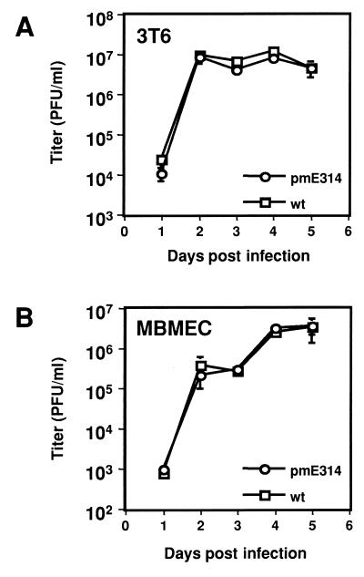
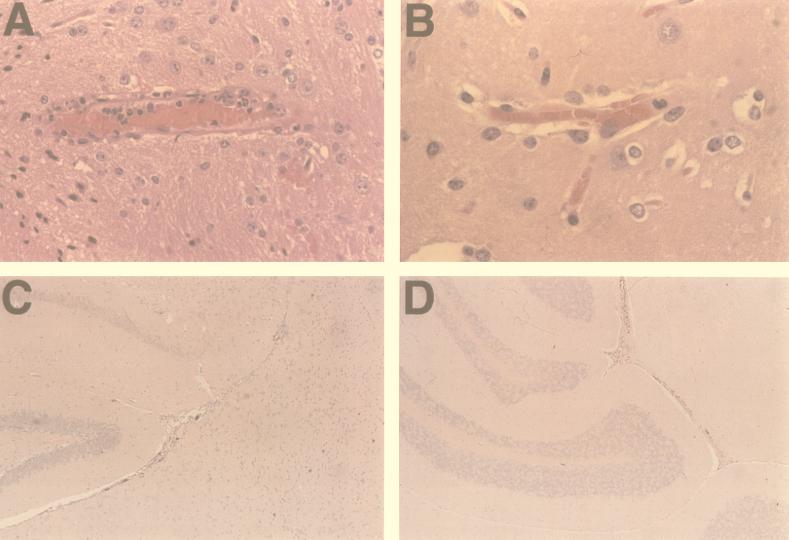
Previous attempts to construct a mouse adenovirus type 1 early region 3 (E3) null mutant by initiator codon mutagenesis were unsuccessful because one of the E3 proteins, gp11K, is synthesized as a fusion protein from a late viral mRNA (A. N. Cauthen and K. R. Spindler, Virology 259:119-128, 1999). Therefore, a different mutagenesis strategy was employed that inserted termination codons into all three reading frames of the E3 proteins. This strategy produced a mutant, pmE314, that was null for the expression of E3 proteins as determined by immunoprecipitation with E3-specific antisera. This mutant grew as well as wild-type (wt) virus in both 3T6 mouse fibroblasts and mouse brain microvascular endothelial cells. However, the 50% lethal dose for pmE314 in adult NIH Swiss outbred mice was approximately 6 log units higher than that of wt virus, indicating that pmE314 was less virulent in mice. In situ hybridization experiments revealed that the absence of the E3 proteins did not alter the tropism of the mutant virus from that of wt virus. When the histopathology was evaluated, the characteristics of the pmE314 infection at both doses administered were strikingly different from those exhibited by wt virus. The central nervous system of wt-infected mice exhibited damage to the endothelium and recruitment of inflammatory cells, whereas the central nervous system of pmE314-infected mice showed no inflammatory response and only mild signs of endothelial damage.


Figures

FIG. 1
FIG. 1
Schematic diagram of E3 mutagenesis. Early E3 mRNA structure is depicted in panel A and the predicted mRNA from which gp11K is also produced at late times is depicted in panel B (5). Lines indicate mRNA, and carets denote spliced introns. Numbers below the introns indicate splice junctions based on the MAV-1 HindIII-C fragment numbering system (1, 18). Boxes indicate protein coding regions where a late protein (likely pVIII) (5) is represented by diagonal hatch marks and the common coding region of E3 is indicated by the open boxes. The unique portions of the E3 proteins are as follows: stippling, gp11K protein; solid black, class 2 protein; horizontal stripes, class 3 protein. The known signal sequence cleavage site is marked by a downward arrow. The mutant virus described here, pmE314, has termination codons inserted into the E3 common coding region around the signal sequence cleavage site. These mutations are predicted to prevent the translation of the E3 protein sequences (noted by large “X”s) downstream of the signal sequence cleavage site from both early (A) and late (B) mRNAs.
FIG. 2
FIG. 2
Immunoprecipitation of MAV-1 proteins. Cells were radiolabeled with [35S]cysteine, and cell lysates were immunoprecipitated with preimmune serum (lanes 2 and 5); α-Eall3 antiserum, which recognizes an epitope common to all three E3 proteins (lanes 1, 3, 4, 6, and 7); and α-E3-1 antiserum, which recognizes the unique portion of gp11K (lanes 8 to 12). E1A protein was immunoprecipitated with AKO 7-147 in lanes 13 and 15. MAV-1 virion proteins were immunoprecipitated with AKO 1-68 in lanes 14 and 16. A protein size standard is noted to the left. Expected sizes of immunoprecipitated proteins are noted to the right.
FIG. 3
FIG. 3
Growth curve analysis of wt- and pmE314-infected mouse cells. 3T6 cells (A) and MBMECs (B) were infected with wt and mutant viruses at an MOI of 5. Infections were harvested, and virus yields were determined by plaque assay on IE3.3 cells.
FIG. 4
FIG. 4
Effects of virus infection of mouse tissues. Histopathology of brain from adult outbred Swiss mice infected with 103 PFU of wt (A) or pmE314 (B) viruses at 8 dpi is shown. Histopathology damage is more extensive in wt-infected mice (see text for details). In situ hybridization of brain samples from adult outbred Swiss mice infected with 104 PFU of wt (C) or pmE314 (D) virus at 6 dpi is shown. Cells positive for in situ hybridization with a MAV-1 E3/pVIII probe in C and D were observed as dark brown staining. Vascular endothelial staining was seen in both wt and mutant virus-infected tissue; greater amounts were seen in wt-infected tissue at this dosage. Magnification, ×316, ×395, ×79, and ×79 (panels A to D, respectively).

Similar articles

Cited by

References

    1. Beard C W, Ball A O, Wooley E H, Spindler K R. Transcription mapping of mouse adenovirus type 1 early region 3. Virology. 1990;175:81–90. - PubMed
    1. Beard C W, Spindler K R. Analysis of early region 3 mutants of mouse adenovirus type 1. J Virol. 1996;70:5867–5874. - PMC - PubMed
    1. Beard C W, Spindler K R. Characterization of an 11K protein produced by early region 3 of mouse adenovirus type 1. Virology. 1995;208:457–466. - PubMed
    1. Cauthen A N, Spindler K R. Construction of mouse adenovirus type 1 mutants. In: Wold W S M, editor. Adenovirus methods and protocols. Totowa, N.J: Humana Press; 1999. pp. 85–103.
    1. Cauthen A N, Spindler K R. Novel expression of mouse adenovirus type 1 early region 3 gp11K at late times after infection. Virology. 1999;259:119–128. - PubMed

Publication types

Substances

LinkOut - more resources