. 1999 Oct;73(10):8696-702.
doi: 10.1128/JVI.73.10.8696-8702.1999.

Carboxypeptidase D is an avian hepatitis B virus receptor

Affiliations

Carboxypeptidase D is an avian hepatitis B virus receptor

S Tong et al. J Virol. 1999 Oct.

Abstract

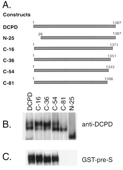
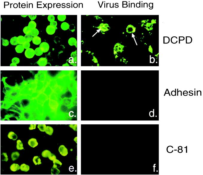
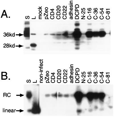
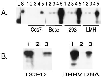
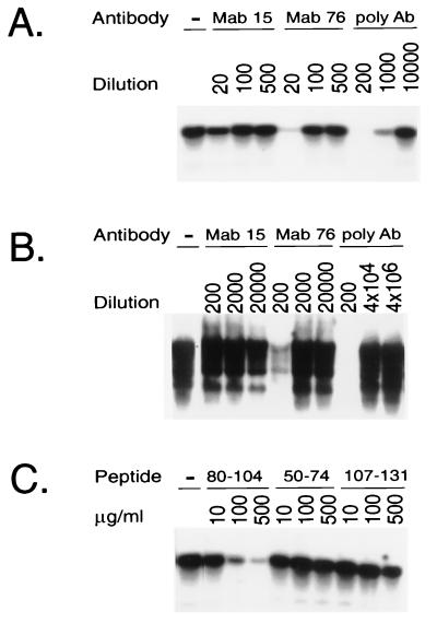
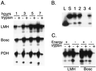
The receptor molecules for human and animal hepatitis B viruses have not been defined. Previous studies have described a 170 to 180 kDa molecule (p170 or gp180) that binds in vitro to the pre-S domain of the large envelope protein of duck hepatitis B virus (DHBV); cDNA cloning revealed the binding protein to be duck carboxypeptidase D (DCPD). In the present study, the DCPD cDNA was transfected into several nonpermissive human-, monkey-, and avian species-derived cell lines. Cells transfected with a plasmid encoding the full-length DCPD protein bound DHBV particles, whereas cells expressing truncated versions of DCPD protein that fail to bind the pre-S protein did not. The DHBV binding to DCPD-reconstituted cells was blocked by a monoclonal antibody that neutralizes DHBV infection of primary duck hepatocytes (PDH) and also by a pre-S peptide previously shown to inhibit DHBV infection of PDH. In addition to promoting virus binding, DCPD expression was associated with internalization of viral particles. The entry process was prevented by incubation of reconstituted cells with DHBV at 4 degrees C and by the addition of energy-depleting agents known to block DHBV entry into PDH. These results demonstrated that DCPD is a DHBV receptor. However, the lack of complete viral replication in DCPD-reconstituted cells suggested that additional factors are required for postentry events in immortalized cell lines.


Figures

FIG. 1
FIG. 1
Illustration of DCPD constructs, protein expression, and affinity for DHBV pre-S envelope protein. (A) Cartoon illustrating the structure of various DCPD constructs used in this study. (B) Expression of DCPD protein and truncation mutants. (C) Affinity with GST–pre-S fusion protein. Transfected Bosc cells were labeled with [35S] methionine, and DCPD proteins present in cell lysates were either immunoprecipitated by a polyclonal antibody (B) or pulled down by a GST–pre-S construct (C). Note that the N-25 and C-81 deletion mutants of DCPD were unable to bind the pre-S protein (C). The failure of the N-25 mutant protein to interact with GST–pre-S constructs may be due to protein degradation, since the N-25 construct migrated faster than the expected size (B).
FIG. 2
FIG. 2
Intracellular localization of transfected proteins and subsequent virus binding properties of the cells. (a, c, and e) Cellular localization and distribution of full-length DCPD (a), sialoadhesin (c), and DCPD truncation mutant C-81 (e) in transfected Bosc cells; (b, d, and f) binding of DHBV particles to Bosc cells transfected with full-length DCPD (b), sialoadhesin (d), and the C-81 mutant of DCPD (f). Sialoadhesin expression was detected by polyclonal rat antiserum provided by D. Sgroi. Bound viral particles were revealed by an antibody raised against the pre-S domain of viral large envelope protein. The different patterns of IF signal between panels b and a was caused by capping and internalization events following virus binding.
FIG. 3
FIG. 3
Binding of DHBV particles to DCPD-reconstituted Bosc cells as revealed by Western and Southern blot analyses. Transfected cells in six-well plates were incubated at 37°C for 8 h with a 1:10 dilution of viremic duck serum. Following a washing step, the cells were mechanically removed. (A) Western blot analysis of viral large envelope protein. Lanes: S, 0.05 μl of the viremic duck serum used for the binding experiment; L, lysate of DHBV-infected duck liver; pZeo, pcDNA3.1/Zeo(−); adhesin, sialoadhesin. Positions of the full-length 36 kDa large envelope protein and the 28-kDa truncated form are indicated. (B) Southern blot analysis of DHBV DNA. Lanes S, DNA corresponding to 0.3 μl of viremic duck serum; L, linear DHBV DNA (20 pg); non-infect, cells not incubated with virus. Positions of the relaxed circular (RC) and linear DHBV DNA forms are indicated.
FIG. 4
FIG. 4
Binding of DHBV virions to several DCPD-reconstituted cell lines. (A) Transiently transfected cells. The transfection/infection profiles of the samples shown in lanes 1 to 5: 1, nontransfected/not infected; 2, nontransfected/infected; 3, pcDNA3.1/Zeo(−) transfected/infected; 4, C-81 mutant transfected/infected; 5, full-length DCPD transfected/infected. Lanes L and S denote 3-kb linear DHBV DNA and duck serum-derived viral DNA, respectively. (B) 293 cells with stable expression of the DCPD protein. Left, expression of DCPD protein in 293 cells by Western blot analysis; right, viral DNA of bound DHBV particles. Virus binding experiment was carried out at 37°C for 6 h, using a 1:5 dilution of viremic duck serum. For both panels, lanes 1 to 3 represent transiently transfected, nontransfected, and stable transfected 293-4 cells, respectively.
FIG. 5
FIG. 5
Inhibition of DHBV binding to the stable DCPD expressing 293-4 cell line by either pre-S antibodies or peptides. (A) Effect of anti-pre-S antibodies (Ab) on virus binding. Cells in six-well plates were incubated with a 1:30 dilution of viremic duck serum for 1 h in the presence of various dilution of pre-S antibodies. After a washing step, cells were scraped off the plates and lysed for Southern blot analysis. (B) Effect of anti-pre-S antibodies on DHBV infection of PDH. Virus infection was carried out in the presence of antibodies and viral replication was measured 7 days later (see Materials and Methods). (C) Effect of pre-S peptides on DHBV binding to 293-4 cell line. The pre-S peptides were expressed as GST fusion protein and purified from the GST moiety following thrombin cleavage (18). A 1:50 dilution of viremic duck serum was used, and binding was at 37°C for 1 h.
FIG. 6
FIG. 6
Internalization of DHBV particles into DCPD-reconstituted cell lines. (A) Total and trypsin-resistant fractions of DHBV DNA at different time points of virus incubation. DCPD-reconstituted Bosc and LMH cells as well as freshly plated PDH were incubated at 37°C with a 1:10 dilution of 60 μl viremic duck serum for 1 to 7 h. Half of the cell pellet was pretreated with trypsin before DNA extraction. (B) DHBV entry at 4°C and at 37°C. Two wells of DCPD-transfected LMH cells were incubated with a 1:10 dilution of viremic duck serum at 4°C for 2 h. After washing, cells from one well (lanes 1 and 3) were removed, and half of the cell pellet (lane 3) was treated with trypsin. Cells in the other well (lanes 2 and 4) were further incubated at 37°C for 2 h and half of the cell pellet (lane 4) was treated with trypsin. Lanes L and S are as in Fig. 4. (C) Energy depletion inhibits DHBV entry. DCPD-reconstituted LMH cells were preincubated for 1 h with 600 μl of medium in the presence (energy −) or absence (energy +) of sodium azide (0.1%) and 2-deoxy-d-glucose (50 mM) and incubated for 3 h following the addition of 60 μl of viremic duck serum. The DCPD-transfected Bosc cells were preincubated with energy depleting agents for 3 h and incubated for 12 h with DHBV. Both total and trypsin-resistant fractions of DHBV DNA were measured.

References

    1. Beasley R P, Hwang L Y, Lin C C, Chien C S. Hepatocellular carcinoma and hepatitis B virus: a prospective study of 22702 men in Taiwan. Lancet. 1981;ii:1129–1133. - PubMed
    1. Breiner K M, Urban S, Schaller H. Carboxypeptidase D (gp180), a Golgi-resident protein, functions in the attachment and entry of avian hepatitis B viruses. J Virol. 1998;72:8098–8104. - PMC - PubMed
    1. Bruns M, Miska S, Chassot S, Will H. Enhancement of hepatitis B virus infection by noninfectious subviral particles. J Virol. 1998;72:1462–1468. - PMC - PubMed
    1. Chassot S, Lambert V, Kay A, Godinot A, Roux C, Trepo C, Cova L. Fine mapping of neutralization epitopes on duck hepatitis B virus (DHBV) pre-S protein using monoclonal antibodies and overlapping peptides. Virology. 1993;192:217–223. - PubMed
    1. Condreay L D, Aldrich C E, Coates L, Mason W S, Wu T T. Efficient duck hepatitis B virus production by an avian liver tumor cell line. J Virol. 1990;64:3249–3258. - PMC - PubMed

Publication types

Associated data