Comparative Study
. 1999 Oct;73(10):8741-9.
doi: 10.1128/JVI.73.10.8741-8749.1999.

Evidence of recombination among enteroviruses

Affiliations
Comparative Study

Evidence of recombination among enteroviruses

J Santti et al. J Virol. 1999 Oct.

Abstract

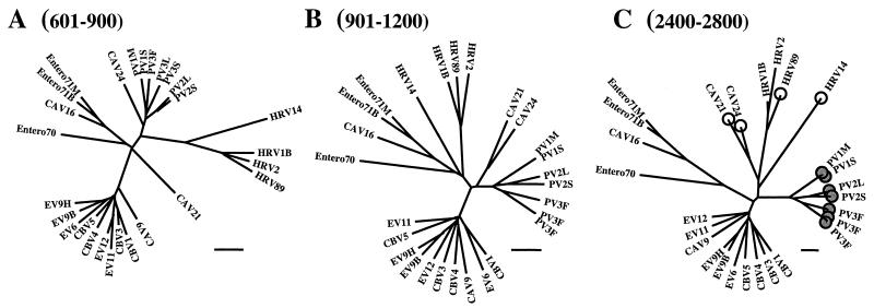
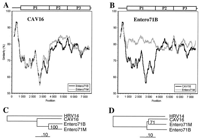
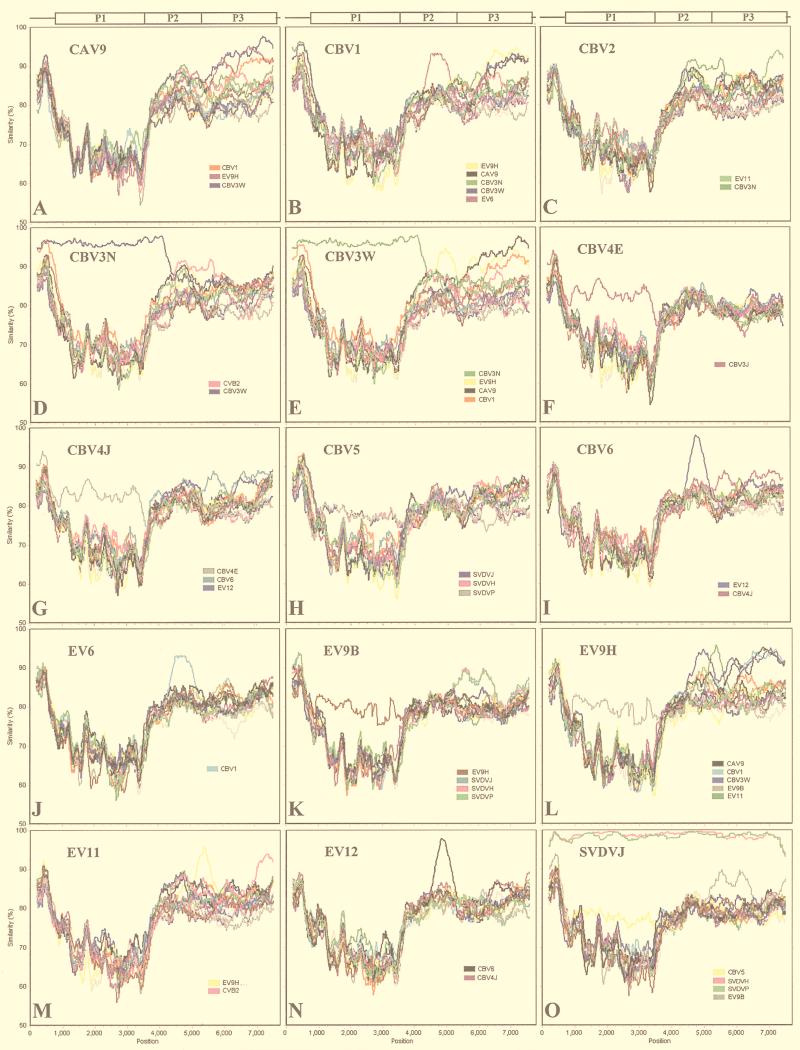
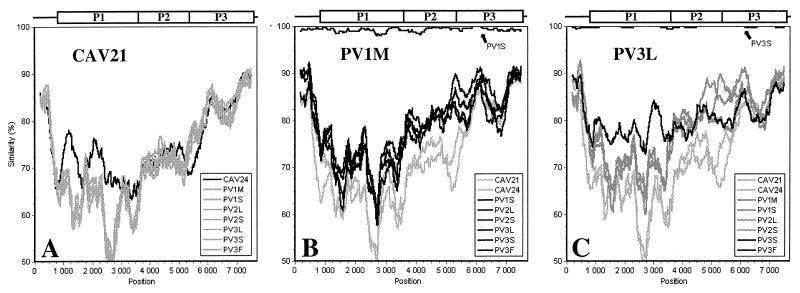
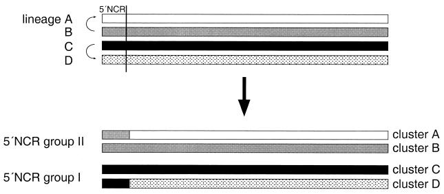
Human enteroviruses consist of more than 60 serotypes, reflecting a wide range of evolutionary divergence. They have been genetically classified into four clusters on the basis of sequence homology in the coding region of the single-stranded RNA genome. To explore further the genetic relationships between human enteroviruses and to characterize the evolutionary mechanisms responsible for variation, previously sequenced genomes were subjected to detailed comparison. Bootstrap and genetic similarity analyses were used to systematically scan the alignments of complete genomic sequences. Bootstrap analysis provided evidence from an early recombination event at the junction of the 5' noncoding and coding regions of the progenitors of the current clusters. Analysis within the genetic clusters indicated that enterovirus prototype strains include intraspecies recombinants. Recombination breakpoints were detected in all genomic regions except the capsid protein coding region. Our results suggest that recombination is a significant and relatively frequent mechanism in the evolution of enterovirus genomes.


Figures

FIG. 1
FIG. 1
(A) Phylogenetic tree illustrating genetic relationships between enterovirus and rhinovirus genomes in the 5′ NCR. Enterovirus 5′ NCR groups I and II are indicated. (B) Bootscanning analysis of groups I (upper panel) and II (lower panel) along the genome was performed by the neighbor-joining method as implemented in the PHYLIP package (see text for details). PHYLIP reports bootstrap frequencies for all clusters found in the bootstrap replicates, which permits plotting of values for clusters which are not found in the consensus tree (areas of low bootstrap values). Bootstrap values are shown at the midpoint of each window.
FIG. 2
FIG. 2
(A) Grouping of enteroviruses and rhinoviruses in the coding region and the 3′ NCR of the genome. The four genetic clusters (A to D) of enteroviruses are shown. (B) Bootscanning of clusters A, B, and C (performed as described in the legend to Fig. 1).
FIG. 3
FIG. 3
Alterations in clustering of enteroviruses and rhinoviruses in the 5′-terminal region of the genome. Trees correspond to nt 601 to 900 (A), 901 to 1200 (B), and 2400 to 2800 (C) of the alignment. Viruses interacting with poliovirus receptor are marked with shaded circles, and those recognizing intercellular adhesion molecule 1 are marked with open circles (C). Bars indicate 10% divergence.
FIG. 4
FIG. 4
Genetic distances between cluster A enteroviruses along the genome as determined by similarity analysis (window, 400 nt; step, 20 nt). Query sequences are noted. Phylogenetic trees correspond to the capsid (P1 [C]) and NS (P2 and P3 [D]) protein coding regions.
FIG. 5
FIG. 5
Genetic distances between cluster B enteroviruses (SimPlot analysis) (window, 400 nt; step, 20 nt). Query sequences are noted.
FIG. 6
FIG. 6
SimPlot analysis of cluster C enteroviruses (window, 400 nt; step, 20 nt). Query sequences are noted.
FIG. 7
FIG. 7
Schematic presentation of proposed recombination events between the 5′ NCR and the rest of the genome during enterovirus evolution which could explain the existence of currently known subgroups. First, four genetic lineages evolved from a common enterovirus progenitor by point mutations. During evolution, the lineage A virus acquired the lineage B virus 5′ NCR by recombination. Alternatively, the lineage B virus could have acquired the lineage A virus 5′ NCR. A similar recombination event may have occurred between lineage C and D viruses. Current species classification of human enteroviruses is based mainly on four genetic lineages (clusters A to D) observed in the coding region and the 3′ NCR. Due to the proposed recombination, only two genetic groups are seen in the 5′ NCR. Viruses containing the 5′ NCR from the other two lineages have disappeared or have not been sampled to date.

References

    1. Abraham G, Colonno R J. Many rhinovirus serotypes share the same cellular receptor. J Virol. 1984;51:340–345. - PMC - PubMed
    1. Cammack N, Phillips A, Dunn G, Patel V, Minor P D. Intertypic genomic rearrangements of poliovirus strains in vaccinees. Virology. 1988;167:507–514. - PubMed
    1. Carr J K, Salminen M O, Albert J, Sanders-Buell E, Gotte D, Birx D L, McCutchan F E. Full genome sequences of human immunodeficiency virus type 1 subtypes G and A/G intersubtype recombinants. Virology. 1998;247:22–31. - PubMed
    1. Drake J W. Rates of spontaneous mutation among RNA viruses. Proc Natl Acad Sci USA. 1993;90:4171–4175. - PMC - PubMed
    1. Evans D J, Almond J W. Cell receptors for picornaviruses as determinants of cell tropism and pathogenesis. Trends Microbiol. 1998;6:198–202. - PubMed

Publication types

LinkOut - more resources