. 1999 Oct;73(10):8750-61.
doi: 10.1128/JVI.73.10.8750-8761.1999.

Vaccinia virus envelope D8L protein binds to cell surface chondroitin sulfate and mediates the adsorption of intracellular mature virions to cells

Affiliations

Vaccinia virus envelope D8L protein binds to cell surface chondroitin sulfate and mediates the adsorption of intracellular mature virions to cells

J C Hsiao et al. J Virol. 1999 Oct.

Abstract

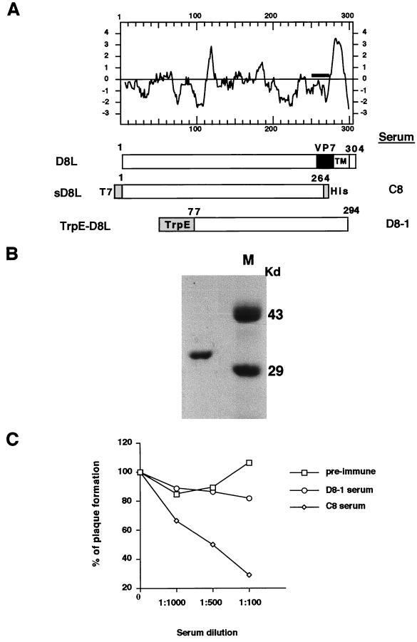
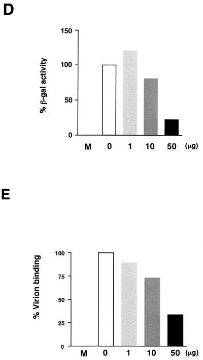
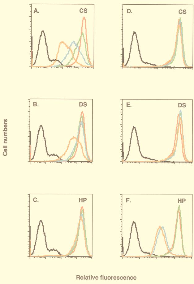
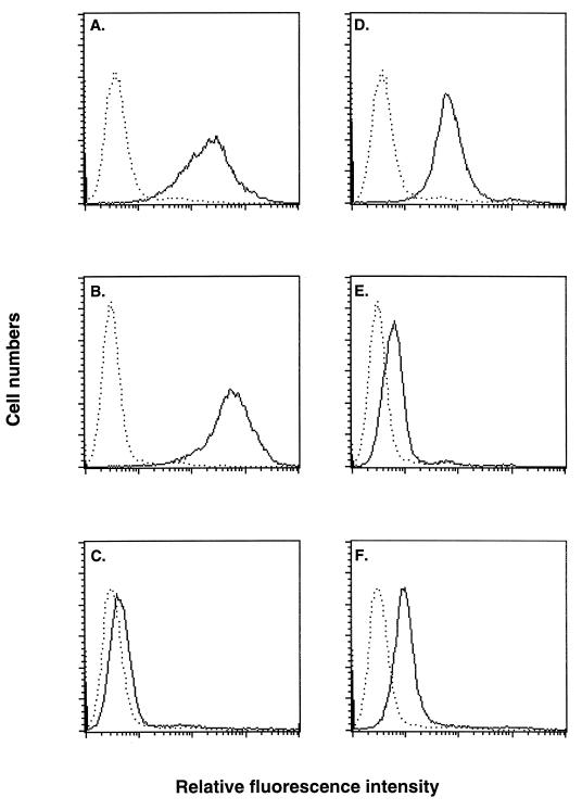
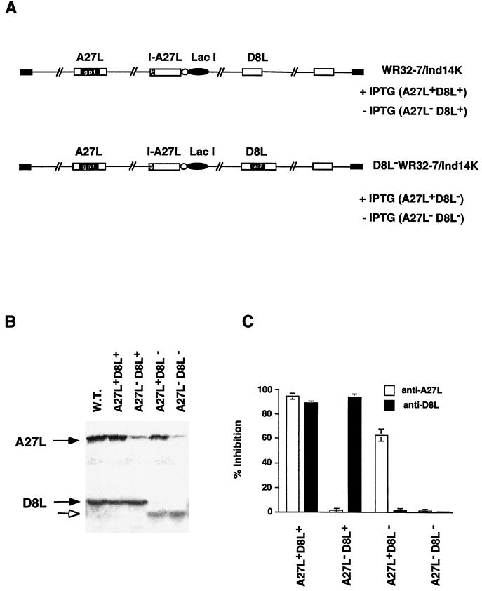
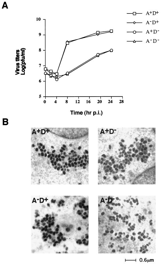
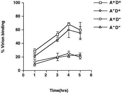
We previously showed that an envelope A27L protein of intracellular mature virions (IMV) of vaccinia virus binds to cell surface heparan sulfate during virus infection. In the present study we identified another viral envelope protein, D8L, that binds to chondroitin sulfate on cells. Soluble D8L protein interferes with the adsorption of wild-type vaccinia virions to cells, indicating a role in virus entry. To explore the interaction of cell surface glycosaminoglycans and vaccinia virus, we generated mutant viruses from a control virus, WR32-7/Ind14K (A27L(+) D8L(+)) to be defective in expression of either the A27L or the D8L gene (A27L(+) D8L(-) or A27L(-) D8L(+)) or both (A27L(-) D8L(-)). The A27L(+) D8L(+) and A27L(-) D8L(+) mutants grew well in BSC40 cells, consistent with previous observations. However, the IMV titers of A27L(+) D8L(-) and A27L(-) D8L(-) viruses in BSC40 cells were reduced, reaching only 10% of the level for the control virus. The data suggested an important role for D8L protein in WR32-7/Ind14K virus growth in cell cultures. A27L protein, on the other hand, could not complement the functions of D8L protein. The low titers of the A27L(+) D8L(-) and A27L(-) D8L(-) mutant viruses were not due to defects in the morphogenesis of IMV, and the mutant virions demonstrated a brick shape similar to that of the control virions. Furthermore, the infectivities of the A27L(+) D8L(-) and A27L(-) D8L(-) mutant virions were 6 to 10% of that of the A27L(+) D8L(+) control virus. Virion binding assays revealed that A27L(+) D8L(-) and A27L(-) D8L(-) mutant virions bound less well to BSC40 cells, indicating that binding of viral D8L protein to cell surface chondroitin sulfate could be important for vaccinia virus entry.


Figures

FIG. 1
FIG. 1
(A) Hydropathy plot of D8L amino acids translated from the DNA sequences (24) and diagrams of D8L, soluble D8L (sD8L), and TrpE-D8L. The D8L diagram shows the open reading frame from aa 1 to aa 304. TM, transmembrane region; VP7, aa sequences with homology to rotavirus glycoprotein VP7 (24). sD8L is the ectodomain of D8L protein fused with T7 and hexahistidine tag (His) sequences, shown as shaded areas at the N and C termini, respectively. TrpE-D8L contains D8L from aa 77 to aa 294 (26). Sera C8 and D8-1 were generated from sD8L and TrpE-D8L proteins, respectively (26). (B) Purified sD8L protein stained with Commassie blue on a sodium dodecyl sulfate–12% polyacrylamide gel. Protein markers (M) are shown. (C) Neutralization of vaccinia virus infections by antiserum C8. BSC40 cells were infected with wild-type vaccinia virus in the absence or in the presence of each serum at various dilutions as shown along the x axis. Cells were then washed, and 1% agar was added for plaque determination. These plaque assays were performed in duplicate, and the averages are presented. The number of plaques obtained from control cells without serum addition was around 150 PFU, which was taken as 100%. (D) sD8L protein blocked vaccinia virus early gene expression. BSC40 cells were mock infected (M) or infected with vMJ360 expressing lacZ at an MOI of 10 PFU per cell in the presence of various amounts of sD8L (0, 1, 10, or 50 μg) and were harvested at 2 h p.i. for β-Gal assays (8). (E) sD8L protein blocked vaccinia virus adsorption to cells. BSC40 cells were mock infected (M) or infected with vaccinia virus at an MOI of 10 PFU per cell at 4°C for 30 min. After a wash, these cells were immediately harvested, and the number of cell-associated virions was determined by plaque assays on BSC40 cells.
FIG. 1
FIG. 1
(A) Hydropathy plot of D8L amino acids translated from the DNA sequences (24) and diagrams of D8L, soluble D8L (sD8L), and TrpE-D8L. The D8L diagram shows the open reading frame from aa 1 to aa 304. TM, transmembrane region; VP7, aa sequences with homology to rotavirus glycoprotein VP7 (24). sD8L is the ectodomain of D8L protein fused with T7 and hexahistidine tag (His) sequences, shown as shaded areas at the N and C termini, respectively. TrpE-D8L contains D8L from aa 77 to aa 294 (26). Sera C8 and D8-1 were generated from sD8L and TrpE-D8L proteins, respectively (26). (B) Purified sD8L protein stained with Commassie blue on a sodium dodecyl sulfate–12% polyacrylamide gel. Protein markers (M) are shown. (C) Neutralization of vaccinia virus infections by antiserum C8. BSC40 cells were infected with wild-type vaccinia virus in the absence or in the presence of each serum at various dilutions as shown along the x axis. Cells were then washed, and 1% agar was added for plaque determination. These plaque assays were performed in duplicate, and the averages are presented. The number of plaques obtained from control cells without serum addition was around 150 PFU, which was taken as 100%. (D) sD8L protein blocked vaccinia virus early gene expression. BSC40 cells were mock infected (M) or infected with vMJ360 expressing lacZ at an MOI of 10 PFU per cell in the presence of various amounts of sD8L (0, 1, 10, or 50 μg) and were harvested at 2 h p.i. for β-Gal assays (8). (E) sD8L protein blocked vaccinia virus adsorption to cells. BSC40 cells were mock infected (M) or infected with vaccinia virus at an MOI of 10 PFU per cell at 4°C for 30 min. After a wash, these cells were immediately harvested, and the number of cell-associated virions was determined by plaque assays on BSC40 cells.
FIG. 2
FIG. 2
Soluble D8L protein bound to BSC40 cell surface CS. Live BSC40 cells were incubated with PBS (black lines) and biotinylated D8L (red lines in panels A through C) or A27L (red lines in panels D through F) protein alone and analyzed with a FACS as described elsewhere (16). Alternatively, biotinylated proteins were mixed with 10, 100, or 1,000 μg of soluble GAGs/ml and analyzed with a FACS, and the cell staining under each condition was shown as a green, blue, or orange line, respectively. The soluble GAGs used in competitions were CS (A and D), DS (B and E), and heparin (C and F).
FIG. 3
FIG. 3
Binding of soluble D8L protein to mutant cells defective in GAG expression. Biotinylated D8L protein (A through C) or A27L protein (D through F) was incubated with L (A and D), gro2C (B and E), or sog9 (C and F) cells as described for Fig. 2, and the surface staining (solid lines) was analyzed with a FACS. Dotted lines, background staining of cells in the absence of biotinylated proteins.
FIG. 4
FIG. 4
Characterization of A27L and D8L mutant viruses. (A) Schematic representations of four phenotypic viruses produced from the parental virus WR32-7/Ind14K and D8L WR32-7/Ind14K virus. (B) Expression of A27L and D8L proteins in virus-infected cells. BSC40 cells were infected with wild-type (WT) vaccinia virus or with either WR32-7/Ind14K or D8L WR32-7/Ind14K virus at an MOI of 10 in the presence (A27L+ D8L+ and A27L+ D8L) or absence (A27L D8L+ and A27L D8L) of 5 mM IPTG. Expression of A27L and D8L proteins was determined by Western blot analyses with sera (1:500) against A27L and D8L proteins, respectively. Solid arrows indicate the positions of A27L and D8L proteins. The open arrow indicates the truncated D8L protein. (C) Neutralization of A27L and D8L mutant viruses. BSC40 cells were infected with each of the mutant viruses shown in panel A in the absence or presence of antiserum (1:100) against A27L or D8L protein. Cells were washed and overlaid with agar, and plaque numbers were determined after 3 days. The plaque count obtained from virus infection without serum, around 150 plaques per 60 mm, was used as a control and taken as 100%. The values on the y axis represent percentages of inhibition and were calculated as [1 − (number of plaques obtained with serum/number of plaques obtained without serum)] × 100. Results shown here are averages from four experiments.
FIG. 5
FIG. 5
(A) One-step growth curve analysis of A27L and D8L mutant viruses. BSC40 cells were infected with each mutant virus at an MOI of 5 PFU per cell, and cells were cultured under appropriate conditions as described in Materials and Methods. Cell lysates were harvested at 0, 2, 4, 8, 19, and 24 h p.i., and virus titers on BSC40 cells in the presence of IPTG were determined. (B) Electron microscopy of ultrathin sections of BSC40 cells infected by mutant viruses at 24 h p.i. A, A27L; D, D8L.
FIG. 6
FIG. 6
Virion binding assays for A27L (A) and D8L (D) mutant viruses. BSC40 cells were infected with each mutant virus at an MOI of 5 PFU per cell at 4°C for various times and then washed with cold PBS, and cell-associated virions were determined by plaque assays on BSC40 cells in the presence of IPTG.

References

    1. Appleyard G, Hapel A J, Boulter E A. An antigenic difference between intracellular and extracellular rabbitpox virus. J Gen Virol. 1971;13:9–17. - PubMed
    1. Banfield B W, Leduc Y, Esford L, Schubert K, Tufaro F. Sequential isolation of proteoglycan synthesis mutants by using herpes simplex virus as a selective agent: evidence for a proteoglycan-independent virus entry pathway. J Virol. 1995;69:3290–3298. - PMC - PubMed
    1. Banfield B W, Leduc Y, Esford L, Visalli R J, Brandt C R, Tufaro F. Evidence for an interaction of herpes simplex virus with chondroitin sulfate proteoglycans during infection. Virology. 1995;208:531–539. - PubMed
    1. Byrne K M, Horohov D W, Kousoulas K G. Glycoprotein B of bovine herpesvirus-1 binds heparin. Virology. 1995;209:230–235. - PubMed
    1. Chakrabarti S, Brechling K, Moss B. Vaccinia virus expression vector: coexpression of β-galactosidase provides visual screening of recombinant virus plaques. Mol Cell Biol. 1985;5:3403–3409. - PMC - PubMed

Publication types

LinkOut - more resources