. 1999 Sep;121(1):61-70.
doi: 10.1104/pp.121.1.61.

Arabidopsis mutants lacking the 43- and 54-kilodalton subunits of the chloroplast signal recognition particle have distinct phenotypes

Affiliations

Arabidopsis mutants lacking the 43- and 54-kilodalton subunits of the chloroplast signal recognition particle have distinct phenotypes

P Amin et al. Plant Physiol. 1999 Sep.

Abstract

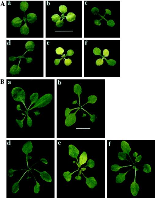
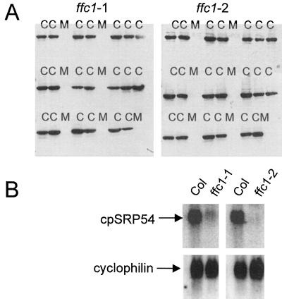
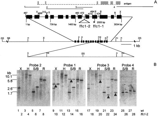
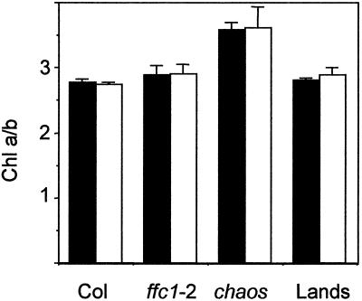
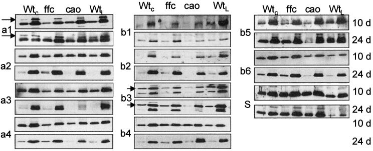
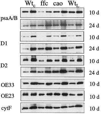
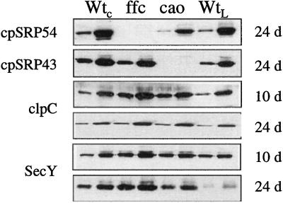
The chloroplast signal recognition particle (cpSRP) is a protein complex consisting of 54- and 43-kD subunits encoded by the fifty-four chloroplast, which encodes cpSRP54 (ffc), and chaos (cao) loci, respectively. Two new null alleles in the ffc locus have been identified. ffc1-1 is caused by a stop codon in exon 10, while ffc1-2 has a large DNA insertion in intron 8. ffc mutants have yellow first true leaves that subsequently become green. The reaction center proteins D1, D2, and psaA/B, as well as seven different light-harvesting chlorophyll proteins (LHCPs), were found at reduced levels in the young ffc leaves but at wild-type levels in the older leaves. The abundance of the two types of LHCP was unaffected by the mutation, while two others were increased in the absence of cpSRP54. Null mutants in the cao locus contain reduced levels of the same subset of LHCP proteins as ffc mutants, but are distinguishable in four ways: young leaves are greener, the chlorophyll a/b ratio is elevated, levels of reaction center proteins are normal, and there is no recovery in the level of LHCPs in the adult plant. The data suggest that cpSRP54 and cpSRP43 have some nonoverlapping roles and that alternative transport pathways can compensate for the absence of a functional cpSRP.


Figures

Figure 1
Figure 1
Visible appearance of Arabidopsis wild type and cpSRP mutants. Seeds were grown on plates lacking Suc under constant illumination. Plants were photographed under identical conditions at 10 d (A) and 28 d (B). a, Landsberg wild type; b, chaos (cpSRP43 mutant); c, ffc1-2::54His (cpSRP54 mutant transformed with cpSRP54 His); d, Columbia wild type; e, ffc1-1 (cpSRP54 mutant); and f, ffc1-2 (cpSRP54 mutant). The scales in panel b of both A and B correspond to 0.5 cm.
Figure 2
Figure 2
Identification of ffc null alleles. A, Cosegregation of the mutant phenotype and the null ffc allele. F2 progeny from backcrosses between ffc1-1 and ffc1-2 to ecotype Columbia were grown under constant illumination on plates lacking both Suc and the selectable marker. From each backcross, seven mutant (M) and 20 wild-type plants (C) were sampled at random, and protein was extracted from the leaf tissue and analyzed by immunoblot analysis using antisera raised against cpSRP54. Cross-reacting proteins were detected by enhanced chemiluminescence. B, ffc transcripts are low or undetectable in ffc mutants. Each lane contained 5 μg of total RNA. Blots were hybridized against a SacI-BglII probe from pNH10 and an EcoRI-BamHI probe from cyclophilin to monitor for equal loading.
Figure 3
Figure 3
Molecular basis of the ffc alleles. A, Restriction map of ffc. Exons used to make recombinant antigen are marked in gray. Probes used for hybridization are numbered 1 to 4. The position of the exons in the ffc gene is indicated by the solid black boxes. Numbers above the gene are arbitrary designations of primers used in the study. Numbers below the gene correspond to the nucleotide sequence. Restriction sites are indicated as follows: X, XhoI; S, SacI; R, EcoRI; B, BglII; H, HindIII; Xb, XbaI; and SB, SacI or BglII site. HindIII sites marked in parentheses have not been mapped relative to the internal restriction sites. The DNA insertion in ffc1-2 in intron 8 is indicated by the triangle and is drawn to scale in the lower restriction map. The asterisk indicates the stop codon in exon 10. B, Southern-blot analysis of the ffc1-2 allele. Each lane contained 10 μg of genomic DNA from either ecotype Columbia (odd lanes) or the ffc1-2 mutant line (even lanes). The first three panels represent the same blot hybridized successively to probes 2, 1, and 3. Blots were not stripped of probes prior to rehybridization. Arrows indicate bands that were detected only upon rehybridization. A new sample was hybridized to probe 4 in the fourth panel.
Figure 4
Figure 4
Chl a/b ratios in wild-type and mutant tissue. Leaves were extracted from ecotype Columbia (Col), ffc1-2, chaos, and ecotype Landsberg (Lands) at 10 and 24 d, and Chl a and b were measured as described in the text.
Figure 5
Figure 5
Immunoblot analysis of LHCP in mutant and wild-type tissue. Proteins were extracted as described in the text. Samples were loaded at low and high concentrations to ensure linearity of the immuno response. Each blot was repeated at least three times. WtC, Wild-type Columbia; ffc, ffc1-2; cao, chaos; WtL, wild-type Landsberg. The antibody dilution used (in parentheses) and the amount of membrane protein loaded in each lane were as follows: Lhca 1 (1:1,000) 10 d, 0.5/2.5 μg, 24 d, 1/3 μg; Lhca2 (1:200) 10 and 24 d, 1/5 μg; Lhca3 (1:1,000) 10 d, 0.5/1.5 μg, 24 d, 0.5/2.5 μg; Lhca4 (1:1,000) 10 d, 7.5/15 μg, 24 d, 10/40 μg; Lhcb1 (1:50) 10 and 24 d, 10/20 μg; Lhcb2 (1:50) 10 and 24 d, 0.5/1.5 μg; Lhcb3 (1:30) 10 and 24 d, 1/3 μg; Lhcb4 (1:100) 10 and 24 d, 2.5/7.5 μg; Lhcb5 (1:100) 10 d, 0.5/1.5 μg, 24 d, 1/3 μg; Lhcb6 (1:100) 10 d, 2/4 μg, 24 d, 2/6 μg; psbS (1:1,000) 10 d, 7.5/15 μg, 24 d, 10/20 μg.
Figure 6
Figure 6
Immunoblot analysis of the reaction center polypeptides OE33, OE23, and CytF in mutant and wild-type tissue. Proteins were extracted and analyzed as described in the legend to Figure 5. The antibody dilution used (in parentheses) and the amount of membrane protein loaded in each lane were as follows: psaA/B (1:1,000) 10 d, 3/6 μg, 24 d, 2.5/5 μg; D1 (1:250) 10 d, 3/6 μg, 24 d, 2.5/7.5 μg; D2 (1:1,000) 10 and 24 d, 2.5/7.5 μg; OE33 (1:10,000) 10 and 24 d, 0.1/0.3 μg; OE23 (1:10,000) 10 and 24 d, 0.5/1.5 μg; CytF (1:500) 10 and 24 d, 2.5/7.5 μg.
Figure 7
Figure 7
Immunoblot analysis of cpSRP, clpC, and SecY in mutant and wild-type tissue. Proteins were extracted and analyzed as described in the legend to Figure 5. The antibody dilution used (in parentheses) and the amount of membrane protein loaded in each lane were as follows: cpSRP54 (1:5,000) 10 and 24 d, 5/25 μg; cpSRP43 (1:2,000) 10 and 24 d, 15/40 μg; clpC (1:5,000) 10 and 24 d, 1/5 μg; SecY (1:2,000) 10 d, 2/5 μg, 24 d, 7.5/15 μg.

Similar articles

Cited by

References

    1. Arnon D. Plant Physiol. 1949;24:1–15. - PMC - PubMed
    1. Barkan A, Miles D, Taylor W. Chloroplast gene expression in nuclear, photosynthetic mutants of maize. EMBO J. 1986;5:1421–1427. - PMC - PubMed
    1. Brock IW, Mills JD, Robinson D, Robinson C. The delta pH-driven, ATP-independent protein translocation mechanism in the chloroplast thylakoid membrane: kinetics and energetics. J Biol Chem. 1995;270:1657–1662. - PubMed
    1. Cline K, Ettinger WF, Theg SM. Protein-specific energy requirements for protein transport across or into thylakoid membranes: two lumenal proteins are transported in the absence of ATP. J Biol Chem. 1992;267:2688–2696. - PubMed
    1. Cline K, Henry R. Import and routing of nucleus-encoded chloroplast proteins. Annu Rev Cell Dev Biol. 1996;12:1–26. - PubMed

Publication types

MeSH terms

Substances

Associated data