. 1999 Sep;121(1):147-52.
doi: 10.1104/pp.121.1.147.

Oligogalacturonic acid and chitosan reduce stomatal aperture by inducing the evolution of reactive oxygen species from guard cells of tomato and Commelina communis

Affiliations

Oligogalacturonic acid and chitosan reduce stomatal aperture by inducing the evolution of reactive oxygen species from guard cells of tomato and Commelina communis

S Lee et al. Plant Physiol. 1999 Sep.

Abstract

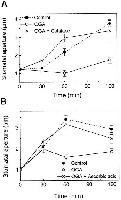
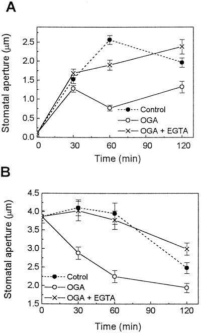
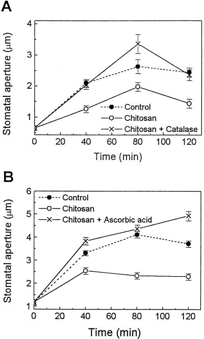
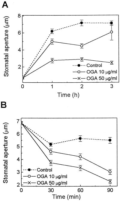
Stomatal opening provides access to inner leaf tissues for many plant pathogens, so narrowing stomatal apertures may be advantageous for plant defense. We investigated how guard cells respond to elicitors that can be generated from cell walls of plants or pathogens during pathogen infection. The effect of oligogalacturonic acid (OGA), a degradation product of the plant cell wall, and chitosan (beta-1,4-linked glucosamine), a component of the fungal cell wall, on stomatal movements were examined in leaf epidermis of tomato (Lycopersicon esculentum L.) and Commelina communis L. These elicitors reduced the size of the stomatal aperture. OGA not only inhibited light-induced stomatal opening, but also accelerated stomatal closing in both species; chitosan inhibited light-induced stomatal opening in tomato epidermis. The effects of OGA and chitosan were suppressed when EGTA, catalase, or ascorbic acid was present in the medium, suggesting that Ca(2+) and H(2)O(2) mediate the elicitor-induced decrease of stomatal apertures. We show that the H(2)O(2) that is involved in this process is produced by guard cells in response to elicitors. Our results suggest that guard cells infected by pathogens may close their stomata via a pathway involving H(2)O(2) production, thus interfering with the continuous invasion of pathogens through the stomatal pores.
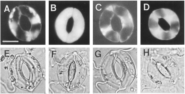

Figures

Figure 1
Figure 1
Effects of OGA on light-induced stomatal opening in tomato leaf epidermis. Note that 3 mg/mL catalase (A) and 5 mm ascorbic acid (B), which reduce the level of ROS, reversed the inhibitory effect of OGA (5 μg/mL) on stomatal opening. Experimental procedures are described in Methods. Results are the averages ± se (n = 120) of four independent experiments.
Figure 2
Figure 2
Involvement of Ca2+ in OGA-induced narrowing of stomata in tomato leaves. In light-induced opening (A) and midday closing (B) experiments, the inhibitory effect of OGA (5 μg/mL) on opening and the enhancing effect of OGA (50 μg/mL) on closing of stomata were reversed by EGTA (2 mm). Experimental procedures are described in Methods. Results are the averages ± se (n = 120) of four independent experiments.
Figure 3
Figure 3
Effect of chitosan on light-induced stomatal opening in tomato leaf epidermis. Note that 3 mg/mL catalase (A) and 10 mm ascorbic acid (B) suppressed the inhibitory effect of chitosan (100 μg/mL in A or 200 μg/mL in B) on stomatal opening. Experimental procedures are described in Methods. Results are the averages ± se (n = 120) of three to four independent experiments.
Figure 4
Figure 4
Effects of OGA on stomatal movements in C. communis. OGA effects on both stomatal opening (A) induced by white light of 500 μmol m−2 s−1 and midday stomatal closing (B) were tested. Note that OGA inhibited stomatal opening (A) and enhanced stomatal closing (B) in a concentration-dependent manner. Results are the averages ± se (n = 120) of four independent experiments.
Figure 5
Figure 5
Chitosan-induced production of H2O2 by guard cells of tomato leaf. Epidermal pieces of tomato leaf without (control) (A and E) or with 30 min of treatment with chitosan alone (B and F), with chitosan and catalase (C and G), or with chitosan and ascorbic acid (D and H) were loaded with 50 μm of H2DCF-DA for 10 min. After a brief wash with 50 mm KCl and 10 mm K+-MES (pH 6.1), photographs were taken for a representative pair of guard cells from each treatment using fluorescence microscopy (A–D) or light microscopy (E–H). The bar in A is 10 μm, and applies to all photographs.

Similar articles

Cited by

References

    1. Adam A, Farkas T, Somlyai G, Hevesi M, Kiraly Z. Consequence of O2− generation during a bacterially induced hypersensitive reaction in tobacco: deterioration of membrane lipids. Physiol Mol Pathol. 1989;34:13–26.
    1. Agrios GN (1997) Plant Pathology, Ed 4. Academic Press, San Diego, pp 46–52
    1. Allan AC, Fluhr R. Two distinct sources of elicited reactive oxygen species in tobacco epidermal cells. Plant Cell. 1997;9:1559–1572. - PMC - PubMed
    1. Apostol I, Heinstein PF, Low PS. Rapid stimulation of an oxidative burst during elicitation of cultured plant cells: role in defense and signal transduction. Plant Physiol. 1989;90:109–116. - PMC - PubMed
    1. Assmann SM. Signal transduction in guard cells. Annu Rev Cell Biol. 1993;9:345–375. - PubMed

Publication types

MeSH terms

LinkOut - more resources