. 1999 Aug;127(8):1769-76.
doi: 10.1038/sj.bjp.0702724.

Pregnancy reduces brain sigma receptor function

Affiliations

Pregnancy reduces brain sigma receptor function

R Bergeron et al. Br J Pharmacol. 1999 Aug.

Abstract

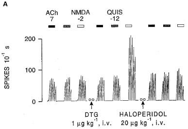
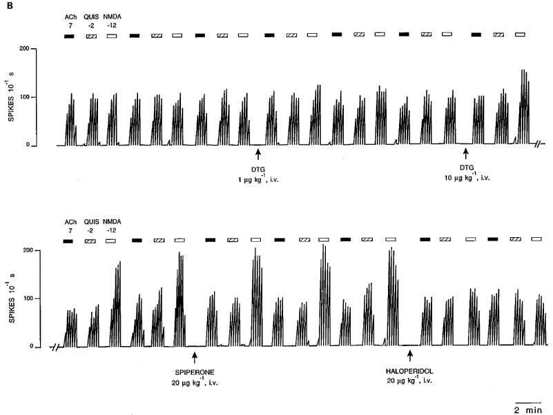
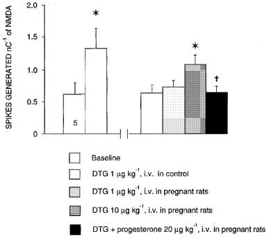
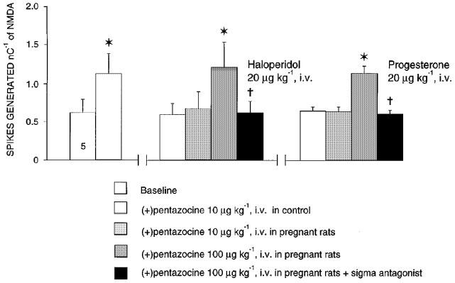
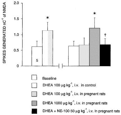
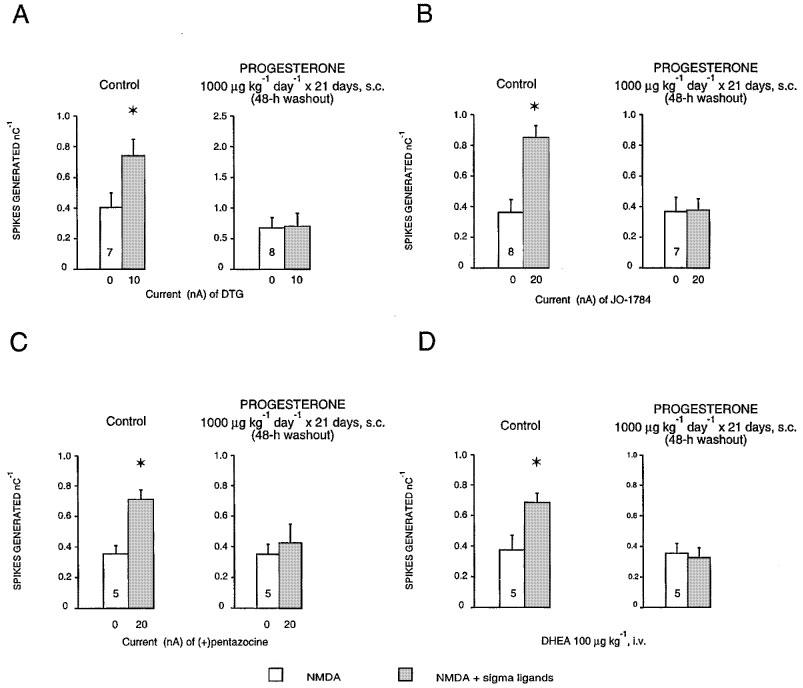
1. Sigma (sigma) receptors have recently been cloned, though their endogenous ligand(s) remain unidentified. However, some neuroactive steroids, such as progesterone, have a high affinity for these receptors. Some sigma ligands, such as DTG, (+)-pentazocine and DHEA, act as sigma 'agonists' by potentiating the neuronal response to NMDA. Others, such as haloperidol, NE-100 and progesterone, act as sigma 'antagonists' by reversing the potentiations induced by sigma 'agonists'. 2. We compared the effects of sigma 'agonists' in four series of female rats: in controls, at day 18 of pregnancy, at day 5 post-partum, and in ovariectomized rats following a 3-week treatment with a high dose of progesterone. 3. In pregnant rats and following a 3-week treatment with progesterone, 10 fold higher doses of DTG, (+)-pentazocine and DHEA were required to elicit a selective potentiation of the NMDA response comparable to that obtained in control females. Conversely, at day 5 post-partum and following the 3-week treatment with a progesterone and after a 5-day washout, the potentiation of the NMDA response induced by the sigma 'agonist' DTG was greater than in control females. 4. The present data suggest that endogenous progesterone acts as an 'antagonist' at sigma receptors. The resulting changes in the function of sigma receptors during pregnancy and post-partum may be implicated in emotional phenomena occurring during these periods.


Figures

Figure 1
Figure 1
Integrated firing rate histograms from extracellular unitary recordings of CA3 dorsal hippocampus pyramidal neurons showing the effects of microionophoretic applications of NMDA, QUIS and ACh. (A) Recording obtained from a control rat before and after the intravenous administration of DTG, and after that of haloperidol. (B) Recording obtained from a pregnant rat at day 18, before and after the intravenous administration of two doses of DTG, followed by spiperone and haloperidol. Bars indicate the duration of applications for which currents are given in nA. Open circles (oo) indicate a 5-min interruption of the histogram. Time reference applies to both histograms.
Figure 1
Figure 1
Integrated firing rate histograms from extracellular unitary recordings of CA3 dorsal hippocampus pyramidal neurons showing the effects of microionophoretic applications of NMDA, QUIS and ACh. (A) Recording obtained from a control rat before and after the intravenous administration of DTG, and after that of haloperidol. (B) Recording obtained from a pregnant rat at day 18, before and after the intravenous administration of two doses of DTG, followed by spiperone and haloperidol. Bars indicate the duration of applications for which currents are given in nA. Open circles (oo) indicate a 5-min interruption of the histogram. Time reference applies to both histograms.
Figure 2
Figure 2
Effects of sigma ligand DTG on the neuronal response to NMDA in control and in 18-day pregnant rats. The responsiveness of CA3 dorsal hippocampus neurons to microionophoretic applications of NMDA is expressed as the number of spikes generated nC−1 (mean±s.e.mean) before and after the intravenous administration of 1 and 10 μg kg−1 of DTG and following the intravenous administration of 20 μg kg−1 of progesterone. The number at the bottom of the first column indicates the number of neurons tested (one neuron per rat in this and subsequent bar chart histograms). *P<0.01, using the Student's t-test, †P<0.01, using variance analysis.
Figure 3
Figure 3
Effects of sigma ligand (+)-pentazocine on the neuronal response to NMDA in control and in 18-day pregnant rats. The responsiveness of CA3 dorsal hippocampus neurons to microionophoretic applications of NMDA is expressed as the number of spikes generated nC−1 (mean±s.e.mean) before and after the intravenous administration of 10 and 100 μg kg−1 of (+)-pentazocine following the intravenous administration of 20 μg kg−1 of haloperidol or 20 μg kg−1 of progesterone. *P<0.01, using the Student's t-test, †P<0.01, using variance analysis.
Figure 4
Figure 4
Effects of neuroactive steroids DHEA on the neuronal response to NMDA in control and in 18-day pregnant rats. The responsiveness of CA3 dorsal hippocampus neurons to microionophoretic applications of NMDA is expressed as the number of spikes generated nC−1 (mean±s.e.mean) before and after the intravenous administration of 100 and 1000 μg kg−1 of DHEA following the intravenous administration of 50 μg kg−1 of NE-100. *P<0.01, using the Student's t-test, †P<0.01, using variance analysis.
Figure 5
Figure 5
Effect of sigma ligand DTG on the NMDA response in control rats and during the post-partum period. The responsiveness of CA3 dorsal hippocampus neurons to microionophoretic applications of NMDA is expressed as the number of spikes generated nC−1 (mean±s.e.mean) before and after the intravenous administration of 1 μg kg−1 of DTG in control female rats, and in rats at days 5, 10 and 15 post-partum. *P<0.01 using paired Student's t-test, †P<0.001 comparing the effect of DTG at day 5 of the post-partum period to that in control females, with analysis of covariance, using the NMDA response before DTG as the regressor.
Figure 6
Figure 6
Responsiveness expressed as the number of spikes generated per nanocoulomb (mean±s.e.mean) of CA3 dorsal hippocampus neurones to microionophoretic applications of NMDA before and after the microionophoretic applications of DTG (A), JO-1784 (B), (+)-pentazocine (C) and the i.v. administration of DHEA (D), in control and in rats treated with 1000 μg kg−1 day−1 of progesterone for 21 days after a 2-day washout. The number at the bottom of the first column indicates the number of neurons tested. *P<0.01, using the Student's t-test.
Figure 7
Figure 7
Responsiveness expressed as the number of spikes generated per nanocoulomb (mean±s.e.mean) of CA3 dorsal hippocampus neurones to microionophoretic applications of NMDA before, after the intravenous administration of DTG, and after the intravenous administration of haloperidol in control rats (A), or in treated rats with 1000 μg kg−1 of progesterone for 21 days after a 5-day washout (B).

Similar articles

Cited by

References

    1. BACKSTROM T., BIXO M., HAMMARBACK S. Ovarian steroid hormones. Effects on mood, behaviour and brain excitability. Acta Obstet. Gynecologica Scand. 1985;130:19–24. - PubMed
    1. BERGERON R., DE MONTIGNY C., DEBONNEL G. Biphasic effects of sigma ligands on the neuronal response to N-methyl-D-aspartate. Naunyn Schmiedebergs Arch. Pharmacol. 1995;351:252–260. - PubMed
    1. BERGERON R., DE MONTIGNY C., DEBONNEL G. Potentiation of neuronal NMDA response induced by dehydroepiandrosterone and its suppression by progesterone: Effects mediated via sigma receptors. J. Neurosci. 1996;16:1193–1202. - PMC - PubMed
    1. BERGERON R., DE MONTIGNY C., DEBONNEL G. Effect of short-term and long-term treatments with σ ligands on the N-methyl-D-aspartate response in the CA3 region of the rat dorsal hippocampus. Br. J. Pharmacol. 1997;120:1351–1359. - PMC - PubMed
    1. CHAKI S., TANAKA M., MURAMATSU M., OTOMO S. NE-100, a novel potent σ ligand, preferentially binds to σ1 binding sites in guinea pig brain. Eur. J. Pharmacol. 1994;251:R1–R2. - PubMed

Publication types

MeSH terms

LinkOut - more resources