. 1999 Sep 1;13(17):2231-41.
doi: 10.1101/gad.13.17.2231.

Activation and centromeric localization of CCAAT/enhancer-binding proteins during the mitotic clonal expansion of adipocyte differentiation

Affiliations

Activation and centromeric localization of CCAAT/enhancer-binding proteins during the mitotic clonal expansion of adipocyte differentiation

Q Q Tang et al. Genes Dev. .

Abstract

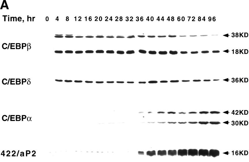
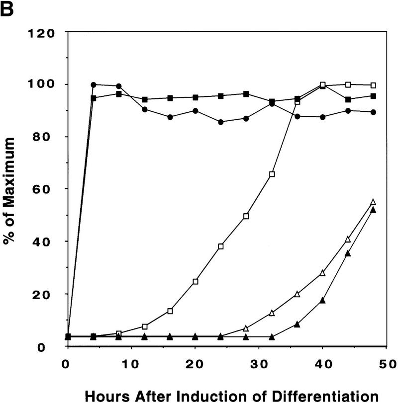
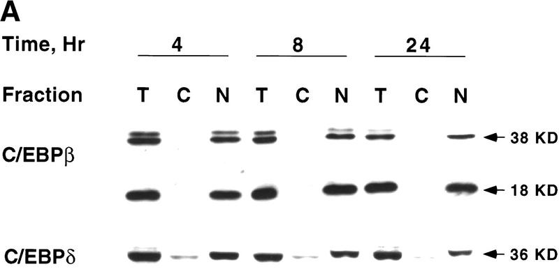
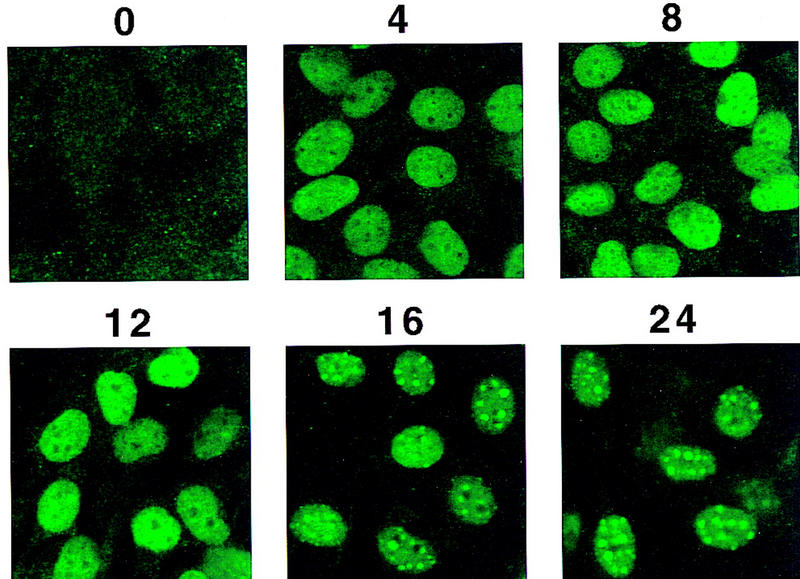
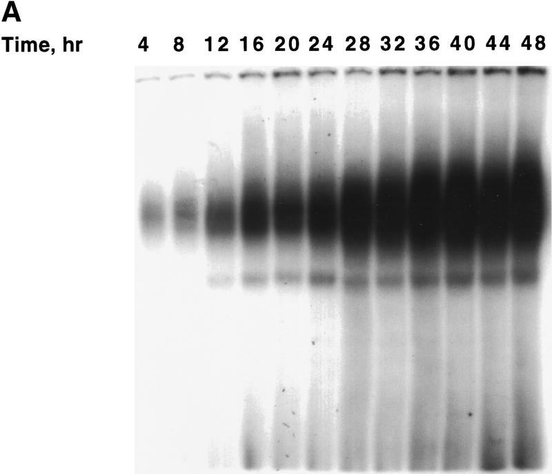
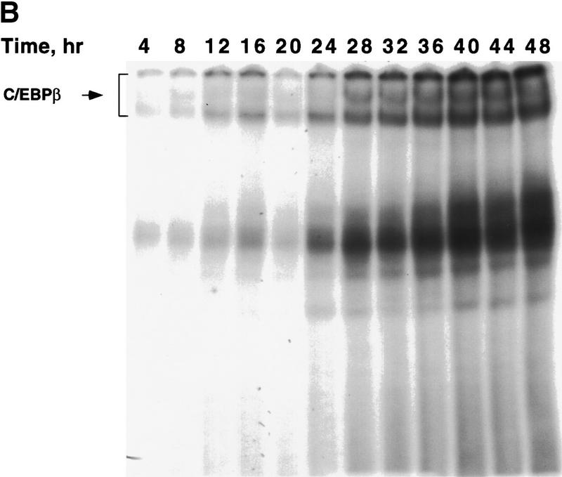
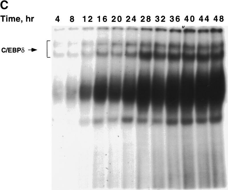
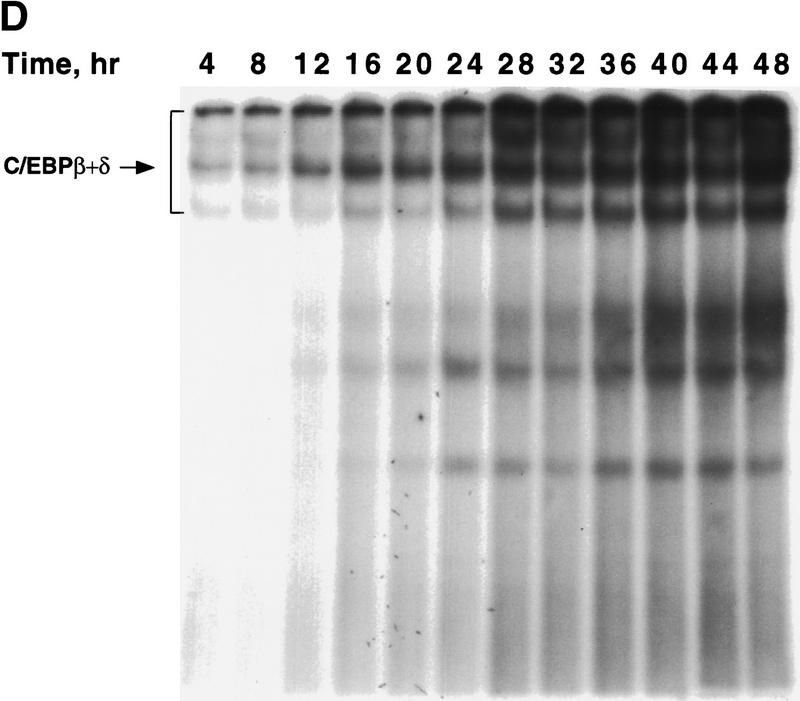
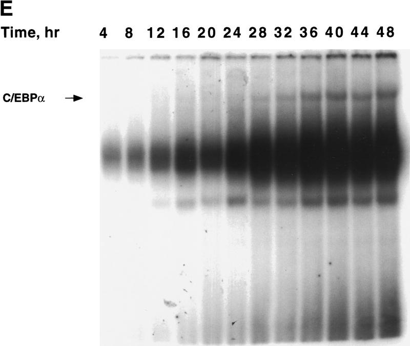
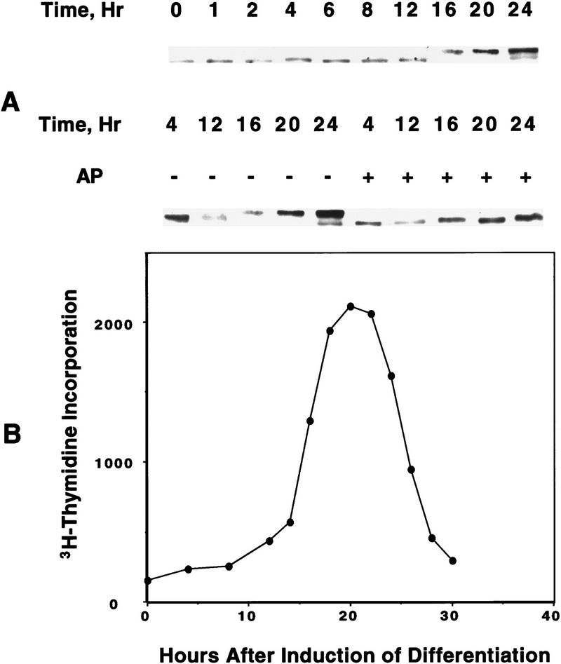
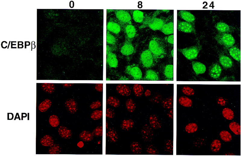
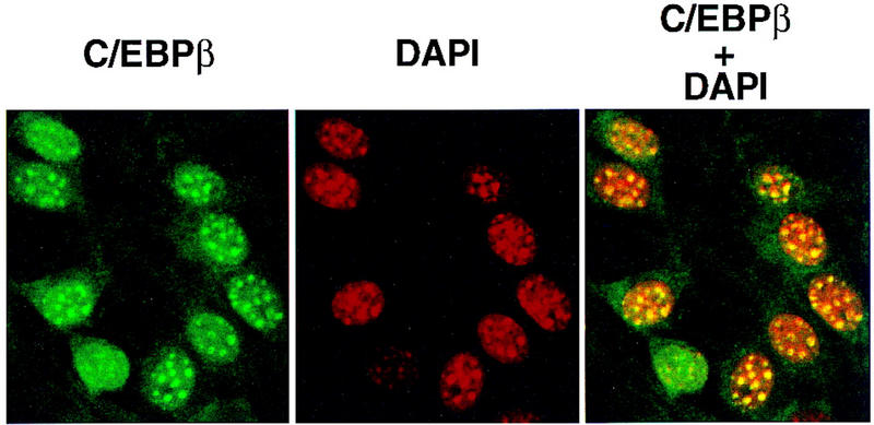
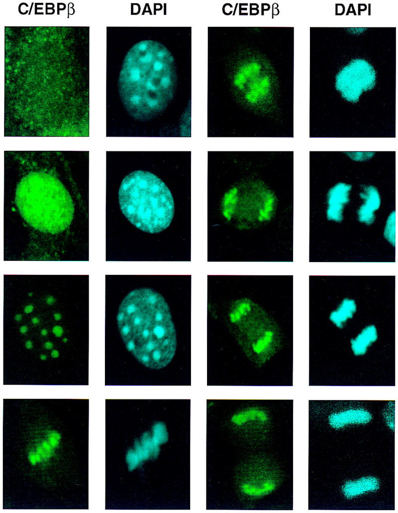
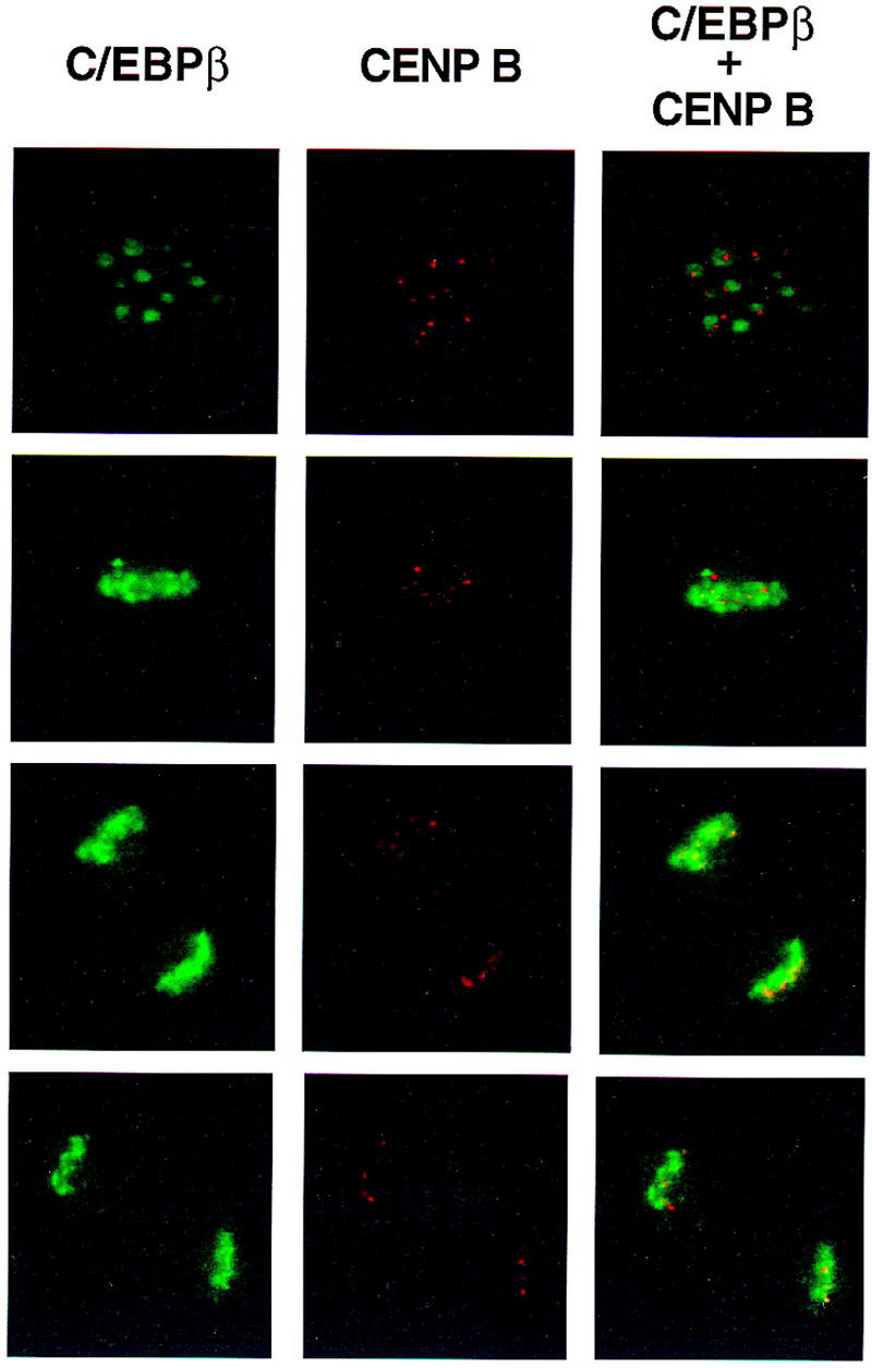
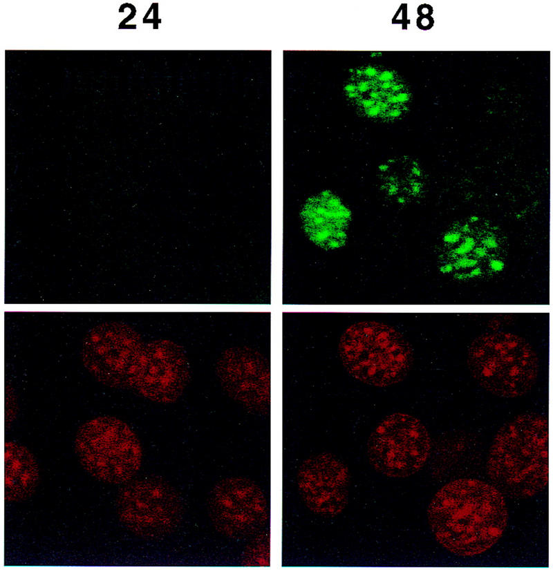
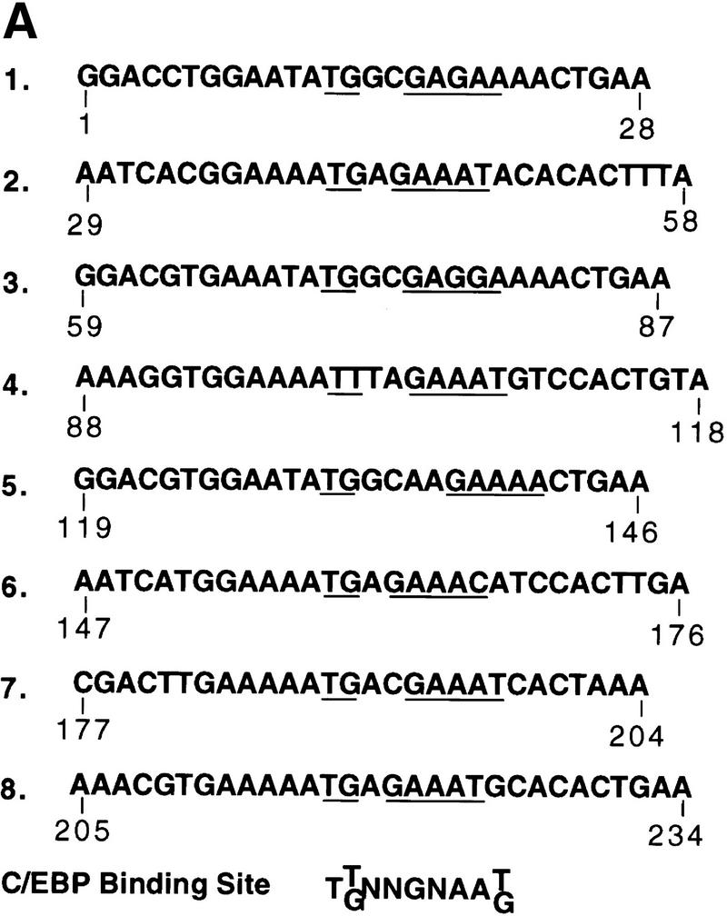
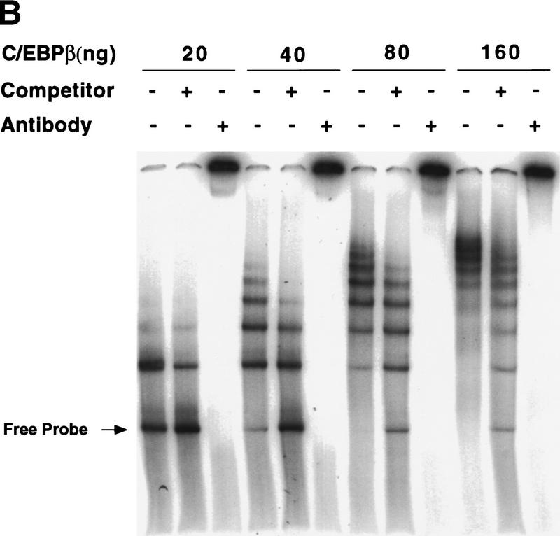
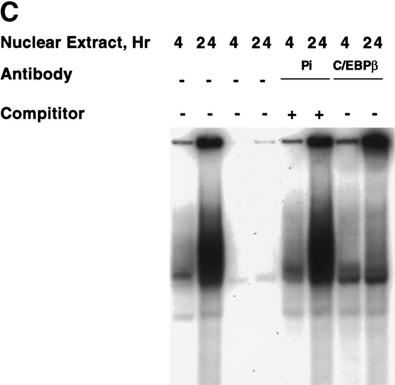
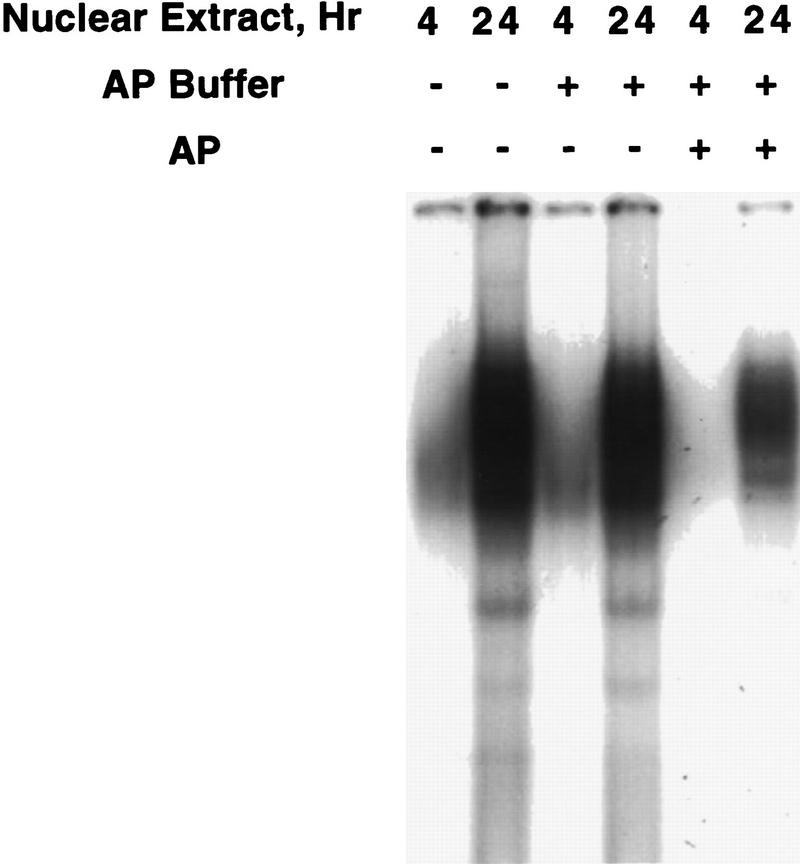
Hormonal induction of 3T3-L1 preadipocytes triggers a cascade of events that initiate differentiation into adipocytes. CCAAT/enhancer-binding proteins beta and delta (C/EBPbeta/delta) are expressed early in the differentiation program, but are not immediately active. After a long lag, C/EBPbeta/delta become competent to bind to the C/EBP regulatory element in the C/EBPalpha gene promoter, C/EBPalpha being a transcriptional activator of numerous adipocyte genes. As C/EBPbeta/delta acquire binding activity, they become localized to centromeres as preadipocytes synchronously enter S phase at the onset of mitotic clonal expansion. Localization to centromeres occurs through C/EBP consensus-binding sites in centromeric satellite DNA. C/EBPalpha, which is antimitotic, becomes centromere-associated much later in the differentiation program as mitotic clonal expansion ceases and the cells become terminally differentiated.


Figures

Figure 1
Figure 1
Expression of C/EBPs and 422/aP2 during differentiation of 3T3-L1 preadipocytes. (A) Day 0 postconfluent 3T3-L1 preadipocytes were induced to differentiate into adipocytes using the standard differentiation protocol. At different times after induction of differentiation, cell lysates were subjected to SDS-PAGE and Western blotting with antibodies directed against C/EBPβ, C/EBPδ, and C/EBPα and 422/aP2. It should be noted that C/EBPβ and C/EBPα each have two isoforms translated from the same mRNA. (B) The results of the Western blots (shown in A) and DNA-binding acitivities for C/EBPβ/δ and C/EBPα (shown in Fig. 3D,E, respectively) were quantitated and the results plotted. The binding activities of C/EBPβ/δ and C/EBPα in nuclear extracts were assessed by EMSA using an oligonucleotide probe corresponding to the C/EBP regulatory element in the promoter of the C/EBPα gene. Results for C/EBPβ/δ protein and C/EBPβ/δ DNA-binding activity are expressed as percent of maximal expression. Results for C/EBPα DNA-binding activity and 422/aP2 protein, which do not achieve their maxima until much later (> 72 hr) are expressed as percent of their half-maximal values. The amount of binding activity for C/EBPβ/δ is based on the amount of label remaining in the EMSA bands attributable to C/EBPβ/δ after removing C/EBPα by supershifting with anti-C/EBPα antibody (data from Fig. 3E). Also, the amount of binding activity due to C/EBPα is based on the the amount of label remaining in the EMSA bands due to C/EBPα after removing C/EBPβ and C/EBPα by supershiting with anti-C/EBPβ/δ antibodies (data from Fig. 3D). (●) C/EBPβ protein; (█) C/EBPδ protein; (□) DNA-binding acitivity of C/EBPβ/δ; (▵) DNA-binding acitivity of C/EBPα; (▴) 422/aP2 protein.
Figure 1
Figure 1
Expression of C/EBPs and 422/aP2 during differentiation of 3T3-L1 preadipocytes. (A) Day 0 postconfluent 3T3-L1 preadipocytes were induced to differentiate into adipocytes using the standard differentiation protocol. At different times after induction of differentiation, cell lysates were subjected to SDS-PAGE and Western blotting with antibodies directed against C/EBPβ, C/EBPδ, and C/EBPα and 422/aP2. It should be noted that C/EBPβ and C/EBPα each have two isoforms translated from the same mRNA. (B) The results of the Western blots (shown in A) and DNA-binding acitivities for C/EBPβ/δ and C/EBPα (shown in Fig. 3D,E, respectively) were quantitated and the results plotted. The binding activities of C/EBPβ/δ and C/EBPα in nuclear extracts were assessed by EMSA using an oligonucleotide probe corresponding to the C/EBP regulatory element in the promoter of the C/EBPα gene. Results for C/EBPβ/δ protein and C/EBPβ/δ DNA-binding activity are expressed as percent of maximal expression. Results for C/EBPα DNA-binding activity and 422/aP2 protein, which do not achieve their maxima until much later (> 72 hr) are expressed as percent of their half-maximal values. The amount of binding activity for C/EBPβ/δ is based on the amount of label remaining in the EMSA bands attributable to C/EBPβ/δ after removing C/EBPα by supershifting with anti-C/EBPα antibody (data from Fig. 3E). Also, the amount of binding activity due to C/EBPα is based on the the amount of label remaining in the EMSA bands due to C/EBPα after removing C/EBPβ and C/EBPα by supershiting with anti-C/EBPβ/δ antibodies (data from Fig. 3D). (●) C/EBPβ protein; (█) C/EBPδ protein; (□) DNA-binding acitivity of C/EBPβ/δ; (▵) DNA-binding acitivity of C/EBPα; (▴) 422/aP2 protein.
Figure 2
Figure 2
Intracellular localization of C/EBPβ/δ early in the differentiation program. (A) At 4, 8, and 24 hr after induction of differentiation 3T3-L1 preadipocytes were lysed and resolved into nuclear and cytoplasmic fractions. The total lysate, cytoplasmic, and nuclear fractions (amounts equivalent to the same number of cells) were subjected to SDS-PAGE, then Western blotted with antibodies against C/EBPβ/δ. (T) Total cell lysate; (C) cytoplasmic fraction; (N) nuclear fraction. (B) 3T3-L1 preadipocytes on coverslips were induced to differentiate. At time 0 and 4, 8, 12, 16, and 24 hr after induction of differentiation the cell monolayers were fixed and subjected to immunofluorescence staining with antibody to C/EBPβ.
Figure 2
Figure 2
Intracellular localization of C/EBPβ/δ early in the differentiation program. (A) At 4, 8, and 24 hr after induction of differentiation 3T3-L1 preadipocytes were lysed and resolved into nuclear and cytoplasmic fractions. The total lysate, cytoplasmic, and nuclear fractions (amounts equivalent to the same number of cells) were subjected to SDS-PAGE, then Western blotted with antibodies against C/EBPβ/δ. (T) Total cell lysate; (C) cytoplasmic fraction; (N) nuclear fraction. (B) 3T3-L1 preadipocytes on coverslips were induced to differentiate. At time 0 and 4, 8, 12, 16, and 24 hr after induction of differentiation the cell monolayers were fixed and subjected to immunofluorescence staining with antibody to C/EBPβ.
Figure 3
Figure 3
Changes in binding of C/EBPβ, C/EBPδ, and C/EBPα to the C/EBP regulatory element in the C/EBPα gene promoter during adipocyte differentiation. EMSA was performed on nuclear extract from 3T3-L1 preadipocytes after induction of differentiation. EMSA was conducted with 10 μg of nuclear extract and a labeled oligonucleotide probe corresponding to the C/EBP regulatory element in the C/EBPα gene promoter. (A) Day 0 postconfluent 3T3-L1 preadipocytes were induced to differentiation into adipocytes using the standard differentiation protocol. Every 4 hr after induction nuclear extracts were prepared and subjected to EMSA as described in Materials and Methods. Supershift experiments were performed as in A with antibodies directed against: C/EBPβ (B); C/EBPδ (C); C/EBPβ and C/EBPδ (D); and C/EBPα (E).
Figure 3
Figure 3
Changes in binding of C/EBPβ, C/EBPδ, and C/EBPα to the C/EBP regulatory element in the C/EBPα gene promoter during adipocyte differentiation. EMSA was performed on nuclear extract from 3T3-L1 preadipocytes after induction of differentiation. EMSA was conducted with 10 μg of nuclear extract and a labeled oligonucleotide probe corresponding to the C/EBP regulatory element in the C/EBPα gene promoter. (A) Day 0 postconfluent 3T3-L1 preadipocytes were induced to differentiation into adipocytes using the standard differentiation protocol. Every 4 hr after induction nuclear extracts were prepared and subjected to EMSA as described in Materials and Methods. Supershift experiments were performed as in A with antibodies directed against: C/EBPβ (B); C/EBPδ (C); C/EBPβ and C/EBPδ (D); and C/EBPα (E).
Figure 4
Figure 4
Changes in phosphorylation state of Rb protein and in [3H]thymidine incorporation into DNA during mitotic clonal expansion of preadipocytes induced to differentiate. (A) Day 0 postconfluent 3T3-L1 preadipocytes were subjected to standard differentiation protocol. At the times indicated cell lysates were prepared, subjected to SDS-PAGE, and immunoblotted with antibody against Rb. Incubation of cell lysates with alkaline phosphatase (AP) caused a shift in the mobility of the slow-moving band to that of the faster moving band. (B) [3H]Thymidine incorporation into cellular DNA during a 15-min incubation was determined at 4- or 2-hr intervals for 30 hr after induction of differentiation of 3T3-L1 preadipocytes.
Figure 5
Figure 5
Colocalization of C/EBPβ, C/EBPα, and CENP-B and DAPI staining during differentiation of 3T3-L1 preadipocytes. 3T3-L1 preadipocytes were induced to differentiate using the standard protocol, fixed, treated with antibodies (and FITC-labeled anti-rabbit IgG), and DAPI at the times indicated. Fluorescence images were obtained by confocal microscopy. (A, top) Immunofluorescence with antibody against C/EBPβ and FITC-labeled anti-rabbit IgG. Similar results were obtained with antibody against C/EBPδ (results not shown). (Bottom) Immunofluorescence imaging of the same field as in A with DAPI. (B) Dual fluorescence imaging of C/EBPβ and DAPI by confocal microscopy of cells 24 hr after induction of differentiation. (C) Fluorescence imaging of 3T3-L1 preadipocytes treated with C/EBPβ antibody and DAPI during the first round of mitotic clonal expansion. These selected images of preadipocytes at 28 hr after induction of differentiation represent various stages of the cell cycle. (D) Fluorescence imaging of 3T3-L1 preadipocytes treated with antibodies against C/EBPβ and CENP-B during the first round of mitotic clonal expansion. These selected images of preadipocytes at 28 hr after induction of differentiation represent various stages of the cell cycle. The column on the right represents dual fluorescence images of the two columns of images to the left. (E) Colocalization of immunofluorescence of C/EBPα antibody and DAPI in the terminal stages of the differentiation program.
Figure 5
Figure 5
Colocalization of C/EBPβ, C/EBPα, and CENP-B and DAPI staining during differentiation of 3T3-L1 preadipocytes. 3T3-L1 preadipocytes were induced to differentiate using the standard protocol, fixed, treated with antibodies (and FITC-labeled anti-rabbit IgG), and DAPI at the times indicated. Fluorescence images were obtained by confocal microscopy. (A, top) Immunofluorescence with antibody against C/EBPβ and FITC-labeled anti-rabbit IgG. Similar results were obtained with antibody against C/EBPδ (results not shown). (Bottom) Immunofluorescence imaging of the same field as in A with DAPI. (B) Dual fluorescence imaging of C/EBPβ and DAPI by confocal microscopy of cells 24 hr after induction of differentiation. (C) Fluorescence imaging of 3T3-L1 preadipocytes treated with C/EBPβ antibody and DAPI during the first round of mitotic clonal expansion. These selected images of preadipocytes at 28 hr after induction of differentiation represent various stages of the cell cycle. (D) Fluorescence imaging of 3T3-L1 preadipocytes treated with antibodies against C/EBPβ and CENP-B during the first round of mitotic clonal expansion. These selected images of preadipocytes at 28 hr after induction of differentiation represent various stages of the cell cycle. The column on the right represents dual fluorescence images of the two columns of images to the left. (E) Colocalization of immunofluorescence of C/EBPα antibody and DAPI in the terminal stages of the differentiation program.
Figure 6
Figure 6
Association of mouse satellite DNA with rC/EBPβ or C/EBPδ in nuclear extract from 3T3-L1 preadipocytes induced to differentiate. (A) Nucleotide sequence of 234 bp of the major mouse satellite DNA. The underlined repetitive sequences represent probable C/EBP-binding sites based on their similarity to the consensus sequence for C/EBP-binding sites (shown at bottom) in numerous gene promoters. (B) EMSA with rC/EBPβ and a 234-bp oligonucleotide probe corresponding to the major mouse satellite DNA. The unlabeled competitor oligonucleotide corresponds to the C/EBP regulatory element in the C/EBPα gene promoter. Similar results (not shown) were obtained with a competitor oligonucleotide corresponding to sequence 7 in A. (C) Nuclear extracts from 3T3-L1 preadipocytes 4 or 24 hr after induction of differentiation were subjected to EMSA with a labeled oligonucleotide probe corresponding to C/EBP-binding site (sequence 7 in the major mouse satellite DNA; see A; Materials and Methods). Supershift experiments were performed with preimmune serum (Pi) or anti-serum to C/EBPβ.
Figure 6
Figure 6
Association of mouse satellite DNA with rC/EBPβ or C/EBPδ in nuclear extract from 3T3-L1 preadipocytes induced to differentiate. (A) Nucleotide sequence of 234 bp of the major mouse satellite DNA. The underlined repetitive sequences represent probable C/EBP-binding sites based on their similarity to the consensus sequence for C/EBP-binding sites (shown at bottom) in numerous gene promoters. (B) EMSA with rC/EBPβ and a 234-bp oligonucleotide probe corresponding to the major mouse satellite DNA. The unlabeled competitor oligonucleotide corresponds to the C/EBP regulatory element in the C/EBPα gene promoter. Similar results (not shown) were obtained with a competitor oligonucleotide corresponding to sequence 7 in A. (C) Nuclear extracts from 3T3-L1 preadipocytes 4 or 24 hr after induction of differentiation were subjected to EMSA with a labeled oligonucleotide probe corresponding to C/EBP-binding site (sequence 7 in the major mouse satellite DNA; see A; Materials and Methods). Supershift experiments were performed with preimmune serum (Pi) or anti-serum to C/EBPβ.
Figure 7
Figure 7
Effect of phosphatase treatment on DNA-binding activity of nuclear extract from “induced” 3T3-L1 preadipocytes. Nuclear extract (10 μg of protein), prepared from 3T3-L1 preadipocytes after induction of differentiation for 4 or 24 hr, was incubated for 30 min at 37°C in an incubation mixture containing 50 mm Tris-HCl (pH 8.5) and 0.1 mm EDTA in the absence or presence of 80 units of Escherichia coli alkaline phosphatase in a total volume of 40 μl. EMSA was then performed with a “fill-in” 32P-labeled oligonucleotide probe corresponding to the C/EBP regulatory element in the C/EBPα gene promoter.

Similar articles

Cited by

References

    1. Baer M, Williams SC, Dillner A, Schwartz RC, Johnson PF. Autocrine signals control CCAAT/enhancer binding protein β expression, localization and activity in macrophages. Blood. 1998;92:4353–4365. - PubMed
    1. Bernlohr DA, Bolanowski MA, Kelly TJ, Lane MD. Evidence for an increase in transcription of specific mRNAs during differentiation of 3T3-L1 preadipocytes. J Biol Chem. 1985;260:5563–5567. - PubMed
    1. Brun RP, Tontonoz P, Forman BM, Ellis R, Jasmine C, Evans RM, Spiegelman BM. Differential activation of adipogensis by multiple PPAR isoforms. Genes & Dev. 1996;10:974–984. - PubMed
    1. Cao Z, Umek RM, McKnight SL. Regulated expression of three C/EBP isoforms during adipose conversion of 3T3-L1 cells. Genes & Dev. 1991;5:1538–1552. - PubMed
    1. Cheneval D, Christy RJ, Geiman D, Cornelius P, Lane MD. Cell-free transcription directed by the 422 adipose P2 gene promoter: Activation by the CCAAT/enhancer binding protein. Proc Natl Acad Sci. 1991;88:8465–8469. - PMC - PubMed

Publication types

MeSH terms