. 1999 Sep 1;13(17):2242-57.
doi: 10.1101/gad.13.17.2242.

Components of an SCF ubiquitin ligase localize to the centrosome and regulate the centrosome duplication cycle

Affiliations

Components of an SCF ubiquitin ligase localize to the centrosome and regulate the centrosome duplication cycle

E Freed et al. Genes Dev. .

Abstract

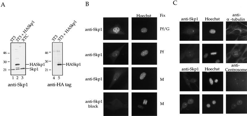
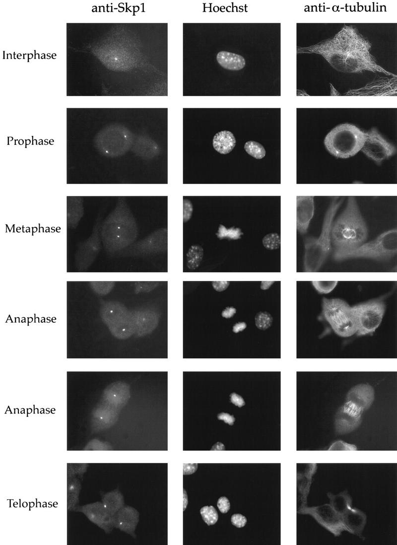
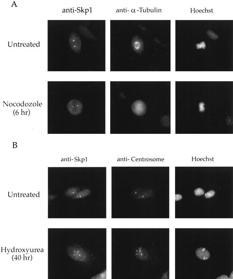
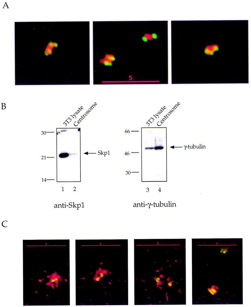
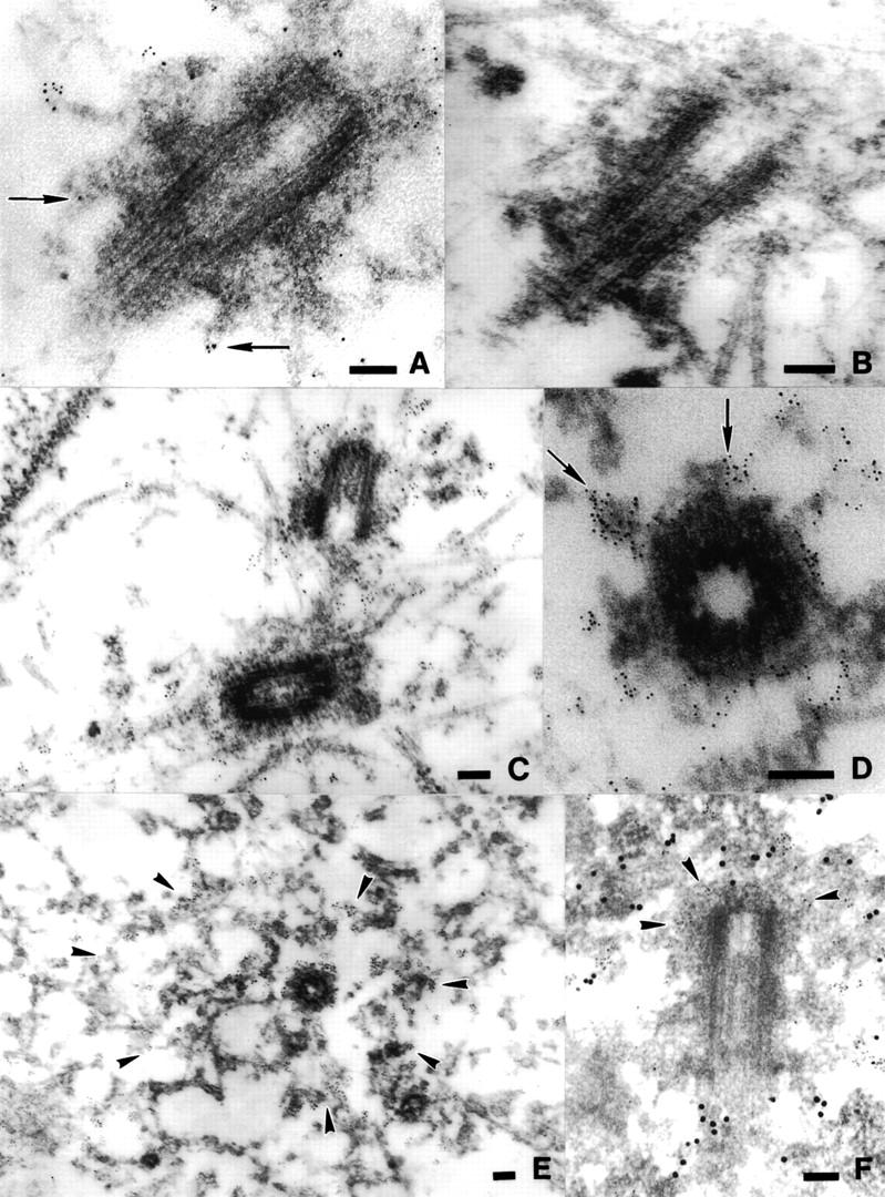
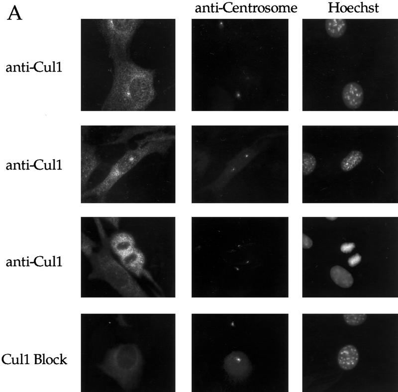
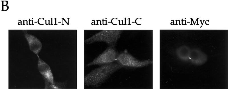
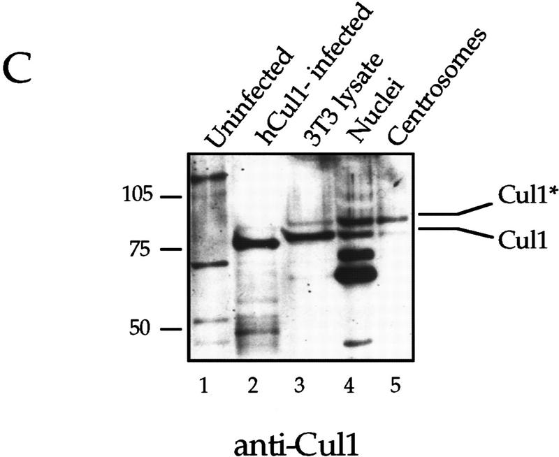
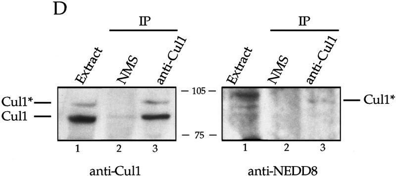
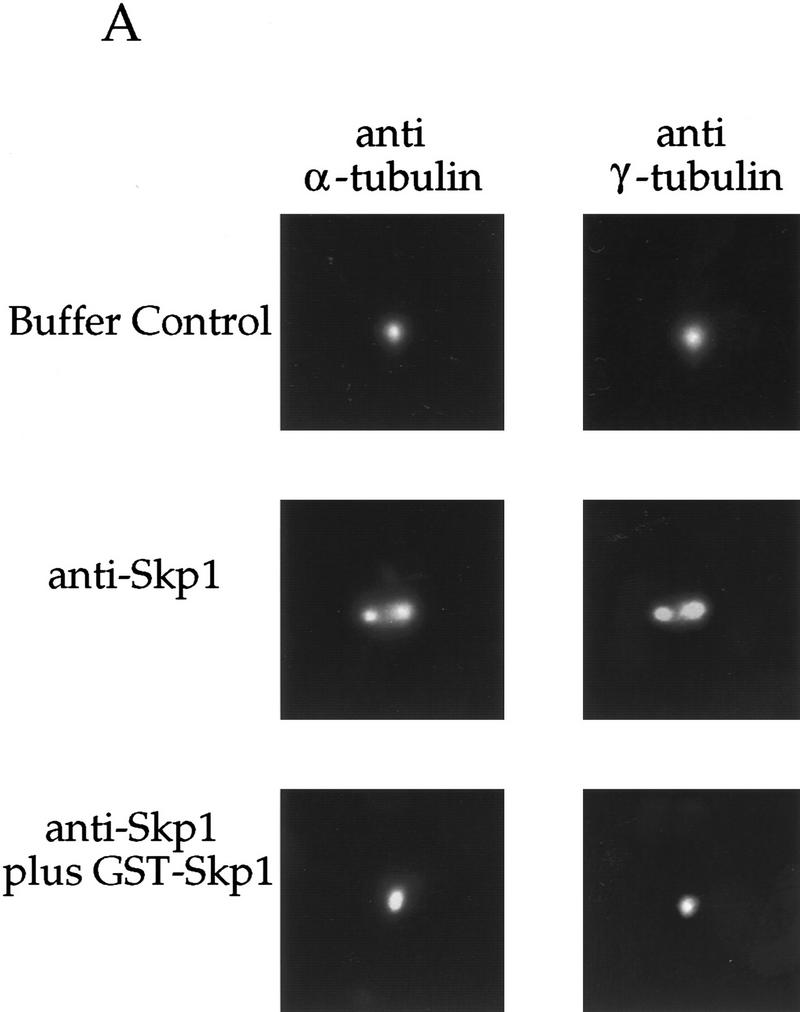
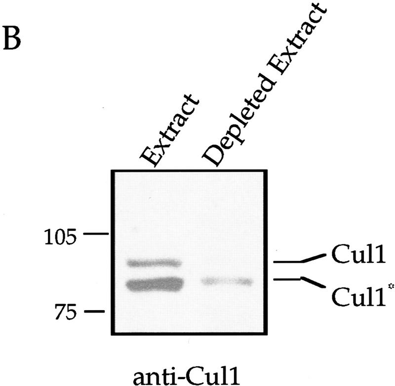
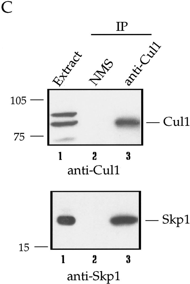
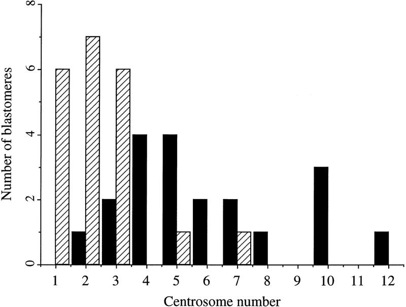
Centrosomes organize the mitotic spindle to ensure accurate segregation of the chromosomes in mitosis. The mechanism that ensures accurate duplication and separation of the centrosomes underlies the fidelity of chromosome segregation, but remains unknown. In Saccharomyces cerevisiae, entry into S phase and separation of spindle pole bodies each require CDC4 and CDC34, which encode components of an SCF (Skp1-cullin-F-box) ubiquitin ligase, but a direct (SCF) connection to the spindle pole body is unknown. Using immunofluorescence microscopy, we show that in mammalian cells the Skp1 protein and the cullin Cul1 are localized to interphase and mitotic centrosomes and to the cytoplasm and nucleus. Deconvolution and immunoelectron microscopy suggest that Skp1 forms an extended pericentriolar structure that may function to organize the centrosome. Purified centrosomes also contain Skp1, and Cul1 modified by the ubiquitin-like molecule NEDD8, suggesting a role for NEDD8 in targeting. Using an in vitro assay for centriole separation in Xenopus extracts, antibodies to Skp1 or Cul1 block separation. Proteasome inhibitors block both centriole separation in vitro and centrosome duplication in Xenopus embryos. We identify candidate centrosomal F-box proteins, suggesting that distinct SCF complexes may direct proteolysis of factors mediating multiple steps in the centrosome cycle.


Figures

Figure 1
Figure 1
Characterization of Skp1 antibodies and localization of Skp1 in cells. (A) Western blotting using affinity-purified anti-Skp1 antibodies. (Lanes 1,4) NIH-3T3 cell lysate; (lanes 2,5) lysate of NIH-3T3 cells transiently transfected with HA-tagged Skp1; (lane 3) XTC cell lysate. The blots were incubated with affinity-purified anti-Skp1 antibodies (lanes 1–3) or with rabbit anti-HA antibodies (lanes 4,5), followed by secondary antibodies, and bands visualized using enhanced chemiluminescence (ECL) Western blotting detection reagents. Positions of molecular weight markers are indicated. (B) Immunofluorescence localization of Skp1. Cells were fixed in paraformaldehyde (Pf), a paraformaldehyde/glutaraldehyde (Pf/G) mixture, or methanol (M) as indicated, and labeled with affinity-purified anti-Skp1 antibodies (top three panels) or anti-Skp1 antibodies blocked with excess GST–Skp1 protein (bottom panel) and with Hoechst dye to label DNA. (C) Costaining confirms centrosomal localization. NIH-3T3 cells were fixed in methanol and stained with affinity-purified anti-Skp1 antibodies and either mAb against α-tubulin or human anti-centrosome antiserum as indicated, followed by secondary antibodies and Hoechst dye.
Figure 2
Figure 2
Skp1 is centrosomal during all phases of the cell cycle in NIH-3T3 cells. NIH-3T3 cells were fixed in methanol and labeled with the indicated antibodies or with Hoechst dye to stain the DNA. Cells at specific stages of the cell cycle are indicated.
Figure 3
Figure 3
Skp1 is centrosomal in nocodazole-treated and ectopically produced centrosomes. (A) Nocodazole treatment of NIH-3T3 cells. Cells were incubated with nocodazole (0.3 μg/ml) or untreated (control), fixed in methanol, and stained with the indicated antibodies and Hoechst dye. (B) Hydroxyurea treatment of CHO cells. Cells were grown on coverslips and incubated with or without hydroxyurea (2 mm). They were fixed in methanol and stained with the indicated antibodies and with Hoechst dye as above. (Bottom panel) An example of a cell containing multiple centrosomes.
Figure 4
Figure 4
Skp1 is pericentriolar in purified centrosomes and in NIH-3T3 cells. Centrosomes were purified from CHO cells and prepared for immunofluorescence as described (see Materials and Methods). (A) Centrosomes spun onto coverslips were fixed in methanol and labeled with mouse monoclonal antibodies against α-tubulin and affinity-purified rabbit antibodies against Skp1, followed by secondary antibodies. Anti-α-tubulin (green); Anti-Skp1 (red). (B) Immunoblotting detects Skp1 in a purified centrosome sample. An NIH-3T3 cell lysate (lanes 1,3) and a centrosome sample (lanes 2,4) were subjected to Western blotting with either affinity-purified anti-Skp1 antibodies (lanes 1,2) or rabbit antibodies to γ-tubulin (lanes 3,4). Secondary antibody was HRP-conjugated donkey anti-rabbit IgG and bands were detected with ECL reagents as above. Skp1 and γ-tubulin are indicated (arrows). Molecular mass markers are as indicated. (C) NIH-3T3 cells fixed in methanol, and labeled using a mouse mAb against γ-tubulin and with rabbit affinity-purified anti-Skp1 antibodies followed by secondary antibodies. Anti-γ-tubulin (green); anti-Skp1 (red). Deconvolution images of centrosomes in cells at different phases of the cell cycle are pictured (left to right).
Figure 5
Figure 5
Immunoelectron microscopy of Skp1 stained centrosomes. NIH-3T3 cells were grown on mylar sheets, fixed in glutaraldehyde/paraformaldehyde (A–C, F), or in methanol (D,E) and labeled with no primary antibody (B), with affinity-purified anti-Skp1 antibodies (A,C–E), or with affinity-purified anti-Skp1 antibodies and a mouse monoclonal antibody against γ-tubulin (F), followed by 5 nm gold-conjugated goat anti-rabbit IgG (A–E) or 5 nm gold-conjugated goat anti-mouse IgG and 15 nm gold-conjugated goat anti-rabbit IgG (F). Samples were prepared for electron-microscopy as described (see Materials and Methods). Positions of centriolar appendages in A and D (arrows), concentrations of gold particles in a broad region surrounding the centriole in E (arrowheads), and concentrations of 5 nm gold particles (γ-tubulin staining) around the centriole in F (arrowheads) are indicated. Space bars, 0.1 μm.
Figure 6
Figure 6
A Nedd8-modified form of Cul1 is present at the centrosome. (A) NIH-3T3 cells were grown on cover slips, fixed in methanol and labeled with affinity-purified antibodies against the amino-terminal portion of human Cul1 and with human anti-centrosome antiserum, followed by secondary antibodies and Hoechst dye. Cul1 staining is shown at the centrosome at different times in the cell cycle (top three panels). Cells were also stained with anti-centrosome antibody in combination with blocked anti-amino-terminal Cul1 antibodies (bottom panel). Cells were visualized and photographed as described (see Materials and Methods). (B) Cul1 localizes to the midbody. (Left and middle) NIH-3T3 cells were grown on coverslips, fixed in methanol, and labeled with affinity-purified rabbit anti-amino-terminal Cul1 antibodies (left) or affinity-purified rabbit anti-carboxy-terminal Cul1 antibodies followed by secondary antibodies(middle). (Right) NIH-3T3 cells expressing Myc-tagged Cul1 were fixed as above and labeled with anti-Myc mAb 9E10 followed by secondary antibodies. (C) Immunoblotting also demonstrates that Cul1 is present at the centrosome. Samples as indicated: (Lane 1) Sf9 cell extract; (lane 2) insect cell extract [Hi5] expressing human Cul1; (lane 3) NIH-3T3 cell lysate; (lane 4) nuclei prepared from CHO cells; and (lane 5) centrosomes purified from CHO cells were subjected to Western blotting with affinity-purified rabbit antibodies to the amino-terminal region of Cul1 followed by HRP-conjugated secondary antibodies. Bands were detected using ECL reagents (Amersham). Two forms of Cul1 detected are indicated (arrows). Positions of migration of molecular mass markers are also indicated. Note also that whole-cell lysate from CHO cells gave a similar result as did 3T3 cell lysate (data not shown). (D) Immunoblotting demonstrates that Cul1 is NEDD8-modified. Rabbit affinity-purified anti-Cul1 (left) and rabbit anti-NEDD8 (right) immunoblots of partially purified Xenopus egg extract (lanes 1), and immunoprecipitates from the same partially purified extract using control normal mouse serum (lanes 2), or mouse anti-Cul1 antibodies (lanes 3). Bands were detected using ECL reagents (Amersham). Two forms of Cul1 and positions of migration of molecular mass markers are indicated.
Figure 6
Figure 6
A Nedd8-modified form of Cul1 is present at the centrosome. (A) NIH-3T3 cells were grown on cover slips, fixed in methanol and labeled with affinity-purified antibodies against the amino-terminal portion of human Cul1 and with human anti-centrosome antiserum, followed by secondary antibodies and Hoechst dye. Cul1 staining is shown at the centrosome at different times in the cell cycle (top three panels). Cells were also stained with anti-centrosome antibody in combination with blocked anti-amino-terminal Cul1 antibodies (bottom panel). Cells were visualized and photographed as described (see Materials and Methods). (B) Cul1 localizes to the midbody. (Left and middle) NIH-3T3 cells were grown on coverslips, fixed in methanol, and labeled with affinity-purified rabbit anti-amino-terminal Cul1 antibodies (left) or affinity-purified rabbit anti-carboxy-terminal Cul1 antibodies followed by secondary antibodies(middle). (Right) NIH-3T3 cells expressing Myc-tagged Cul1 were fixed as above and labeled with anti-Myc mAb 9E10 followed by secondary antibodies. (C) Immunoblotting also demonstrates that Cul1 is present at the centrosome. Samples as indicated: (Lane 1) Sf9 cell extract; (lane 2) insect cell extract [Hi5] expressing human Cul1; (lane 3) NIH-3T3 cell lysate; (lane 4) nuclei prepared from CHO cells; and (lane 5) centrosomes purified from CHO cells were subjected to Western blotting with affinity-purified rabbit antibodies to the amino-terminal region of Cul1 followed by HRP-conjugated secondary antibodies. Bands were detected using ECL reagents (Amersham). Two forms of Cul1 detected are indicated (arrows). Positions of migration of molecular mass markers are also indicated. Note also that whole-cell lysate from CHO cells gave a similar result as did 3T3 cell lysate (data not shown). (D) Immunoblotting demonstrates that Cul1 is NEDD8-modified. Rabbit affinity-purified anti-Cul1 (left) and rabbit anti-NEDD8 (right) immunoblots of partially purified Xenopus egg extract (lanes 1), and immunoprecipitates from the same partially purified extract using control normal mouse serum (lanes 2), or mouse anti-Cul1 antibodies (lanes 3). Bands were detected using ECL reagents (Amersham). Two forms of Cul1 and positions of migration of molecular mass markers are indicated.
Figure 7
Figure 7
A requirement for Skp1, Cul1, and an SCF complex in centriole separation in vitro. (A) Centrosomes used in the centriole separation assay were pelleted onto coverslips as described (see Materials and Methods), and labeled with antibodies to both α- and γ-tubulin. Rabbit antibodies to Skp1 were used as part of the assay. The staining was done with rat anti-α-tubulin mAb and mouse anti-γ-tubulin mAb (see Materials and Methods). Events were counted and the data tabulated as described in Table 1. (B) Immunodepletion with rabbit anti-amino-terminal Cul1 antiserum was performed (see Materials and Methods), and an aliquot of extract before (lane 1) and after (lane 2) immunodepletion subjected to Western blotting with rabbit anti-amino-terminal Cul1 antibodies. Arrows indicate molecular mass markers and two forms of Cul1. (C) Immunoprecipitation with control normal mouse serum (lanes 2) or mouse anti-Cul1 antiserum (lane 3) was performed using extract prepared as for the centriole separation assay (lane 1) (see Materials and Methods). Starting extract and resultant immunoprecipitates were subjected to immunoblotting with affinity-purified rabbit anti-Cul1 antibodies (top) or affinity-purified anti-Skp1 antibodies (bottom). Bands were detected using ECL reagents. Positions of migration of Cul1, Skp1, and molecular mass markers are indicated.
Figure 7
Figure 7
A requirement for Skp1, Cul1, and an SCF complex in centriole separation in vitro. (A) Centrosomes used in the centriole separation assay were pelleted onto coverslips as described (see Materials and Methods), and labeled with antibodies to both α- and γ-tubulin. Rabbit antibodies to Skp1 were used as part of the assay. The staining was done with rat anti-α-tubulin mAb and mouse anti-γ-tubulin mAb (see Materials and Methods). Events were counted and the data tabulated as described in Table 1. (B) Immunodepletion with rabbit anti-amino-terminal Cul1 antiserum was performed (see Materials and Methods), and an aliquot of extract before (lane 1) and after (lane 2) immunodepletion subjected to Western blotting with rabbit anti-amino-terminal Cul1 antibodies. Arrows indicate molecular mass markers and two forms of Cul1. (C) Immunoprecipitation with control normal mouse serum (lanes 2) or mouse anti-Cul1 antiserum (lane 3) was performed using extract prepared as for the centriole separation assay (lane 1) (see Materials and Methods). Starting extract and resultant immunoprecipitates were subjected to immunoblotting with affinity-purified rabbit anti-Cul1 antibodies (top) or affinity-purified anti-Skp1 antibodies (bottom). Bands were detected using ECL reagents. Positions of migration of Cul1, Skp1, and molecular mass markers are indicated.
Figure 8
Figure 8
Injection of proteasome inhibitors into Xenopus blastomeres blocks centrosome duplication. Animal pole cells from frog embryos were injected with DMSO alone (solid bars) or DMSO containing CLBL (hatched bars), and with FITC-conjugated dextran and subsequently incubated in cycloheximide, fixed and stained with antibodies to α-and γ-tubulin. Centrosomes in individual injected cells were counted and the data represented in histogram form.

Similar articles

Cited by

References

    1. Agard DA, Hiraoka Y, Shaw P, Sedat JW. Fluorescence microscopy in three dimensions. In: Taylor DL, Wang Y-l, editors. Fluorescence microscopy of living cells in culture. San Diego, CA: Academic Press; 1989. pp. 353–377. - PubMed
    1. Bai C, Sen P, Hofmann K, Ma L, Goebl M, Harper JW, Elledge SJ. SKP1 connects cell cycle regulators to the ubiquitin proteolysis machinery through a novel motif, the F-box. Cell. 1996;86:263–274. - PubMed
    1. Balczon R, Bao L, Zimmer WE, Brown K, Zinkowski RP, Brinkley BR. Dissociation of centrosome replication events from cycles of DNA synthesis and mitotic division in hydroxyurea-arrested Chinese hamster ovary cells. J Cell Biol. 1995;130:105–115. - PMC - PubMed
    1. Bornens M, Paintrand M, Berges J, Marty M-C, Karsenti E. Structural and chemical characterization of isolated centrosomes. Cell Mobility Cytoskeleton. 1987;8:238–249. - PubMed
    1. Bouckson-Castaing V, Moudjou M, Ferguson DJ, Mucklow S, Belkaid Y, Milon G, Crocker PR. Molecular characterization of ninein, a new coiled-coil protein of the centrosome. J Cell Sci. 1996;109:179–190. - PubMed

Publication types

MeSH terms

Substances