. 1999 Sep 1;13(17):2258-70.
doi: 10.1101/gad.13.17.2258.

Synapsis and chiasma formation in Caenorhabditis elegans require HIM-3, a meiotic chromosome core component that functions in chromosome segregation

Affiliations

Synapsis and chiasma formation in Caenorhabditis elegans require HIM-3, a meiotic chromosome core component that functions in chromosome segregation

M C Zetka et al. Genes Dev. .

Abstract

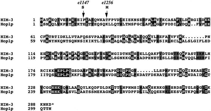
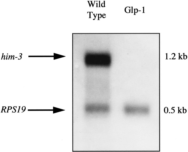
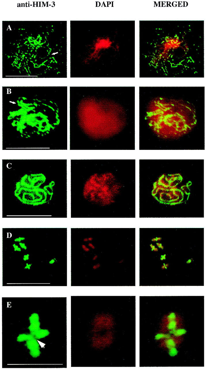
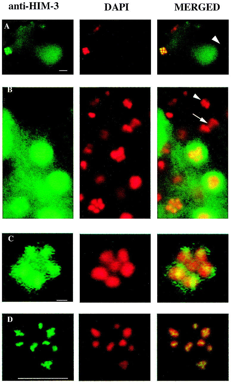
Meiotic chromosomes are organized about a proteinaceous core that forms between replicated sister chromatids. We have isolated a Caenorhabditis elegans gene, him-3, which encodes a meiosis-specific component of chromosome cores with some similarity to the yeast lateral element protein Hop1p. Antibodies raised against HIM-3 localize the protein to condensing chromosomes in early prophase I and to the cores of both synapsed and desynapsed chromosomes. In RNA interference experiments, chromosomes appear to condense normally in the absence of detectable protein but fail to synapse and form chiasmata, indicating that HIM-3 is essential for these processes. Hypomorphs of him-3, although being synapsis proficient, show severe reductions in the frequency of crossing-over, demonstrating that HIM-3 has a role in establishing normal levels of interhomolog exchange. Him-3 mutants also show defects in meiotic chromosome segregation and the persistence of the protein at the chromosome core until the metaphase I-anaphase I transition suggests that HIM-3 may play a role in sister chromatid cohesion. The analysis of him-3 provides the first functional description of a chromosome core component in a multicellular organism and suggests that a mechanistic link exists between the early meiotic events of synapsis and recombination, and later events such as segregation.


Figures

Figure 1
Figure 1
The predicted amino acid sequence of the him-3 cDNA (GenBank accession no. AF172351) in an alignment with the first 302 amino acids of Hop1p (Hollingsworth et al. 1990), showing identical amino acids in black boxes and conserved amino acids in gray boxes. The positions of the missense mutations associated with the e1147 (amino acid 16) and e1256 (amino acid 24) are shown by arrows (see text for details).
Figure 2
Figure 2
him-3 expression is germ line specific. Poly(A)+ RNA was isolated from wild-type and Glp-1 hermaphrodites, a Northern blot was prepared, and it was probed with digoxigenin-labeled him-3 RNA. glp-1(q224ts) homozygotes shifted to the restrictive temperature are defective in the signal required for germ-line proliferation and produce only six to eight germ cells (Austin and Kimble 1987). The him-3 mRNA is detectable only in wild type, which possess about 1500 germ cells as adults. The transcript of the ribosomal protein gene RPS19 was used as a control for the amount of mRNA loaded in each lane.
Figure 3
Figure 3
DAPI staining of RNAi nuclei shows that him-3 is essential for synapsis and chiasma formation. (A) Oocyte nuclei at the pachytene stage from wild-type hermaphrodites showing synapsed homologous chromosomes. (B) Nuclei from the pachytene zone of the germ line of hermaphrodites injected with him-3 RNA. Arrowheads indicate examples of regions of unsynapsed chromosomes. (C) The six bivalents observed in wild-type oocyte diakinesis representing 12 chromosomes joined by chiasmata. (D) RNA oocytes at diakinesis showing 12 univalents, indicating an absence of chiasmata. Arrow shows a signal from two overlapping univalents, located on different focal planes. Scale bars, 1 μm.
Figure 4
Figure 4
Expression of HIM-3 within the germ lines is meiosis specific. Merged images of microdissected gonad arms and an L3 larva stained with anti-HIM-3 (green) and DAPI (red). (A) Localization of HIM-3 during oogenesis. The adult hermaphrodite germ line has five distinct zones of differentiation (see text for details). Oocytes pause at diakinesis of prophase I in the proximal region of the gonad arm before ovulation. (B) Localization of HIM-3 during male spermatogenesis. The first four zones of differentiation are similar to those observed in adult hermaphrodites; however, the nuclei progress through the meiotic divisions. (C) Localization of HIM-3 during hermaphrodite spermatogenesis. An L3 larva showing HIM-3 expression only in pachytene nuclei (arrows). HIM-3 staining is not detected in the germ line nuclei undergoing mitotic divisions or in the somatic nuclei of the gonad (large arrowhead in A) or in mature sperm (large arrowhead in B). Scale bar, 10 μm.
Figure 5
Figure 5
HIM-3 associates with the chromosome core. (A–C) Immunofluorescence micrographs of squashed meiotic prophase chromosomes assembling lateral elements and SC stained with anti-HIM-3. (A) Arrow shows the formation of a stretch of lateral element early in prophase that becomes contiguous in B as the chromosomes continue to condense. The diffuse appearance of the DAPI staining is probably the result of a variation in the fixation time. (C) HIM-3 colocalizes with the synaptonemal complex of the pachytene chromosomes. Scale bars, 10 μm. (D–E) Chromosomes at diakinesis stained with anti-HIM-3 showing that HIM-3 remains at the chromosome core after desynapsis. Scale bar, 10 μm. (E) A magnification of a single bivalent where HIM-3 is associated with the chromosome core and is not present at the junction of the bivalent occupied by the chiasma (arrowhead). Scale bar, 1 μm.
Figure 6
Figure 6
HIM-3 remains associated with the chromosome core until the metaphase I–anaphase I transition. Immunofluorescence micrographs of oocyte nuclei at metaphase and spermatocyte nuclei undergoing the meiotic divisions stained with anti-HIM-3. (A) Fertilized embryo showing that HIM-3 stains the oocyte metaphase I nucleus (left) but not the sperm nucleus that has completed meiosis (right, arrowhead). Scale bar, 10 μm. (B) Spermatocyte nuclei of males undergoing meiotic divisions. The X chromosome is centrally located in a ring formed by the autosomal bivalents at metaphase I (Albertson and Thomson 1993) in the lower left corner. The single arrow shows the lagging X chromosome, which moves to one pole at anaphase I (Albertson and Thomson 1993). HIM-3 is associated with the cores of metaphase I chromosomes but abruptly dissociates by anaphase I and is not detectable on any of the chromosomes at this time or at the second meiotic division (arrowhead). Other nuclei at earlier stages stain with anti-HIM-3, indicating that the gonad is permeable to the antibody. Scale bar, 10 μm. (C) A magnification of the metaphase I nucleus in B showing that HIM-3 is associated with the ring of five autosomal bivalents, but is not detectable above background levels on the single X chromosome. Scale bar, 1 μm. (D) Oocyte of him-3(e1256) showing that HIM-3 associates with the cores of univalents. Scale bar, 10 μm.

References

    1. Albertson DG, Thomson JN. Segregation of holocentric chromosomes at meiosis in the nematode, Caenorhabditis elegans. Chromosome Res. 1993;1:15–26. - PubMed
    1. Albertson DG, Rose AM, Villeneuve AM. Chromosome organization, mitosis, and meiosis. In: Riddle DL, Blumenthal T, Meyer BJ, Priess JR, editors. C. elegans II. Cold Spring Harbor, NY: Cold Spring Harbor Laboratory Press; 1997. pp. 47–78. - PubMed
    1. Altschul SF, Gish W, Miller W, Myers EW, Lipman DJ. Basic local alignment search tool. J Mol Biol. 1990;215:403–410. - PubMed
    1. Aravind L, Koonin EV. The HORMA domain: A common structural denominator in mitotic checkpoints, chromosome synapsis and DNA repair. Trends Biochem. 1998;32:284–286. - PubMed
    1. Austin J, Kimble, J J. glp-1 is required in the germ line for regulation of the decision between mitosis and meiosis in Caenorhabditis elegans. Cell. 1987;51:589–599. - PubMed

Publication types

Substances

Associated data

LinkOut - more resources