. 1999 Sep 1;13(17):2271-83.
doi: 10.1101/gad.13.17.2271.

Fission yeast condensin complex: essential roles of non-SMC subunits for condensation and Cdc2 phosphorylation of Cut3/SMC4

Affiliations

Fission yeast condensin complex: essential roles of non-SMC subunits for condensation and Cdc2 phosphorylation of Cut3/SMC4

T Sutani et al. Genes Dev. .

Abstract

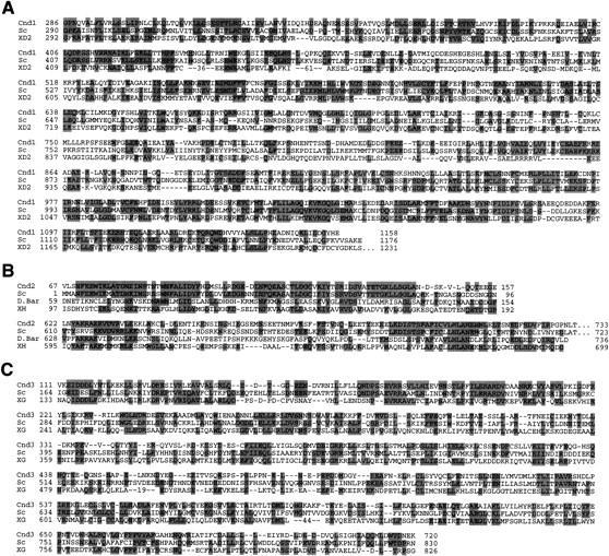
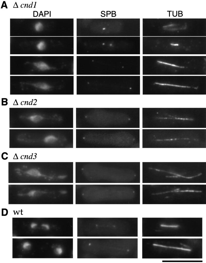
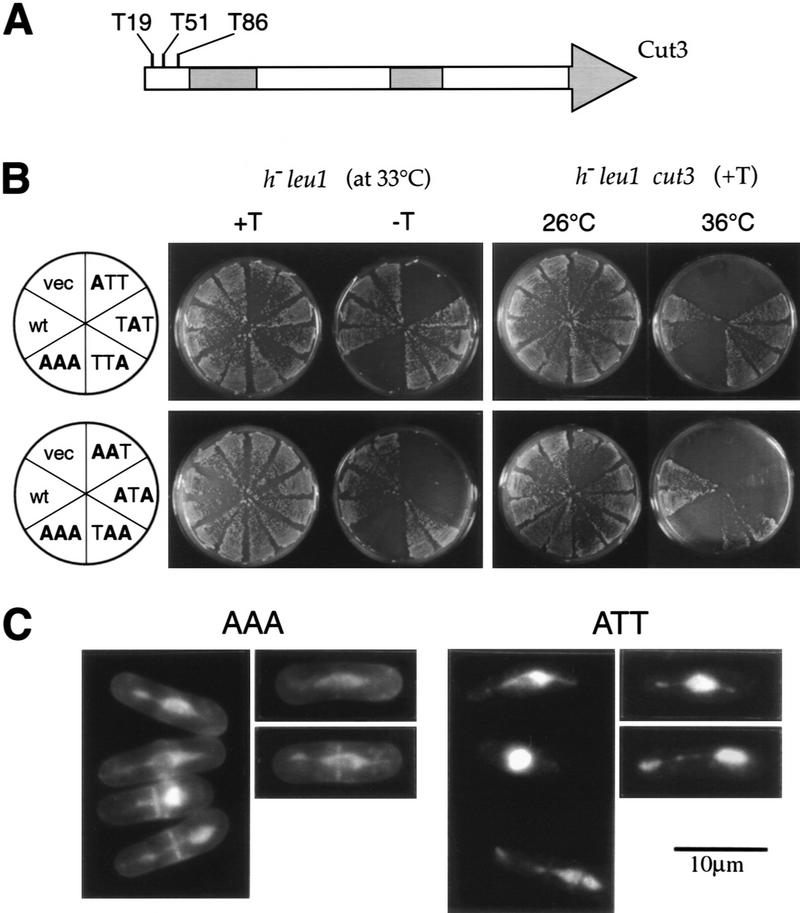
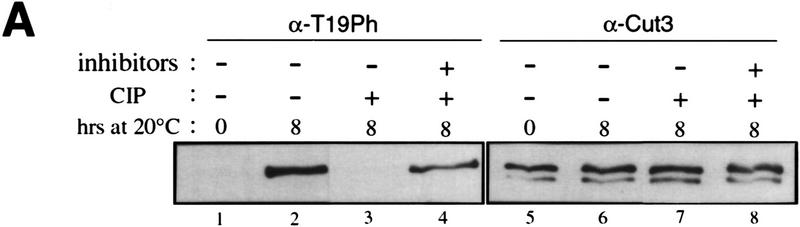
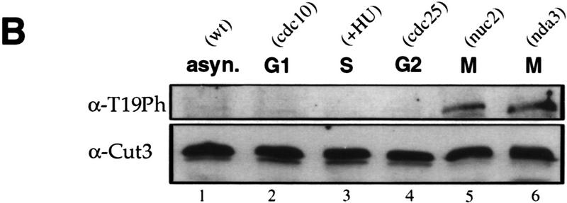
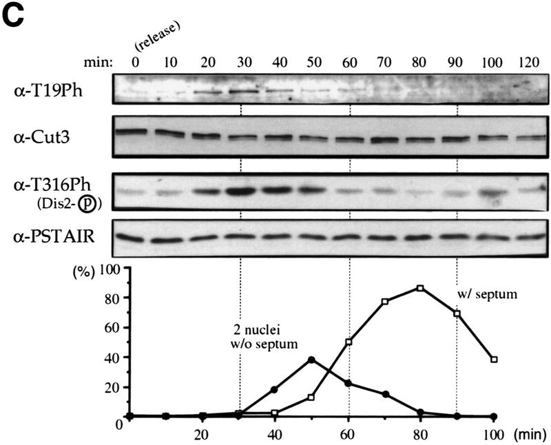
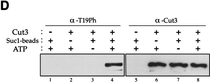
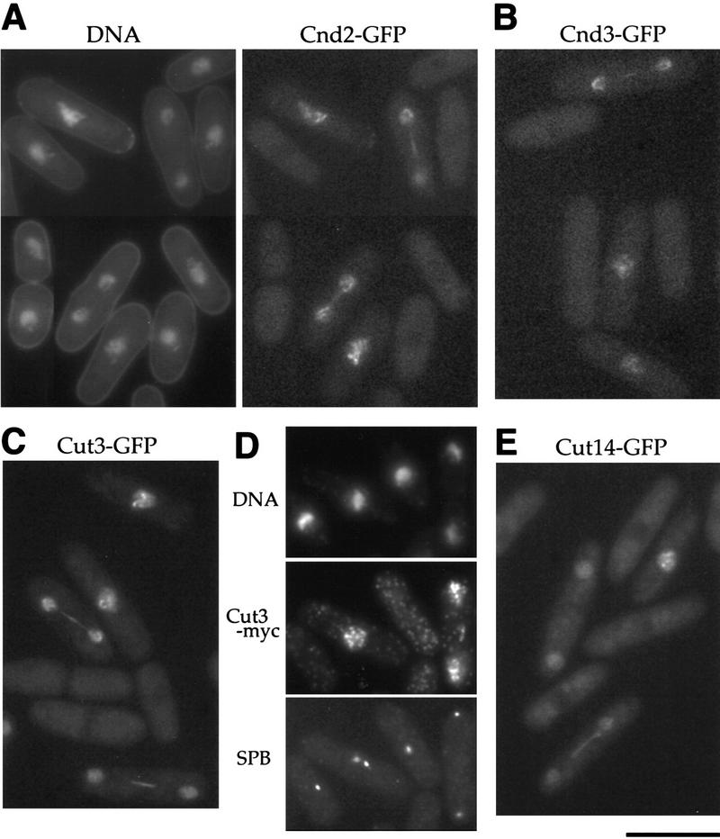
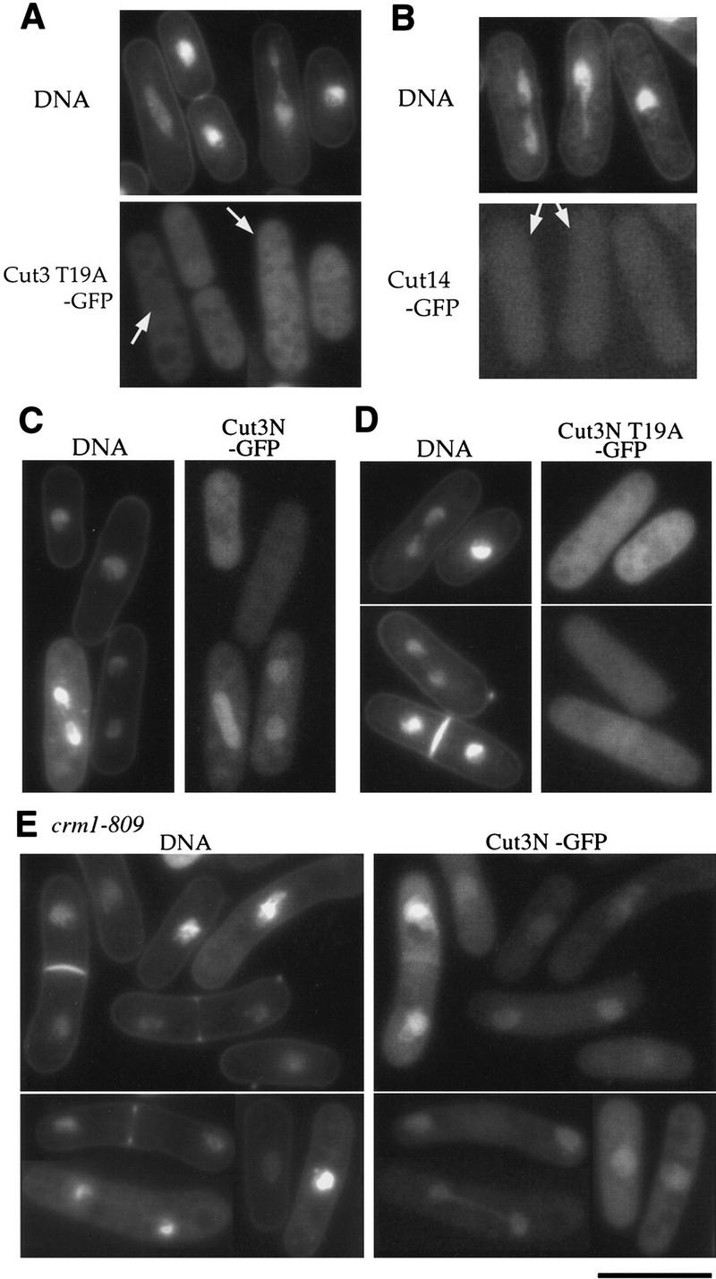
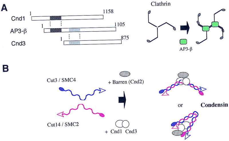
The condensin complex in frog extracts, containing two SMC (structural maintenance of chromosomes) and three non-SMC subunits, promotes mitotic chromosome condensation, and its supercoiling activity increases during mitosis by Cdc2 phosphorylation. Here, we report that fission yeast has the same five-member condensin complex, each of which is essential for mitotic condensation. The condensin complex was purified and the subunits were identified by microsequencing. Cnd1, Cnd2, and Cnd3, three non-SMC subunits showing a high degree of sequence conservation to frog subunits, are essential for viability, and their gene disruption leads to a phenotype indistinguishable from that observed in cut3-477 and cut14-208, known mutations in SMC4 and SMC2-like subunits. Condensin subunits tagged with GFP were observed to alter dramatically their localization during the cell cycle, enriched in the nucleus during mitosis, but cytoplasmic during other stages. This stage-specific alteration in localization requires mitosis-specific phosphorylation of the T19 Cdc2 site in Cut3. The T19 site is phosphorylated in vitro by Cdc2 kinase and shows the maximal phosphorylation in metaphase in vivo. Its alanine substitution mutant fails to suppress the temperature-sensitive phenotype of cut3-477, and shows deficiency in condensation, probably because Cut3 T19A remains cytoplasmic. Therefore, direct Cdc2 phosphorylation of fission yeast condensin may facilitate its nuclear accumulation during mitosis.


Figures

Figure 1
Figure 1
Isolation of condensin subunits. (A) 3× HA+6× His-tagged cut3+ gene was integrated into the chromosome as schematized. Plasmid carrying the carboxy-terminal portion of the tagged cut3+ and the ura4+ marker was introduced into a cut3-477 mutant, which was Ts/Ura. The mutation site was located near the carboxyl-terminus (indicated by ×). Ts+/Ura+ integrants were thus obtained. The same strategy was applied for the Cut14–3HAH6 gene. (B) Immunoblotting of Cut3–3HAH6 (lane 1) and Cut14–3HAH6 (lane 2) integrants, and wild-type 972 (lane 3) was done using anti-HA antibodies. Bands at the expected molecular mass positions were obtained. (C) Silver-stained immunoprecipitation patterns of wild type 972 (lane 1) and the integrant Cut3-3HAH6 by anti-HA antibody. (D) Silver-stained immunoprecipitation patterns of wild-type 972, as well as the integrants of Cut3–3HAH6 and Cut14–3HAH6, by anti-HA antibodies. Assignments of individual bands are shown. The bands indicated by asterisks are not present in the 972 wild-type strain (see text). Cut3* is the amino-terminally cleaved form.
Figure 1
Figure 1
Isolation of condensin subunits. (A) 3× HA+6× His-tagged cut3+ gene was integrated into the chromosome as schematized. Plasmid carrying the carboxy-terminal portion of the tagged cut3+ and the ura4+ marker was introduced into a cut3-477 mutant, which was Ts/Ura. The mutation site was located near the carboxyl-terminus (indicated by ×). Ts+/Ura+ integrants were thus obtained. The same strategy was applied for the Cut14–3HAH6 gene. (B) Immunoblotting of Cut3–3HAH6 (lane 1) and Cut14–3HAH6 (lane 2) integrants, and wild-type 972 (lane 3) was done using anti-HA antibodies. Bands at the expected molecular mass positions were obtained. (C) Silver-stained immunoprecipitation patterns of wild type 972 (lane 1) and the integrant Cut3-3HAH6 by anti-HA antibody. (D) Silver-stained immunoprecipitation patterns of wild-type 972, as well as the integrants of Cut3–3HAH6 and Cut14–3HAH6, by anti-HA antibodies. Assignments of individual bands are shown. The bands indicated by asterisks are not present in the 972 wild-type strain (see text). Cut3* is the amino-terminally cleaved form.
Figure 2
Figure 2
Isolation of the cnd1+, cnd2+, and cnd3+ genes. Partial sequences of the cloned genes are shown. (A) Three amino acid sequences (S. pombe Cnd1, Cnd1; S. cerevisiae YLR272c, Sc; frog XCAP-D2, XD2) are aligned. Identical amino acids are boxed. (B) Amino acid sequence comparison (S. pombe, Cnd2, Cnd2; S. cerevisiae YBL097w, Sc; Drosophila Barren, D. Bar; frog XCAP-H, XH). (C) Amino acid sequence comparison (S. pombe, Cnd3, Cnd3; S. cerevisiae YDR325w, Sc; frog XCAP-G, XG).
Figure 3
Figure 3
Gene disruption phenotypes of cnd1+, cnd2+, and cnd3+. Gene-disrupted cells were observed after germination by DAPI for DNA, SPB, and TUB antibodies. Mitotic cells were distinguished by the presence of the spindle and the two SPBs. (A) Gene disruption of cnd1+; (B) gene disruption of cnd2+; (C) gene disruption of cnd3+. Chromosome condensation was defective and the spindle was elongated. (D) Wild-type mitotic cells. Bar, 10 μm.
Figure 4
Figure 4
Dissection of putative Cdc2 kinase sites in Cut3. (A) Cut3 has three Cdc2 kinase sites in the amino terminus. The shaded areas represent homologous regions among SMC proteins: the amino-terminal ATP binding, the central hinge, and the carboxy-terminal DA box. (B) Plasmid carrying the wild-type cut3+ (wt) or one of the substitution mutant genes (see text) placed under the inducible promoter REP81 were introduced into the wild type (h leu1): resulting transformants were plated at 33°C in the presence of thiamine (+T, the promoter repressed) or the absence of thiamine (−T, the promoter induced). The same plasmids were introduced into cut3–477 mutant and plated at 26°C and 36°C under the repressed promoter condition (+T). (C) Wild-type cells overexpressing triple AAA or single ATT substitution mutant protein under the REP81 promoter were stained by DAPI. Phenotypes were indistinguishable from those of cut3 or cut14 mutant. Bar, 10 μm.
Figure 5
Figure 5
Phosphorylation at T19 identified by phosphopeptide antibodies. (A) Cells of cold-sensitive nda3–KM311 containing the integrated Cut3–3HAH6 gene were grown at 33°C (0 hr) and then arrested in mitosis by shifting to 20°C for 8 hr. Immunoprecipitation was done for extracts by anti-HA antibodies. Precipitates were immunoblotted using phosphopeptide antibodies (α-T19Ph) raised against chemically synthesized phosphopeptide PSIVDVT(PO3H2) PDRGER or polyclonal anti-Cut3 antibodies (α-Cut3). A band corresponding to the full-length Cut3 position recognized by α-T19Ph was obtained only in mitotically arrested cells. α-Cut3 detected the full-length and amino-terminally cleaved forms. Immunoprecipitates prepared above were phosphatase treated (CIP, +) in the presence or the absence of phosphatase inhibitors. The band detected by α-T19Ph disappeared after phosphatase treatment; the band recognized by α-Cut3 remained at the same intensity. (B) Immunoblotting of extracts prepared from exponentially growing wild-type (asyn. wt), G1-arrested cdc10 mutant, HU-treated wild type blocked in S phase, G2-arrested cdc25 mutant, and M-phase-arrested nuc2 and nda3 mutant cells using α-T19Ph and α-Cut3 antibodies. Only M-phase-arrested cell extracts showed the T19-phosphorylated Cut3 band. (C) Synchronous culture by block and release of cdc25 mutant. G2-arrested cdc25–22 cells cultured at 36°C for 4.25 hr were released to 26°C, and cells were collected every 10 min. Extracts were immunoblotted using α-T19Ph, α-Cut3, α-T316Ph (against phosphorylated Dis2 at T316, Ishii et al. 1996) and α-PSTAIR (against Cdc2). The frequencies of cells with the septum and containing two nuclei without the septum were also measured. T19 phosphorylation peaked at metaphase. (D) Purified Cut3 protein (Sutani and Yanagida 1997) was incubated with Cdc2 kinase bound to Suc1 beads in the presence or absence of ATP, and detected by α-T19Ph or α-Cut3 antibodies. Cut3 was in vitro phosphorylated by Cdc2 kinase as detected by α-T19Ph.
Figure 5
Figure 5
Phosphorylation at T19 identified by phosphopeptide antibodies. (A) Cells of cold-sensitive nda3–KM311 containing the integrated Cut3–3HAH6 gene were grown at 33°C (0 hr) and then arrested in mitosis by shifting to 20°C for 8 hr. Immunoprecipitation was done for extracts by anti-HA antibodies. Precipitates were immunoblotted using phosphopeptide antibodies (α-T19Ph) raised against chemically synthesized phosphopeptide PSIVDVT(PO3H2) PDRGER or polyclonal anti-Cut3 antibodies (α-Cut3). A band corresponding to the full-length Cut3 position recognized by α-T19Ph was obtained only in mitotically arrested cells. α-Cut3 detected the full-length and amino-terminally cleaved forms. Immunoprecipitates prepared above were phosphatase treated (CIP, +) in the presence or the absence of phosphatase inhibitors. The band detected by α-T19Ph disappeared after phosphatase treatment; the band recognized by α-Cut3 remained at the same intensity. (B) Immunoblotting of extracts prepared from exponentially growing wild-type (asyn. wt), G1-arrested cdc10 mutant, HU-treated wild type blocked in S phase, G2-arrested cdc25 mutant, and M-phase-arrested nuc2 and nda3 mutant cells using α-T19Ph and α-Cut3 antibodies. Only M-phase-arrested cell extracts showed the T19-phosphorylated Cut3 band. (C) Synchronous culture by block and release of cdc25 mutant. G2-arrested cdc25–22 cells cultured at 36°C for 4.25 hr were released to 26°C, and cells were collected every 10 min. Extracts were immunoblotted using α-T19Ph, α-Cut3, α-T316Ph (against phosphorylated Dis2 at T316, Ishii et al. 1996) and α-PSTAIR (against Cdc2). The frequencies of cells with the septum and containing two nuclei without the septum were also measured. T19 phosphorylation peaked at metaphase. (D) Purified Cut3 protein (Sutani and Yanagida 1997) was incubated with Cdc2 kinase bound to Suc1 beads in the presence or absence of ATP, and detected by α-T19Ph or α-Cut3 antibodies. Cut3 was in vitro phosphorylated by Cdc2 kinase as detected by α-T19Ph.
Figure 6
Figure 6
Cell cycle stage-specific nuclear location of condensin subunits. (A) Living cells expressing integrated Cnd2-GFP (right). The nuclear signal was found in mitotic cells from early mitosis to late anaphase. DNA was stained by Hoechst 33342 (left). (B) cnd3-null cells containing plasmid with the cnd3+–GFP gene under the repressed REP81 promoter in the presence of thiamine. (C) Living wild-type cells expressing Cut3–GFP under the native promoter. (D) Fixed wild-type cells expressing integrated Cut3–Myc. DNA and SPB were stained by DAPI and SPB antibodies, respectively. Anti-myc antibodies were used to detect Cut3–Myc. (E) Living wild type cells expressing integrated Cut14–GFP. Bar, 10 μm.
Figure 7
Figure 7
Cell cycle stage-specific nuclear accumulation depends on T19 of Cut3 and Crm1. (A) Living wild-type cells expressing T19A Cut3 under the REP81 promoter in the absence of thiamine. Cut3 T19A was cytoplasmic. DNA was stained by Hoechst 33342 in A–E. (B) Localization of Cut14–GFP in cells overproducing T19A Cut3 mutant protein under the REP81 promoter. Cut14–GFP was cytoplasmic. (C) The amino-terminal fragment of Cut3 tagged with GFP was expressed in wild-type cells. The nuclear signal was found in mitotic cells. (D) The amino-terminal fragment of Cut3 substituted with alanine at T19 and tagged with GFP was expressed in wild-type cells. GFP signal was cytoplasmic. (E) The amino-terminal fragment of Cut3 tagged with GFP was expressed in crm1-809 cultured at the restrictive temperature (20°C) for 6 hr. Crm1 is required for nuclear export. GFP signal accumulated in both interphase and mitotic nuclei. Bar, 10 μm.
Figure 8
Figure 8
A hypothesis for condensin assembly. (A) Weak homology exists between AP3-β and Cnd1 or Cnd3 (hatched). AP3-β assists in the assembly of clathrin (Schmid 1997), which contains rod and hinge regions. (B) Cnd1 and Cnd3 might assist in the assembly of Cut3, Cut14, and Cnd2 proteins into the five-member condensin complex.

References

    1. Adachi Y, Luke M, Laemmli UK. Chromosome assembly in vitro: Topoisomerase II is required for condensation. Cell. 1991;64:137–148. - PubMed
    1. Altschul SF, Madden TL, Schaffer AA, Zhang J, Zhang Z, Miller W, Lipman DJ. Gapped BLAST and PSI-BLAST: A new generation of protein database search programs. Nucleic Acids Res. 1997;25:3389–3402. - PMC - PubMed
    1. Bak AL, Zeuthen J, Crick FH. Higher-order structure of human mitotic chromosomes. Proc Natl Acad Sci. 1977;74:1595–1599. - PMC - PubMed
    1. Basi G, Schmid E, Maundrell K. TATA box mutations in the Schizosaccharomyces pombe nmt1 promoter affect transcription efficiency but not the transcription start point or thiamine repressibility. Gene. 1993;123:131–136. - PubMed
    1. Bhat MA, Philp AV, Glover DM, Bellen HJ. Chromatid segregation at anaphase requires the barren product, a novel chromosome-associated protein that interacts with Topoisomerase II. Cell. 1996;87:1103–1114. - PubMed

Publication types

MeSH terms

Associated data